

 PDF
PDF ePub
ePub Citation
 Print
Print


Abstract
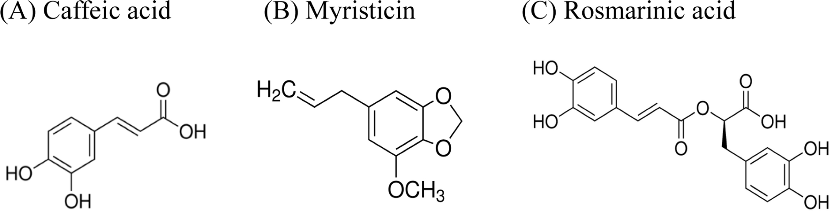
Perilla frutescens was empirically used for controlling airway inflammatory diseases in folk medicine. We investigated whether caffeic acid, myristicin and rosemarinic acid derived from Perilla frutescens significantly affect the gene expression and production of mucin from airway epithelial cells. Confluent NCI-H292 cells were pretreated with caffeic acid, myristicin or rosemarinic acid for 30 min and then stimulated with phorbol 12-myristate 13-acetate (PMA) for 24 h. The MUC5AC mucin gene expression and production were measured by reverse transcription – polymerase chain reaction (RT-PCR) and enzyme-linked immunosorbent assay (ELISA), respectively. Additionally, we examined whether caffeic acid, myristicin or rosemarinic acid affects MUC5AC mucin production indued by epidermal growth factor (EGF) and tumor necrosis factor-α (TNF-α), the other two stimulators of production of airway mucin. The results were as follows: (1) Caffeic acid, myristicin and rosemarinic acid inhibited the gene expression and production of MUC5AC mucin induced by PMA from NCI-H292 cells, respectively; (2) Among the three compounds derived from Perilla frutescens, only rosemarinic acid inhibited the production of MUC5AC mucin induced by EGF or TNF-α, the other two stimulators of production of airway mucin. These results suggest that rosemarinic acid derived from Perilla frutescens can regulate the production and gene expression of mucin, by directly acting on airway epithelial cells and, at least in part, explains the traditional use of Perilla frutescens as remedies for diverse inflammatory pulmonary diseases.
Go to : 
References
(1). Voynow J.A.; Rubin, B.K. Chest. 2009. 135:505–512.
(2). Heo H. J.., Kim C.., Lee H. J.., Kim Y. S.., Kang S. S.., Seo U. K.., Kim Y. H.., Park Y. C.., Seok J. H.., Lee C. J.Phytother. Res. 2007. 21:462–465.
(3). Heo H. J.., Lee S. Y.., Lee M. N.., Lee H. J.., Seok J. H.., Lee C. J.Phytother. Res. 2009. 23:1458–1461.
(4). Lee H. J.., Lee S. Y.., Lee M. N.., Kim J. H.., Chang G.T.., Seok J. H.., Lee C. J.Phytother. Res. 2011. 25:1196–1200.
(5). Jang I. M.Treatise on asian herbal medicines; Haksul-pyunsu-kwan in Research institute of natural products of Seoul National University: Korea,. 2003. , p. 2847.
(6). Rocha J.., Eduardo-Figueira M.., Barateiro A.., Fernandes A.., Brites D.., Bronze R.., Duarte C. M.., Serra A. T.., Pinto R.., Freitas M.., Fernandes E.., Silva-Lima B.., Mota-Filipe H.., Sepodes B.Basic Clin. Pharmacol. Toxicol. 2015. 116:398–413.
(7). Lee J. Y.., Park W.Molecules. 2011. 16:7132–7142.
(8). Genaro-Mattos T. C.., Maurício Â. Q.., Rettori D.., Alonso A.., Hermes-Lima M.PLoS One. 2015. 10:e0129963.
(9). Coelho V. R.., Vieira C. G.., de Souza L. P.., Moysés F.., Basso C.., Papke D. K.., Pires T. R.., Siqueira I. R.., Picada J. N.., Pereira P.Life Sci. 2015. 122:65–71.
(10). Rogers D. F.., Barnes P. J.Ann. Med. 2006. 38:116–125.
(11). Li J. D.., Dohrman A. F.., Gallup M.., Miyata S.., Gum J. R.., Kim Y. S.., Nadel J. A.., Prince A.., Basbaum C. B.Proc. Natl. Acad. Sci. USA. 1997. 94:967–972.
(12). Takeyama K.., Dabbagh K.., Shim J. J.., Dao-Pick T.., Ueki I. F.., Nadel J. A. J.Immunol. 2000. 164:1546–1552.
(13). Shao M. X.., Ueki I. F.., Nadel J. A.Proc. Natl. Acad. Sci. USA. 2003. 100:11618–11623.
(14). Hong D. H.., Petrovics G.., Anderson W. B.., Forstner J.., Forstner G.Am. J. Physiol. 1999. 277:G1041–G1047. . Am. J. Physiol. 1999 277.
(15). Hewson C. A.., Edbrooke M. R.., Johnston S. L. J.Mol. Biol. 2004. 344:683–695.
(16). Park S. J.., Kang S. Y.., Kim N. S.., Kim H. M.Immunopharmacol. Immunotoxicol. 2002. 24:211–226.
(17). Kim K. D.., Lee H. J.., Lim S. P.., Sikder A.., Lee S. Y.., Lee C. J.Phytother. Res. 2012. 26:1301–1307.
(18). Fischer B. M.., Rochelle L. G.., Voynow J. A.., Akley N. J.., Adler K. B.Am. J. Respir. Cell Mol. Biol. 1999. 20:413–422.
(19). Song K. S.., Lee W. J.., Chung K. C.., Koo J. S.., Yang E. J.., Choi J. Y.., Yoon J. H. J.Biol. Chem. 2003. 278:23243–23250.
(20). Takeyama K.., Dabbagh K.., Lee H. M.., Agustí C.., Lausier J. A.., Ueki I. F.., Grattan K. M.., Nadel J. A.Proc. Natl. Acad. Sci. USA. 1999. 96:3081–3086.
(21). Cohn L.., Whittaker L.., Niu N.., Homer R. J.Novartis. Found. Symp. 2002. 248:201–213.
Go to :
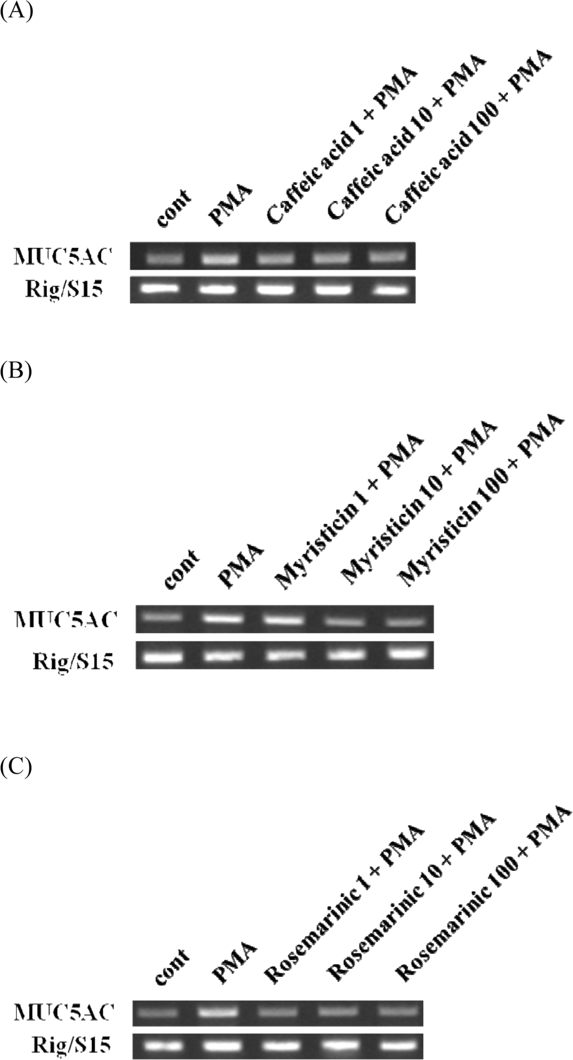 | Fig. 2.Effect of caffeic acid, myristicin or rosemarinic acid on PMA-induced MUC5AC gene expression from NCI-H292 cells. NCI-H292 cells were pretreated with varying concentrations of caffeic acid, myristicin or rosemarinic acid for 30 min and then stimulated with PMA (10 ng/mL) for 24 h. MUC5AC gene expression was measured by RT-PCR. Three independent experiments were performed and the representative images were shown. (cont: control, concentration unit is µM.). |
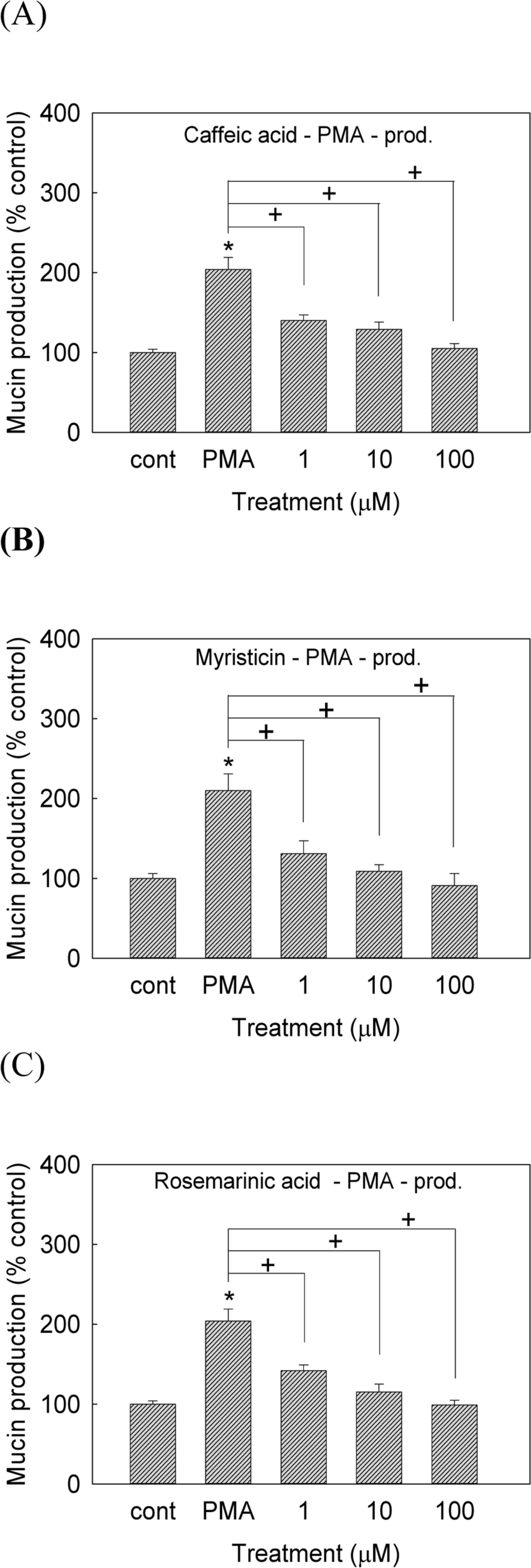 | Fig. 3.Effect of caffeic acid, myristicin or rosemarinic acid on PMA-induced MUC5AC mucin production from NCI-H292 cells. NCI-H292 cells were pretreated with varying concentrations of caffeic acid, myristicin or rosemarinic acid for 30 min and then stimulated with PMA (10 ng/mL) for 24 h. Cell lysates were collected for measurement of MUC5AC mucin production by ELISA. Each bar represents a mean ± S.E.M. of 3 culture wells in comparison with that of control set at 100% (A, B, C). Three independent experiments were performed and the representative data were shown. ∗significantly different from control (p < 0.05). +significantly different from PMA alone (p < 0.05). (cont: control, concentration unit is µM.) |
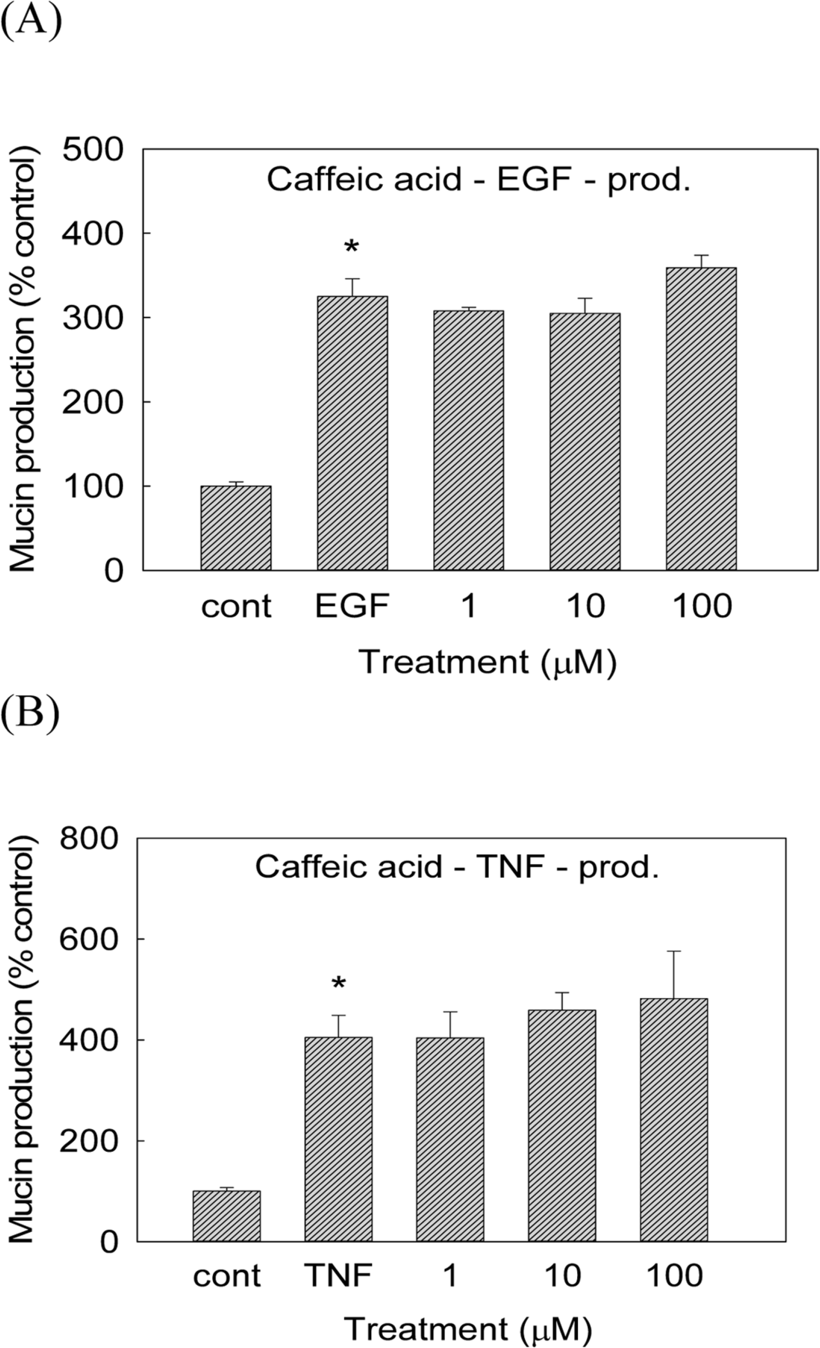 | Fig. 4.Effect of caffeic acid on EGF- or TNF-α-induced MUC5AC mucin production from NCI-H292 cells. NCI-H292 cells were pretreated with varying concentrations of caffeic acid for 30 min and then stimulated with EGF (25 ng/mL) or TNF-α (0.2 nM, 10 ng/mL) for 24 h. Cell lysates were collected for measurement of MUC5AC mucin production by ELISA. Each bar represents a mean ± S.E.M. of 3 culture wells in comparison with that of control set at 100% (A, B). Three independent experiments were performed and the representative data were shown. ∗significantly different from control (p < 0.05). (cont: control, concentration unit is µM.) |
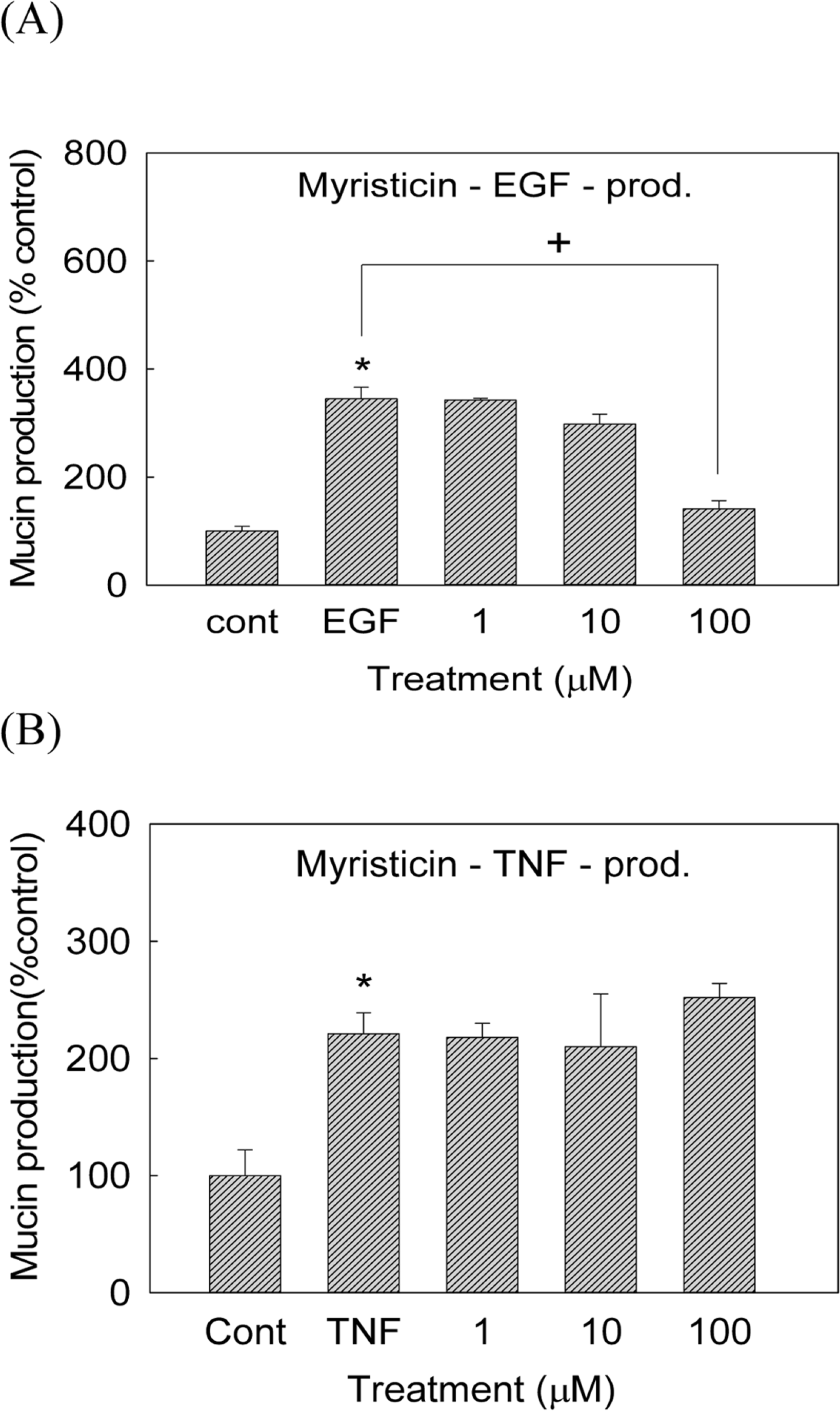 | Fig. 5.Effect of myristicin on EGF- or TNF-α-induced MUC5AC mucin production from NCI-H292 cells. NCI-H292 cells were pretreated with varying concentrations of myristicin for 30 min and then stimulated with EGF (25 ng/mL) or TNF-α (0.2 nM, 10 ng/mL) for 24 h. Cell lysates were collected for measurement of MUC5AC mucin production by ELISA. Each bar represents a mean ± S.E.M. of 3 culture wells in comparison with that of control set at 100% (A, B). Three independent experiments were performed and the representative data were shown. ∗significantly different from control (p < 0.05).+ significantly different from EGF alone (p < 0.05).(cont: control, concentration unit is µM.) |
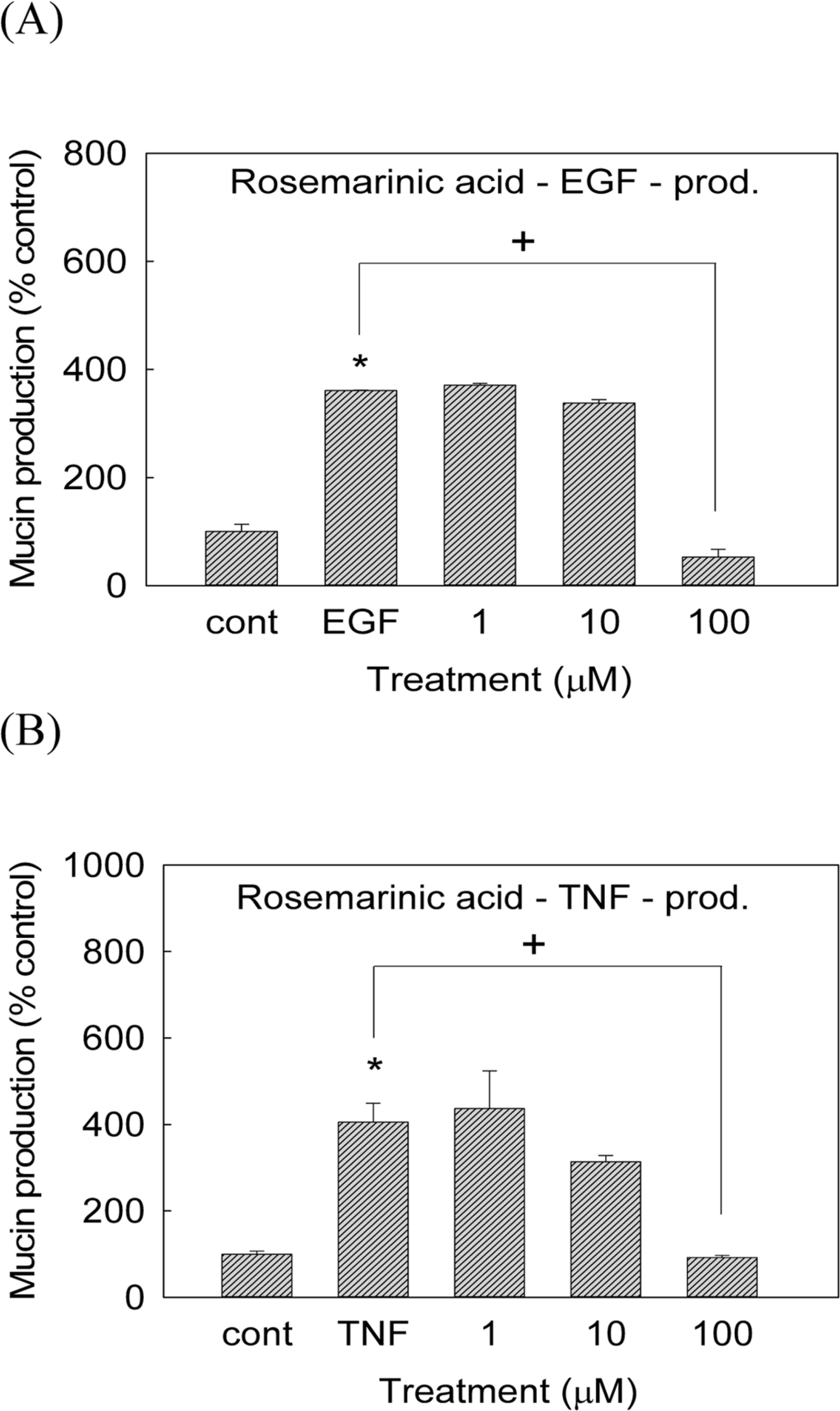 | Fig. 6.Effect of rosemarinic acid on EGF- or TNF-α-induced MUC5AC mucin production from NCI-H292 cells. NCI-H292 cells were pretreated with varying concentrations of rosemarinic acid for 30 min and then stimulated with EGF (25 ng/mL) or TNF-α (0.2 nM, 10 ng/mL) for 24 h. Cell lysates were collected for measurement of MUC5AC mucin production by ELISA. Each bar represents a mean ± S.E.M. of 3 culture wells in comparison with that of control set at 100% (A, B). Three independent experiments were performed and the representative data were shown. ∗significantly different from control (p < 0.05). +significantly different from EGF or TNF-α alone (p<0.05). (cont: control, concentration unit is µM.) |
 XML Download