

 PDF
PDF ePub
ePub Citation
 Print
Print


Inferior alveolar nerve block anesthesia (IANBA) is commonly used in dental anesthesia while performing minor mandibular surgeries, such as third molar extraction. It is also essential in dental conservative treatments, including endodontic treatment. However, the widespread use of IANBA in dental treatment has resulted in various complications. It may cause diplopia, facial paralysis, and other complications when injected into blood vessels [123]. This report documents a case of facial blanching symptoms occurring immediately after IANBA.
CASE REPORT
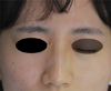
A 25-year-old woman visited our hospital for left mandibular third molar extraction. She had no notable medical history. Before surgery, she complained of gastrointestinal discomfort after taking prophylactic antibiotics and anti-inflammatory drugs, but her symptoms had improved. To extract the third molar, IANBA was performed on the left side. For local anesthesia, 1.8 cm3 of lidocaine with epinephrine in a 1:80000 ratio (Huons Lidocaine HCL and Epinephrine Inj. [1:80,000], Huons co. LTD, Sungnam, Republic of Korea) was used. Immediately after anesthetic injection in the left mandibular foramen area, the left side of the patient's face turned pale white, and she complained of pain and discomfort in the left facial area (Fig. 1). Aspiration was not performed during injection.
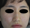
Thereafter, she complained of pain and discomfort extending from the middle left facial region to the left orbital region, including the eye. After evaluating facial nerve function, we confirmed that the symptoms were not related to any complication caused by anesthetic injection into the facial nerve. Although the patient chiefly complained of eye discomfort, she showed no signs such as the inability to close the eye or weakness of the frontalis associated with facial nerve paralysis (Fig. 2). The patient was observed in a supine position. Signs indicating blanching were observed from the outside of the nose, upper lip, central facial region, and the left zygomatic area. The patient complained of discomfort accompanying pain in the region with blanching, especially around the eye. The symptoms had occurred immediately after anesthetic injection and became severe within 1–2 min. The symptoms remained stable, but they improved after more than 10 min (Fig. 3). The patient's pain symptoms also improved, but the ischemic sign in the left facial region did not improve completely, especially in the lateral nasal area.
As the patient did not complain of severe symptoms and showed improvement, her current condition was explained to her, and surgical extraction of the left mandibular third molar was performed. After extraction, her progress was monitored until her symptoms completely improved approximately 40 min after the initial facial blanching sign. She was informed again that it was a complication of anesthesia. We decided to conduct additional examinations and treatment if the symptoms reappeared after discharge. No notable additional symptoms were observed.
DISCUSSION
This report documents a case of facial blanching that occurred immediately after IANBA was performed. As a primary consideration to reduce complications, an anesthetic must be injected into the mandibular foramen region and not into other anatomically important structures such as blood vessels. Therefore, knowing the location of the mandibular foramen is important. Studies on the mandibles of cadavers and three-dimensional analyses using computed tomography (CT) data have been performed to determine the location of the mandibular foramen [45].
Kang et al. conducted a three-dimensional analysis using CT data to determine the location of the mandibular foramen during IANBA [5]. During IANBA, the insertion angle of the injection needle and the distance from the premolar or molar on the opposite side of anesthesia to the mandibular foramen were examined. Knowing the anatomical location may not necessarily reduce complications and guarantee successful IANBA because the shape and size of the mandible as well as the shape and location of the mandibular foramen differ among individuals. However, understanding the typical anatomical structure of the mandible and the anatomical location of the mandibular foramen is critical for IANBA.
In our case, the anesthetic agent evaded the location of the mandibular foramen and entered the maxillary artery, causing contraction of the blood vessels running through the facial region and reducing blood flow, which resulted in temporary blanching in the facial region [67]. Although the amount of anesthetic agent to be used cannot be clearly defined, a minimal amount is desirable for IANBA because of the toxicity of anesthetics and the undesirable side effects of injection. However, because the anesthetic is not directly injected into the nerve during IANBA, additional amount of anesthetic is required. In general, 1.8 cm3 of anesthetic is used for IANBA.
In Kang et al.'s study, the height of the mandibular foramen was 3.8 mm from the mandibular occlusal plane and 22.9 mm from the front of the ascending ramus [5]. When the ascending ramus was used as a reference, the opposite side premolar formed a 38.7° angle with the mandibular foramen and the opposite side molar formed a 43.8° angle. The distance to the mandibular foramen was 82.1 mm from the opposite side premolar and 80.0 mm from the opposite side molar [5].
Delivering an anesthetic to the mandibular foramen is difficult when the injection site is further away from the mandibular foramen; therefore, IANBA can lead to failures and cause complications. When the anesthetic is injected into the inner side at an inaccurate angle, the parotid gland and facial nerve become anesthetized; this can lead to xerophthalmia, which prevents the eyelids from being closed. In such cases, immediate care is needed to prevent dryness of the eyes until the facial nerve paralysis disappears and the eyelids can be moved. In our case, the anesthetic entering the maxillary artery might have caused ischemia of the facial artery and discomfort accompanying facial pain on one side. It may be resulted from the injection further out of angle towards upside and inside from mandibular foramen. In our case, additional IANBA was not needed for third molar extraction; therefore, we thought that the anesthetic was not directly injected into the vessel.
In our patient, the symptoms of discomfort improved after a few minutes, but the facial blanching did not improve completely. The patient chiefly complained of discomfort in the eye. However, no sign associated with facial nerve paralysis was noted. The condition was diagnosed as an IANBA complication caused by the direct injection of epinephrine and anesthetic into the maxillary artery.
Facial blanching after IANBA can be caused by anesthetic injection into the maxillary artery area, affecting the infraorbital artery. Studies have suggested that peripheral vasoconstriction of the facial arterioles supplied by the infraorbital artery occurs because of the effect of the α-receptor agonist [789]. In the current case, facial blanching resulted from the injection of epinephrine and anesthetic into the maxillary artery area.
The blanching signs were observed in the lips, lateral nose, and midface area in the current case. The pain was likely caused by the sudden contraction of blood vessels in the region supplied by the maxillary artery and the subsequent reduction of blood supply. Anesthetics contain epinephrine; it reduces the metabolism of the anesthetic and enhances the anesthetic effect. Infiltration anesthesia to the surgical site can reduce hemorrhage caused by the contraction of blood vessels. Although epinephrine is included in anesthetics for this purpose, epinephrine can act on the maxillary artery and cause complications as in our case; this factor should be considered when using local anesthetics containing epinephrine.
In the current case, aspiration to prevent intravascular injection was not performed. The incidence of intravascular needle entrance during IANBA is relatively high [1011]. Performing aspiration during IANBA injection is important to prevent intravascular needle entrance.
Recently, the use of CT for evaluating the mandible has become widespread. Owing to the popularity of cone-beam CT in dentistry, CT images of the mandible can be reconstructed three-dimensionally. Using three-dimensional images of the mandible can help obtain information such as the shape of the mandible and the location of the mandibular foramen in each patient. However, using three-dimensional images of the mandible is not easy because of the large amounts of soft tissues inside the mouth, such as the oral mucosa and tongue. Analyzing the teeth can help determine the shape of the mandible and identify the location of the mandibular foramen by comparing the three-dimensional images of the mandible with oral images. Such advancements in augmented-reality techniques can help increase the success rate of IANBA and reduce complications [12]. The navigation methods and augmented-reality methods, such as computer-assisted techniques, not only help in clinical treatment but also in dental education. Digital techniques can also be used to verify whether the various angles and depths of injection determined using previously known methods of referencing oral anatomical structures used for IANBA could lead to complications and failure of anesthesia. Furthermore, these digital techniques enable the performance of IANBA using more abundant imaging information, which could help train dentists who are skilled in IANBA.
Routine performance of IANBA can still lead to complications due to variations in the course of the maxillary artery. If such complications repeatedly occur in a patient, examination of the maxillary artery by using angiography might be necessary. Both the patient and doctor should be aware of such a peculiar maxillary artery when such a patient undergoes surgical treatment associated with the mandible and mandibular foramen.
Complications can also occur because of the incorrect use of the needle or incorrect injection of the anesthetic by a surgeon unskilled in IANBA. Even if the surgeon is skilled, complications can still occur because of the limitations of conventional anesthesia wherein the anatomical reference points used in IANBA cannot be used to accurately determine the shape of the mandible or the location of the mandibular foramen.
The anesthetic must be injected taking into account that the distance from the proximal plane between the second premolar and the first molar to the mandibular foramen is approximately 80 mm, as reported by Kang et al., and only after confirming that the injection needle is not inserted into the incorrect structure when the injection is very deep [5]. To avoid careless injection of anesthetic without verifying the location, clinicians must be aware of the confirmation process after needle insertion and execute it. This should also be taught during dentist training.
When assistants are present during IANBA, they can view the angle and depth of IANBA injection from a different position. Hence, when the injection needle is inserted differently from a known anatomical reference point, the assistants should inform the surgeon about IANBA complications and help the surgeon-in-charge provide quality treatment. In addition, the assistants should monitor whether the doctor-in-charge is following the IANBA protocol and help by participating in the treatment to minimize complications. However, in many cases of IANBA in dental treatment, the doctor-in-charge performs IANBA alone and therefore a verification process is required. In the future, we believe that computer-assisted systems will determine the three-dimensional relationships between the structure of the mandible, the location of the mandibular foramen, and the injection needle, thereby helping simplify the IANBA process.

 XML Download