

 PDF
PDF ePub
ePub Citation
 Print
Print


During the induction of general anesthesia, difficult airway management, such as difficult mask ventilation (DMV) and difficult intubation, is still a challenge for the anesthesiologists, especially, mask ventilation which is the primary technique of airway management before endotracheal intubation or insertion of any airway device [1]. In addition, it is also a rescue technique when the endotracheal intubation fails or an unanticipated difficult intubation is encountered. Therefore, mask ventilation is an essential step in any difficult airway algorithm [2]. Here, we report a case of difficult airway management in a patient who was being treated for a huge orocutaneous fistula.
CASE REPORT
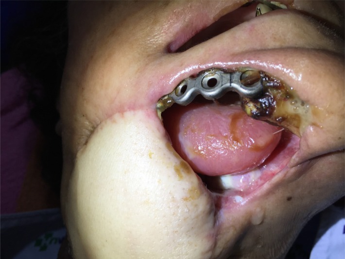
A 58-year-old woman (150 cm, 48 kg) was admitted to the hospital for surgery to remove a squamous cell carcinoma on her right mandible. She had a wide excision, marginal mandibulectomy, and supraomohyoid neck dissection (SOHND). Eight months later, she visited the hospital due to pus formation in the right mandible lesion, and was diagnosed with cancer recurrence. She then underwent segmental mandibulectomy, reconstruction with reconstruction plate and a pectoralis major myocutaneous flap. After the surgery, necrotic tissue was formed on the floor of the mouth, which enlarged in size gradually. Three weeks later, a huge orocutaneous fistula had developed, and the reconstruction plate was exposed. The oral cavity could be seen through the exterior of the submandibular lesion. Therefore, she was scheduled to undergo wide excision, reconstruction with reconstruction plate, and SOHND on the left side and an anterolateral thigh flap under general anesthesia.
Her past medical history included hypertension and arthritis, and she had received chemotherapy. Her glucose level had not been well controlled after the previous operation, but the other laboratory findings, electrocardiogram, and chest X-ray were all normal. The Mallampati classification was class II, but it was expected that the huge orocutaneous fistula might lead to difficult mask ventilation due to the insufficient sealing of the oral cavity (Fig. 1). Therefore, maintenance of spontaneous breathing was essential for the patient during induction. Since difficult intubation was not predicted, we obtained informed consent from the patient to perform awake intubation under conscious sedation.
Routine monitoring by pulse oximetry, ECG, non-invasive blood pressure measurements, bispectral index (BIS), and capnography was performed. Baseline oxygen saturation, blood pressure, heart rate, and BIS were 99%, 140/80 mmHg, 80 beats/min, and 94, respectively. Glycopyrrolate (0.2 mg) was injected intramuscularly as a premedication to reduce saliva secretion. After the tongue was extruded and depressed with a tongue depressor, the oropharynx was anesthetized with a 10% lidocaine pump spray. For conscious sedation, a loading dose of 1 µg/kg dexmedetomidine was infused for 10 min and followed by a 0.6 µg/kg/hr infusion for maintenance. The BIS was found to be 70–80, and the patient was pre-oxygenated with 100% oxygen before the induction. After infusion of the loading dose of dexmedetomidine, 0.1 µg/kg/min remifentanil was also infused for analgesia, and a north pacing nasal endotracheal tube (Portex® tube; Smiths Medical International, Hythe, UK) of 6.0 mm internal diameter was inserted via the right nasal cavity. A video laryngoscope was also inserted via the oral cavity and a Cormack-Lehane grade 1 view was obtained. Although she had mild gagging and coughing, the nasotracheal tube was inserted fairly smoothly. The end-tidal CO2 waveform was checked, and 30 mg of rocuronium was injected intravenously. The dexmedetomidine infusion was then stopped, and the patient was mechanically ventilated with desflurane 6 vol % and 3 L/min of fresh gas flow with 50% oxygen in the air. The operation was continued for 9 hrs without any problems. After the surgery, spontaneous breathing recovered, but the nasotracheal intubation was maintained due to the anticipation of airway edema. On the next day, the patient was successfully extubated. For this case presentation, we received consent from the patient.
DISCUSSION
There is no standard definition for DMV. However, in 2003, the American Society of Anesthesiologists Task Force defined it as the clinical situation that develops when it is not possible for the anesthesiologist to provide adequate mask ventilation due to one or more of the following problems: inadequate mask seal, excessive gas leak, or excessive resistance to the ingress or egress of gas [1]. A large facial defect as seen in the patient in our case can hamper mask ventilation during induction because of inadequate mask seal and excessive gas leak. In 2004, Han et al. [3] suggested a grading scale for the risk stratification associated with mask ventilation. According to Han's scale, grade 1 patients are those who can be ventilated comfortably, and grade 4 patients are those who cannot be mask ventilated. Grade 3 and 4 patients are expected to be at an increased risk of inadequate ventilation after anesthesia induction. Especially, Grade 4 patients are at a high risk of impossible mask ventilation and cannot be ventilated in spite of applying the corrective measures for ventilation. Therefore, preoperative assessment of the DMV grade is positively necessary.
In our case, we conducted awake intubation due to the predicted DMV. Awake intubation may be conducted when DMV or tracheal intubation is expected. The awake fiberoptic intubation (AFOI) is the most commonly used method for awake intubation. However, AFOI is not widely used due to a number of factors associated with its failure, such as inappropriate sedation, inability to view landmarks, and tracheal tube impingement during railroading [4]. In addition, fiberoptic intubation requires additional special training, and some studies reported a high incidence of desaturation caused by it [5]. We used a video laryngoscope for awake intubation. Video laryngoscopy is becoming established as a valuable technique for the management of difficult direct laryngoscopy. It has many advantages such as similarities to the conventional laryngoscopy, and more familiarity to psychomotor skills [6]. A recent study reported that the C-MAC video laryngoscope offers an advantage over the flexible fiberoptic scope with respect to the time required to obtain a glottic view and successful placement of the tracheal tube in patients with cervical spine immobilization [7]. In addition, Shirgoska and Netkovski [8] suggested that video laryngoscopy can be easily learned and should be used for managing difficult airway situations in the operating room, as well as in the emergency department. Therefore, video laryngoscopy for awake intubation may be easier, faster, and safer than AFOI in patients with an anticipated difficult airway.
For successful awake intubation, a number of conditions (conscious sedation, maintenance of a patent airway, and adequate spontaneous ventilation) are required. For conscious sedation, several sedative and analgesic drugs are available [9]. We used dexmedetomidine for sedation, which is a widely used sedative for awake intubation. It has many favorable properties such as anxiolysis, anterograde amnesia, analgesia, and minimal respiratory depression. In addition, dexmedetomidine reduces salivary secretion through the sympatholytic and vagomimetic effects, which aids in getting a fine laryngeal view during awake intubation [1011]. Therefore, dexmedetomidine is the optimal sedative drug for awake intubation because it facilitates the conscious sedation and maintenance of spontaneous breathing.
In conclusion, we performed an awake intubation due to predicted DMV in a patient with a huge orocutaneous fistula. Preoperative evaluation of the DMV grade is indispensable to secure the airway during induction. In addition, awake intubation using a video laryngoscope may be the alternative to AFOI.
 XML Download