

 PDF
PDF ePub
ePub Citation
 Print
Print


Pain and sensory disorder resulting from injury to peripheral nerves of the face and jaws are a major source of patient dissatisfaction and suffering. Although there has been a reduction in nerve injury from traffic accidents, the greater availability of oral and maxillofacial operations has, ironically, increased the risk of concomitant iatrogenic nerve injury. Often, attempts by oral and maxillofacial surgeons to help nerve injury patients have been hampered by medicolegal and disability considerations that surround this problem. A better understanding of the nature and effects of sensory nerve injuries and the recent development of microsurgical techniques offer new possibilities for improved treatment of neuropathies.
The majority of patients who sustain injuries to the peripheral sensory nerves of the face and jaws experience a slow but orderly return of sensation that is functional and tolerable in quality, if not normal (i.e., return of pre-injury levels of function and sensation). For some patients, however, the posttraumatic symptoms become pathological and painful. The predominant pain components are (1) numbing anesthesia dolorosa pain, (2) triggered neuralgiaform pain, (3) burning and aching causalgiaform pain, and (4) phantom pain [12].
Of these components, the most serious clinical problem is a triggered pain in the form of trigeminal neuralgia, similar to convulsions [3]. This study discusses the case of a geriatric patient with cerebral infarction who had undergone dental extraction and alveoloplasty in a broad range, and his postoperative pain in the form of posttraumatic trigeminal neuralgia was treated with intravenous (I.V.) infusion and oral administration of carbamazepine, resulting in effective analgesia and desirable prognosis.
CASE REPORT
A 74-year-old man was admitted to Wonju Severance Christian Hospital for extraction of his remaining maxillary teeth and mounting of a removable full denture after alveoloplasty. He had suffered a stroke due to a cerebral infarction three months ago and was on medication while hospitalized and under the care of the Neurology Department. Although he was undergoing treatment at the rehabilitation department, a combined examination revealed that dental surgery likely would be uneventful.
On the basis of an oral examination and radiography results showing progressive periodontitis, it was deemed necessary to extract the remaining maxillary teeth (#15, #17, and #25) for mounting of a removable full denture; alveoloplasty of teeth #12-#15 and #27 and the surrounding regions was required where sharp margins of the remaining alveolar bones were present.
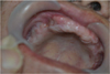
The three teeth extractions and the alveoloplasty procedures were performed under local anesthesia by use of 2% lidocaine HCl with 1:100,000 epinephrine on August 17, 2015. Wound closure was uneventful, and sutures were removed at postoperative day 7. On day 10 postoperatively, he was urgently admitted to our Department of Dentistry for paroxysmal throbbing pain in regions 12, 13, 14, 15, and 17, comprising the sites of tooth extraction and alveoloplasty. His vital signs were normal, and there were no abnormal findings on oral examination and radiography (Fig. 1).
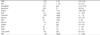
Neuritis owing to systemic weakening was diagnosed; posttraumatic pain syndrome was caused by tooth extraction and alveoloplasty. The authors determined the pain to be trigeminal neuralgia pain, and clinical tests were performed accordingly (Table 1). As therapy for acute neuroinflammation, an I.V. infusion (normal saline 1,000 cc I.V., clindamycin 600 mg I.V., diazepam [half ampule mixed with 10 cc distilled water] was administered. As a result, the pain was decreased but did not disappear completely. Therefore, a cephalosporin oral antibiotic, anti-inflammatory drugs (streptokinase, acetaminophen), an anti-seizure drug (carbamazepine 300 mg/day), and a digestive drug (simethicone) were administered orally three times a day for one week.
The pain was decreased further and improved to a tolerable level on August 31, 2015, and the medication dosing regimen was reduced to twice daily. Thereafter, the patient recovered completely on September 14, 2015, after two weeks of treatment, and he was transferred to the prosthetic dentistry department for mounting of a removable full denture.
DISCUSSION
When diagnosing maxillofacial neurological problems, the examination of the neurological history should begin with a question on the systemic diseases and environmental conditions that have neurological effects on the maxillofacial region. Patients should then be questioned about past history of major neurological and psychiatric disorders. Once the systemic, environmental, primary neurological, and psychiatric disease factors have been taken into consideration, the patient should be questioned on the nature and incidence of lesions in the maxillofacial tissues themselves [16]. On the basis of the neurological history, it may be possible to make a tentative pathological diagnosis, such as an infectious, degenerative, arterial or demyelinating condition, or a tentative etiologic diagnosis such as a posttraumatic, odontogenic, psychogenic, or diabetic condition. Because the patient in this case also had metabolic disorders including anemia, among other systemic environmental factors, and the major neurological disorder was neuralgia, which occurred after tooth extraction during dental surgery and alveoloplasty, he was diagnosed with traumatic neuralgia.
In addition, clinicians should examine specific and local sources of acute neuritis in detail using routine dental diagnostic skills and aids such as inspection, palpitation, radiography, and electrodiagnosis. In summary, a differential diagnosis is made after reviewing the neurological history and examining a patient with maxillofacial neurological disorders. The four disease elements (symptoms, pathology, location, and etiology) should be ascertained in each case. In particular, it is necessary to differentiate posttraumatic neuropathy after oral maxillofacial surgery from the existing masticatory myofascial pain, trigeminal neuralgia, and systemic neuropathy.
In clinical dentistry, neuralgia is defined as paroxysmal, intense, and intermittent pain that usually is confined to specific nerve branches of the head and neck. Currently, studies have shown that paroxysmal spurts of maxillofacial pain may share a common histopathology, which is caused by disruption of the insulating mechanism between axons without destroying them [29]. This primary condition may occur in peripheral nerve branches, sensory ganglion tissues, or posterior roots. Various study results have shown that these peripheral lesions could cause pain by creating afferent imbalances and setting up abnormal pools of secondary central neurons in the trigeminal descending tract nuclei, possibly the epileptogenic foci type [89].
An injured nerve undergoes segmental dymyelination, Wallerian degeneration, and degeneration processes such as dying-back neuropathy, neurotrophic effect, and normal or abnormal regeneration; among these, the biggest clinical problem is posttraumatic pain syndromes due to abnormal regeneration from injury during surgery [26]. Unfortunately, many other factors may delay the return of proper functioning in regenerated peripheral nerves, resulting in abnormal nerve regeneration. Poorly myelinated tubular zones, called neuromas in continuity, may occur in the regenerated nerve.
The nerve tissues of the peripheral neuromas rarely mature and myelinate, and thus their stimulation may result in bursts of intermittent pain and bizarre paresthesias (e.g., "pins and needles" sensations). This phenomenon may be explained by setting up artificial synapses, in which impulses in one demyelinated fiber may excite neighboring demyelinated fibers, resulting in an abnormal chain reaction to the original stimulus. The concept of an artificial synapse occurring in pathological peripheral nerve zones may be a common explanation for many trigeminal neuralgias, including multiple sclerosis lesions and paroxysmal shocking pain of the tic douloureux. A similar explanation can be applied for the deep burning pain of posttraumatic causalgia, which may be caused by the excitation of demyelinated sensory nerve segments by adjacent unmyelinated sympathetic fibers.
In addition to neuroma formation, other potential regenerations can occur. Relocation of the growth cone fibers in the distal Schwann cell passages reportedly is largely a nonspecific selection process, and the identical matching of new regenerating fibers with their former tissue receptors may not occur. Fibers may innervate the wrong tissues, potentially disrupting motor controls in reinnervated skeletal muscle and glands; because regenerated fibers rarely attain their original diameters, distances between the nodes of Ranier may decrease in regenerated nerves. These two factors can lead to disproportionate reduction of nerve conduction velocities [10]. According to gate control theories of pain and sensory modulation, imbalance in afferent fiber diameters could lead to sensory abnormalities such as hyperpathia.
In addition to the imbalance induced by histopathological conditions in the peripheral nerve fibers, serious imbalances also can be caused by selective effects on the nerve cell bodies themselves. Moreover, various studies have suggested that trigeminal ganglion cell bodies can be lost selectively as a result of various life and disease processes. Trauma, metabolic disease, and viral infection can result in neuronal necrosis. Peripheral nerves subsequently may undergo Wallerian degeneration, and central nerves may disintegrate. This can damage the functional synaptic connection with secondary transmission, reflex, and integration centers in the central nervous system.
Trigeminal deafferentation (i.e., loss of peripheral fibers and synaptic contacts that normally reach the primary synaptic regions) can induce both morphological and physiological changes in the nuclei of the descending trigeminal tract [411]. Deafferentation conduct patterns in brain stem regions are similar to that of initial epilepsy response, showing electrical characteristics called epileptogenic foci. It has been postulated that these epileptogenic firing patterns may represent the physiological change responsible for paroxysmal and atypical neuralgia states. Deafferentation effects eventually may explain the cause of poorly understood conditions such as trigeminal neuralgia, postherpetic neuralgia, and phantom pain. Bell et al. grouped deafferentation pain states as a syndrome classified by (1) posttraumatic pain, (2) traumatic neuroma, (3) reflex sympathetic dystrophy, (4) neuritic neuralgia, and (5) phantom pain [46].
As in the present case, a stabbing, flashing pain may be experienced within the first several weeks following a nerve injury, and thus clinicians should always consider the possibility of a secondary source of mechanical irritation or inflammation in the still-intact nerve trunk. Clinicians should search for entrapped or pinched nerves, in order to eliminate the acute sources of neuritis including foreign bodies, mobile bone fragments, and infection. Triggered pain lasts for more than a few weeks or emerges after a delayed period of anesthesia and is usually accompanied by sensory neuropathy. This pain component is like conventional tic douloureux in that fine tactile or mild heat stimuli evoke brief hyperesthesias and sharp "tingling" or stabbing sensations. These pains may be due to either peripheral neuromas or secondary deafferentation pathology in the brain stem. In these cases, clinical examination is of considerable importance because pain triggered by manual palpation at specific points along the neurovascular bundle is suggestive of neuroma.
However, in cases of tooth extraction or alveoloplasty that have no possibility of neuroma as in the present case, posttraumatic trigeminal neuralgia can be induced by deafferentation pathology phenomenon. In other words, spontaneous or random spasms of pain are probably linked more to the central neuropathology. Peripheral microsurgery is highly beneficial for patients who have neuromas, whereas centrally acting pharmacological agents are more appropriate for patients with central neuropathology [112].
Hence, the patient in this case was administered oral carbamazepine, which is effective for the treatment of trigeminal neuralgia to reduce excessive pain in the form of posttraumatic neuralgia. At the same time, I.V. infusion was accompanied with antibiotics, anti-inflammatory drugs, and sedatives to improve systemic symptoms and neuritis conditions, with good clinical results.
The neuritis mentioned here means "inflammation of the nerve" and pertains to acute reversible irritations to maxillofacial nerves. Neuritis occurs in the sensory, motor, or autonomic nerves and results from peripheral pathology that infect, compress, entrap, or erode the adjacent nerves. Neuritis is significant because it is an indication of an acute pathological condition; once it persists, it induces degenerative and irreversible neuropathy. Sensory neuritis-characterized by a decline in pain thresholds-is almost always manifested as pain, but its characteristics depend on the location and nature of the primary lesion. Neuritis etiology includes trauma, infection, paranasal sinusitis, otalgia, salivary gland disorders, mucosal disorder, motor neuritides, and myofascial dysfunction [12].
In conclusion, even after routinely performed tooth extraction or alveoloplasty in clinical dentistry, neuritis can occur in the trigeminal nerve, as can posttraumatic pain syndrome (e.g., trigeminal neuralgia, neuropathic pain, phantom pain). Therefore, continuous monitoring and management are required until the wound has completely healed.
 XML Download