

 PDF
PDF ePub
ePub Citation
 Print
Print


INTRODUCTION
Warming blood products before administration can prevent hypothermia caused by the massive transfusion of cold blood to neonates. If appropriate fluid-warming devices are unavailable, a hand-made water chamber can be used as a simple and inexpensive method to warm cold blood. However, excessive warming of red blood cells (RBCs) during rapid transfusion causes thermal damage, thereby resulting in hemolysis [1]. Irradiated and leukoreduced RBCs are frequently transfused to critically ill neonates and small children to prevent graft-versus-host disease [2], febrile nonhemolytic transfusion reactions, alloimmunization, and postcardiopulmonary bypass lung injury, but may increase the risk of hemolysis through increased osmotic fragility, cell lysis, and a reduction in cell survival [3]. Hemolyzed RBCs that contain high levels of potassium (K+) and free hemoglobin (Hb) increase the potential for serious adverse effects in transfused patients, especially if these effects occur over a short period of time [4]. It has been reported that cardiac arrest may occur following the massive transfusion of RBCs that contain high K+ concentrations, particularly in infants and small children [56]. The aim of this study was to determine, through the measurement of in vitro plasma K+ and free Hb concentrations, whether rapid warming of irradiated and leukoreduced RBCs enhances hemolysis and to determine the safe range for the temperature of water in the water chamber.
METHODS
1. Materials
Forty-four units of RBCs stored in citrate phosphate dextrose were used in this study. RBCs were leukoreduced using leukocyte-reducing filters (RCXL2; Pall Corporation, East Hills, NY) in accordance with the manufacturer's instructions. Units in additive solution were gamma-irradiated with 25 Gy using a blood irradiator with a 137Cs source. The irradiated leukoreduced RBCs were stored in a refrigerator at 2–6℃.
2. Study design
The Institutional Review Board waived the requirement to conduct the study and report its findings. After carefully mixing the irradiated leukoreduced RBCs, each unit was stored at 2–6℃, and connected in sequence to a blood transfusion set (DooWon Meditec Corp., Gimje si, Korea), 6-inch-long extension tubing, and a 3-way stopcock at the end of the tubing. Before commencing this study, we collected baseline blood samples from the 3-way stopcock using a 10-ml syringe after the RBCs were passed through the transfusion set at a rate of 1 ml/sec. Following baseline sampling, the transfusion set and tubing (length = 120 cm) were warmed for 5 min in a 50℃ water chamber. To confirm the exact temperature, 20-ml aliquots were gently withdrawn into 20 ml syringes (without an attached needle), and a 10-ml blood sample was obtained. Rapid warming of the RBCs, as performed in clinical settings, was simulated using a manually controlled hand-held syringe. However, a sampling rate of 1 ml/sec was achieved using the minimum amount of pressure required to pass blood through the tubing. Free Hb and K+ were analyzed in each plasma sample. The same procedure was repeated using transfusion sets that were maintained at 60℃ and 70℃.
3. Measuring Hb and K+
RBCs were separated from the supernatant within 15 minutes by centrifugation at 2,000 g for 10 minutes. The separated supernatant was centrifuged again at 2,000 g for 10 minutes. The supernatant was analyzed to determine the concentrations of extracellular Hb and K+. Free Hb concentrations were determined using spectrophotometry (Hitachi U-3210 spectrophotometer; Hitachi, San Jose, CA). Absorbance was measured at 380, 415, and 450 nm. K+ concentrations were measured using an ion-selective electrode (Toshiba 200FR Neo Chemistry autoanalyzer; Toshiba Medical Systems Co., Ltd., Tokyo, Japan).
4. Statistical analysis
In our study, P values < 0.05 were considered significant. Normal distributions were determined using the Shapiro–Wilk test. Statistical analyses were performed using nonparametric methods if the variables were not normally distributed. We used the one-way repeated-measures analysis of variance with the Bonferroni post hoc test to compare values determined at different warming temperatures. Linear associations between two variables were ascertained using Pearson or Spearman correlations. We determined that a minimum of 44 samples were needed to detect differences in the mean potassium level (5.0 mmol/L with an expected standard deviation [SD] of 7.0 mmol/L) between different temperatures (at 80% power and P < 0.05). Results are expressed as the mean ± SD or median values (range or interquartile range), as appropriate. Statistical analyses were performed using SPSS 12.0 software (SPSS, Chicago, IL).
RESULTS
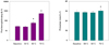
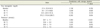
The mean storage duration of the irradiated leukoreduced RBCs was 8 days [range, 3–20]. The warming of RBCs to 60℃ and 70℃ induced significant increases in the free Hb levels (60.5 mg/dl [interquartile range = 34.9–101.4] and 570.2 mg/dl1 [115.6–2289.7], respectively) in comparison with baseline and (27.4 mg/dl [19.9–38.2] and 24.1 mg/dl [17.4–46.8], respectively) (Fig. 1). At 70℃, the plasma K+ concentration (31.4 ± 7.6 mmol/L) was significantly higher in comparison with baseline (29.7 ± 7.1 mmol/L; P = 0.029) (Fig. 1). After warming to 50℃ and 60℃, however, the plasma K+ level did not significantly increase in comparison with baseline. There was a significant correlation between the storage duration and plasma K+ concentration after warming to 50℃ and 60℃ (Table 1).
DISCUSSION
We conclude rapid warming of irradiated leukoreduced RBCs in a 50℃ water chamber does not significantly increase hemolysis. RBCs warmed to 60℃ did not demonstrate an increase in plasma K+ levels, but the free Hb in the plasma was markedly increased at this temperature, which may suggest excessive hemolysis. In addition, a longer storage duration was found to correlate with increases in the plasma K+ level after warming, even at 50℃.
The irradiation of RBCs with gamma rays reduces the lymphocyte concentration, which can result in transfusion-associated graft-versus-host disease [2]. However, irradiation may also affect nonlymphoid blood cells. The properties of some components are remarkably altered when RBCs are irradiated [7] which induces changes in membrane integrity and decreases in cell elasticity and deformability, indicating an acceleration of hemolysis and K+ release [38]. Additionally, these changes increase with storage duration and irradiation energy [8]. Accordingly, the shelf life of irradiated RBCs is only 28 days [9].
Leukocyte-reducing filtration reportedly accelerates hemolysis in RBCs [1011] as the filters and micropores used may mechanically damage the cells [12]. Moreover, filtration-associated hemolysis increases with storage duration [10]. However, prestorage leukoreduction may prevent RBC damage caused by the degradation of white blood cells and significantly reduce the deleterious effects of irradiation in terms of hemolysis [13]. In our study, K+ levels were not found to be significantly different from those at 50–60℃, which may be due to the effects of prestorage leukoreduction.
In packed RBCs, the concentration of Hb (130 g/L) is much higher than that of potassium (3.9 g/L). The packed RBCs unit has low baseline free Hb concentration levels while potassium is high. The larger amount of Hb compared to potassium in RBCs, and the lower baseline concentration of free Hb compared to potassium in packed RBCs units may result in statistically significant increases of Hb during hemolysis whereas potassium would have lower (or no) statistical significance. This may explain our study result of non-significant increases in potassium levels by hemolysis at 60℃, in contrast to Hb levels.
The massive transfusion of cold blood products can induce or worsen hypothermia, particularly in pediatric patients with a small blood volume capacity. Hypothermia can cause coagulopathy, increased oxygen consumption, fatal tachyarrhythmia, and metabolic acidosis. To prevent hypothermia during a massive transfusion, in-line warming using a water chamber is still a common surrogate in lieu of a countercurrent water bath or dry heat warmer, if these devices are unavailable. However, RBC membranes exposed to high temperatures can become damaged, leading to changes in their viscosity, fluidity, deformity, permeability, and osmotic fragility [1]. To ensure safety during the rapid transfusion of irradiated leukoreduced RBCs, it is important to verify the high temperature limits of the water chamber. Heating to 45℃ may not lead to RBC damage [14], and mixing with saline and heating to 60℃ does not increase hemolysis [15]. However, RBCs exposed to temperatures between 46–51℃ undergo denaturing thermal transitions at the cytoskeletal protein level [1]. Our study findings demonstrate that a higher warming temperature of 60℃ induces a marked increase in the free Hb level, indicating gross hemolysis. Additionally, RBCs warmed to 70℃ demonstrated increased plasma K+ levels.
The transfusion of hemolyzed blood can detrimentally affect neonates and small children [416]. The K+ concentration in the supernatant of hemolyzed RBCs is frequently much higher than that in normal human plasma. Neonates and small children have smaller circulating volumes, immature renal function and K+ handling, and differences in autonomic tone [1718]. Hence, neonatal patients are at risk of hyperkalemia- associated cardiac arrest during a massive RBC transfusion. Furthermore, hemolysis causes the release of free Hb and plasma discoloration. Hemoglobinemia occurs when the binding capacity of haptoglobin is exceeded. Since free Hb is considered harmful to many organs, particularly the kidneys, prompt removal is necessary to prevent adverse effects [19]. Hb does not characteristically appear in the urine until plasma levels reach > 100 mg/dl. In practice, eliminating free Hb using haptoglobin demonstrates better patient tolerance because of the rapid formation and metabolic turnover of haptoglobin [3]. However, the United States Food and Drug Administration recommends a maximum permissible limit of 1% hemolysis for RBCs. According to our results, grossly hemolyzed RBCs following rapid warming should not be used to prevent clinical complications.
We did not consider catheter size-related effects, infusion rate, and pressure in our present study, and further investigations are needed before applying our findings to a clinical setting. In addition, we measured in vitro plasma Hb and K+ concentrations to assess the extent of hemolysis but these variables are not representative of in vivo extracellular Hb and K+ concentrations in the blood. Finally, we did not determine the effects of filtration or RBC irradiation on warming temperature-related hemolysis.
CONCLUSIONS
Rapid warming of irradiated leukoreduced RBCs to 50℃ in a water chamber can be used as a surrogate procedure without resulting in gross hemolysis. However, irradiated leukoreduced RBCs—particularly older units that contain high levels of free Hb and K+ in plasma—should be infused with caution, and the authors recommend the use of fresh whole blood for neonates requiring massive transfusion, if it is feasible.
 XML Download