

 PDF
PDF ePub
ePub Citation
 Print
Print


INTRODUCTION
Papillary thyroid carcinoma (PTC) is commonly accompanied by cervical lymph node metastasis. Regional lymph node involvement shows a frequency of 30~80% and nearly always occurs in the central compartment and lateral regions of the neck along the internal jugular chain, whereas metastases to the retropharyngeal lymph nodes (RPN) and parapharyngeal lymph nodes are extremely rare.(1) Radioactive iodine (RAI) ablation is a major modality for the detection and treatment of DTC.(23) However, in the case of iodine non-avid DTC, which does not concentrate RAI, the diagnostic value is insufficient and therefore affects further therapeutic planning.(3) F-18 Fluorodeoxyglucose (FDG) positron emission tomography/ computed tomography (PET/CT) performed concurrently with I-131 treatment can help in detecting additional lesions in DTC patients.(2) We report a case with papillary thyroid carcinoma showing rare retropharyngeal lymph node involvement, which were not detected perioperatively due to the fact that there was no uptake on the I-131 scan, that was only found on FDG PET/CT. We also address the characteristic radiologic features of these metastatic retropharyngeal lymph nodes and the impact of this clinical manifestation on the management of PTC.
CASE REPORT
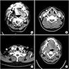
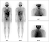
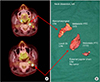
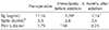
A 45-year-old female patient with no previous medical history presented to our hospital with a thyroid nodule detected on neck ultrasonography performed for a health examination. Neck ultrasonography showed two nodules (mid 1.4 cm/lower 0.6 cm) that implied malignancy on the left thyroid, and the diagnosis of papillary thyroid cancer was made with fine needle aspiration cytology (FNAC). In the neck CT obtained for preoperative staging, there were multiple enlarged lymph nodes on the lateral neck area, suggesting metastasis (Fig. 1A~C). Thus, an additional FNAC was performed at level II, and the pathologic result was reported as metastatic lesion of papillary thyroid cancer. With the diagnosis of papillary thyroid cancer and left lateral cervical lymph node metastasis, we performed total thyroidectomy with central cervical lymph node dissection and ipsilateral modified radical neck dissection (levels IIa, III, IV, and Vb) through a transcervical approach. The postoperative course was uneventful, and the patient was discharged six days after surgery. Definitive histologic examination revealed three nodules of conventional PTC in the both thyroid with invasion of the strap muscle and several metastatic lymph nodes were also identified. Three months after the operation, high-dose (5.55 GBq) RAI ablation using I-131 was performed. On a post-therapeutic I-131 scan, only remnant thyroid activity was seen at the thyroid bed, and there was no other abnormal I-131 uptake to suggest metastasis of PTC (Fig. 2). In contrast, FDG PET/CT, performed just before the I-131 treatment according to the protocol of our hospital, revealed two enlarged lymph nodes with increased FDG uptake in the left level II (maximum standardized uptake value (SUVmax) of 11.1) and retropharyngeal space (SUVmax of 17.6) (Fig. 3). However, the thyroglobulin (Tg) level was low (2.39 ng/ml) at the time of I-131 ablation with a TSH serum level of 100.0 IU/ml. Because the serum Tg measurement and RAI whole-body scan are considered to be more sensitive than 18F-FDG PET/CT for detecting metastasis of welldifferentiated thyroid cancer, clinical follow-up, rather than further work-up and treatment, was performed. Even though, the serum Tg level stayed low at less than 0.2 ng/mL during the follow-up period, there was a suspicious lymph node at level II detected by neck ultrasonography performed one year after the operation (Table 1). Fine needle aspiration cytology was performed, and washout Tg was simultaneously measured. The pathologic result was reactive hyperplasia, but the Tg value exceeded 500 ng/ml, which suggested metastasis. A neck CT obtained for diagnostic work up also detected similar, well enhanced lymph nodes at the left level II, which were found to be equivalent to those identified on previous neck CTs. Also, a retropharyngeal lymph node was still present on an initial neck CT and PET/CT taken before RAI ablation (Fig. 1D). It was regarded as a remnant metastatic lymph node that was not identified during the initial operation rather than the possibility of a second malignancy of the head and neck origin or recurrence. Thus, a second operation was planned in light of these two issues. In the second operation, we determined that an approach through the previous incision would be difficult because of postoperative adhesions; therefore, a Mcfee incision was performed through the left upper neck. Selective lymph node dissection was first performed on the level IIb area in the superior lateral portion of the spinal accessory nerve, and then retropharyngeal lymph nodes were removed by approaching the posterior pharyngeal space after identifying the common carotid artery and internal jugular vein and removing retropharyngeal lymph nodes. The pathologic result was metastatic papillary thyroid carcinoma. The patient was discharged without complication five days after surgery and is currently in the process of outpatient follow-up.
1. Protocol for iodine-131 whole-body scan and 18F-FDG PET/CT
At our institution, 18F-FDG PET/CT is performed just before RAI ablation on the same day in order to maximize the effect of TSH stimulation.
1) Patients undergoing I-131 treatment were instructed to discontinue replacement of L-thyroxine (T4) for four weeks before the radioiodine treatment and received replacement L-triiodothyronine (T3) for the first two weeks of this period. 2) In addition, all patients followed a low-iodine diet for two weeks before the I-131 treatment. 3) On the day of I-131 treatment, serum TSH, thyroglobulin, and anti-thyroglobulin antibody levels were measured, and FDG PET/CT scanning using Biograph mCT (Siemens Healthcare) was performed just before the administration of I-131. Patients were instructed to fast for at least six hours before injection of FDG, and 6.3 MBq/kg of FDG was intravenously administered one hour prior to the PET/CT scan. 4) After blood tests and FDG PET/CT scanning, a therapeutic dose of I-131 was orally administered. 5) Post-therapeutic I-131 scans were obtained at 2~3 days and 7~9 days after I-131 treatment using a large field-of-view, dual-head gamma camera (Symbia E, Siemens Healthcare) equipped with a medium-energy parallel-hole collimator.
DISCUSSION
We examined three important aspects of the presented case.
First, papillary thyroid carcinoma has the highest incidence of cervical lymph node metastasis. In particular, the ratio of simultaneously detecting lateral neck metastasis in diagnosis varies greatly depending on the report, but the frequency is in the range of 15~30%. Therapeutic lateral cervical lymph node dissection should be performed when there is clinical evidence in addition to biopsy-proven metastatic lymphadenopathy.(4) Additionally, determination of the appropriate extent of lateral neck dissection uses various methods and is most significantly affected by clinical judgments (5) and is thereby determined according to the preference of surgeons. Lateral cervical lymph node metastasis of PTC most often occurs in the neck at levels IIa, III, and IV along the internal jugular chain.(5) At level V, surgery is performed if metastasis is clinically verified. Data on the basis of level IIb dissection remains debatable.(6) Neck level II is divided into IIa and IIb based on the spinal accessory nerve as the boundary. It has been reported that lymph node metastasis of level IIb is not common in PTC and is thus generally not included in the scope of neck dissection even if the surgeon routinely omits level IIb dissection. However, several previous studies have recommended performing a level IIb dissection if lymph node metastasis is confirmed in level IIa or if multilevel lymph nodes metastasis is expected. However, the surgical space is confined and the approach to level IIb is more limited when using the conventional cervical approach with the extended collar incision. In addition, although the rates of spinal accessory nerve injury in expert hands is low, aggressive dissection around the nerve is not easy due to the possibility of postoperative morbidity.(57) Level IIb dissection should have been considered more aggressively in the presented case as there was a metastatic lymph node at level IIa at the time of the initial operation.(8)
Second, the retropharyngeal space is located behind the pharynx and between vertebra fascia and visceral fascia, from the skull base to thorax level. This is where tumors from the parotid gland most commonly occur in head and neck cancers. In PTC, metastasis to retropharyngeal lymph nodes (RPN) and parapharyngeal lymph nodes is rarely reported (~0.5%).(9) Retropharyngeal lymph nodes are observed on the inner side of the internal carotid artery and sympathetic chain at the level of C1. The route in which thyroid cancer spreads to retropharyngeal lymph nodes may be identified. One pathway is via jugular chain lymphatics, and the other is the direct postero-superior lymphatic trunk that connects the upper pole of the thyroid to retropharyngeal lymph nodes.(10) Retropharyngeal lymph node metastasis of thyroid cancer is difficult to diagnose with neck ultrasonography and is verified through neck CT or MRI. Operative approaches to the posterior pharyngeal space vary according to location and type of tumor. In most cases, this space is accessible through a cervical approach, and mandibulotomy may also be performed if visualization is insufficient. If there is only retropharyngeal lymph node metastasis, there is a direct lymph node flow from the thyroid to posterior pharyngeal space. Thus, if the lateral cervical lymph node is N0 at the time of diagnosis and there is no clinical evidence, neck dissection for levels II~IV is not considered. Moreover, since it is known that retropharyngeal lymph node metastasis of thyroid cancer does not affect the overall survival rate, long-term survival can be expected when performing RAI ablation along with surgery. In this case, a retropharyngeal lesion was already present when the patient was diagnosed with PTC; if detected earlier, it would have been necessary to modify the proper treatment strategy in consideration of the possibility of retropharyngeal lymph node metastasis. Earlier recognition may allow for more acceptable surgical outcomes.
Third, most thyroid cancers are well differentiated tumors of follicular cell origin. The therapeutic strategy of DTC is to perform RAI ablation using I-131 after total thyroidectomy.(2) RAI ablation is known to have the advantage of reducing cancer relapse and survival rate. However, there are rare cases in which the tumor ability to trap iodine is deteriorated. In the case of iodine-negative tumor cells, I-131 treatment has little therapeutic effect, and the diagnostic value of iodine scan will also decrease. Moreover, these tumors show more aggressive clinical behavior than tumors that can concentrate iodine, showing poor prognosis. Therefore, early diagnosis is necessary for iodine-negative DTC, and another therapeutic approach must be considered other than I-131 therapy, such as surgical resection, external beam radiotherapy (EBRT), tyrosine kinase inhibitor, or re-differentiating agents.(2) Serum thyroglobulin level is a useful indicator for verifying the response to treatment and the presence of relapse in patients with differentiated thyroid carcinoma.(11) However, there are exceptional cases in which small-volume metastatic lesions are not properly measured or in which variant thyroglobulin is created in the Tg synthesis process and thus are not detected by conventional radioimmunoassay.(1213) In general, well differentiated thyroid cancer cells show high iodine uptake, whereas dedifferentiated thyroid cancer cells with progressive features show gradually decreasing iodine uptake with a loss of avidity for iodine but increasing FDG uptake (so-called Flip-Flop phenomenon).(2) Since accurate anatomical localization is difficult using only iodine scan, SPET/CT has recently been performed to remedy these shortcomings. However, since the equipment is costly, there are limitations to the utilization of this technology. FDG PET/CT performed concurrently with I-131 therapy has been shown to be helpful in finding additional relapse or metastatic lesions not detected on I-131 scan, which will help to establish further therapeutic plans.(2)
In the presented case, a metastatic lymph node was not removed during the initial operation stage due to insufficient dissection of the level IIb area. However, since this metastatic lesion was not seen on the post-therapy iodine scan and had also low serum thyroglobulin level, there was minimal clinical significance immediately after the initial operation. We performed concurrent FDG PET/CT imaging before the iodine ablation, which showed high FDG uptake and showed signs implying metastasis. Thus, a therapeutic approach to retropharyngeal lymph node metastasis could be possible for remaining metastatic lymph nodes in level IIb. For postoperative tracking of patients with differentiated thyroid carcinoma, most cases undergo only serum thyroglobulin level measurement and neck ultrasonography. However, it is not easy to detect retropharyngeal lymph nodes in conventional neck ultrasonography. Furthermore, DTC that do not concentrate RAI, as in this case, are not easy to detect, which makes it more difficult to identify recurrence in early stages.
Lymph node metastasis of PTC is common. If lateral cervical lymph node metastasis is clinically verified, neck dissection should be performed for therapeutic purposes. However, it is important to perform an appropriately extensive neck dissection to minimize complications related to the surgery and to increase oncologic outcomes by conducting sufficient consideration of preoperative evaluation. Moreover, even though retropharyngeal lymph node metastasis is rare, it is necessary to check for such signs using various diagnostic tools such as CT or MRI not only before the operation, but also during the postoperative follow-up period. Generally, differentiated thyroid carcinoma shows iodine-avid features. While the sensitivity of iodine scan is known to be high, FDG PET/CT is more helpful for the diagnosis of rare iodine-negative DTC.
 XML Download