Journal List > Korean J Endocr Surg > v.16(4) > 1060157

Yun, Kim, Kim, Kim, Lee, Chang, and Park: Adrenal Schwannoma: Rare Entity of Adrenal Incidentaloma

Abstract

Purpose

Adrenal schwannomas are very rare and are usually incidentally found on autopsy and imaging. The aim of this study was to describe our experience of adrenal schwannoma and review the literature regarding this rare tumor.

Methods

To identify patients with adrenal schwannoma, the MEDLINE database was searched via the major electronic database PubMed using the medical subject heading terms “adrenal” and “schwannoma”. Thirty-nine adrenal schwannoma cases, including two from our institution were included.

Results

The mean age at diagnosis was 47.95 years (range, 11~89 years). The female:male ratio was 21:17. Sixteen patients had tumors on the right side, 19 on the left, while one patient had bilateral tumors. The mean tumor size was 6.12 cm (range, 0.6~14.5 cm), and the mean tumor weight was 161.3 g (range, 31.5~600.0 g). Sixteen tumors were solid, four cystic, one solid and cystic, and 18 were not described. Histologically, 14 cases were Antoni A, two were Antoni B, and 12 had concomitant Antoni A and B.

Conclusion

Adrenal schwannoma is usually an incidental finding, and the clinician must have a high index of suspicion to recognize it when imaging reveals suggestive features.

INTRODUCTION

Adrenal incidentaloma is defined as an adrenal lesion found incidentally on autopsy or imaging. The differential diagnosis of adrenal incidentaloma ranges from benign adrenocortical adenoma and pheochromocytoma to malignant lesions including neuroblastoma, ganglioneuroma, and adrenocortical carcinoma.(1)
Schwannoma is a benign nerve sheath tumor composed of Schwann cells in peripheral, motor, sympathetic, or cranial nerves of the head and neck region and upper and lower extremities. Schwannoma arising from visceral organs is very rare and occasionally may represent incidental report.(2) Schwannoma originated from adrenal gland is rare, and a few dozen cases have been described in the literature.(123456789101112131415161718192021222324252627282930)
The aim of this study was to review the schwannoma arising from the adrenal gland including two cases of our institution.

METHODS

To identify patients with adrenal schwannoma, the MEDLINE database was searched via the major electronic database PubMed on January 27, 2015 using the medical subject heading (MeSH) terms “adrenal” and “schwannoma”.
Review articles and editorial comments without new cases were excluded. We manually searched the reference lists of identified articles to find additional eligible reports. No language restriction was imposed.
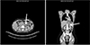
Data including the publication year, country, age, sex, clinical presentation, diagnostic imaging modality, site, tumor size, weight, cystic or solid, Antoni A or B, surgical procedure, and immunohistochemical (IHC) staining results were extracted. Two patients diagnosed in our hospital were included (Fig. 1).
This study was approved by the Institutional Review Board of Gangnam Severance Hospital, Yonsei University College of Medicine, and was conducted according to the principles of the Helsinki Declaration (IRB No.3-2015-0201).

RESULTS

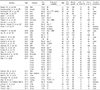
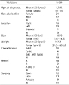
A total of 39 adrenal schwannoma cases, including two treated at our hospital, were enrolled in this study. The patients' clinicopathologic characteristics are listed in Tables 1 and 2.
The mean age at diagnosis was 47.95 years (range, 11~89 years), and the female:male ratio was 21:17. Among the 39 patients, 25 underwent surgical excision; 12 had open surgery, and 13 underwent laparoscopic surgery. Three cases were found on autopsy, and the excision method was not described for 11 cases.
Tumors were located on the right and left sides in 16 and 19 cases, respectively, while one patient had bilateral tumors. The mean tumor size was 6.12 cm (range, 0.6~14.5 cm), and the mean tumor weight was 161.3 g (range, 31.5~600.0 g).
With respect to tumor characteristics, 16 were solid, 4 cystic, 1 was solid and cystic, and 18 were not described. Expression of Antoni A was 14, Antoni B was 2, and concomitant Antoni A and B were 12.

DISCUSSION

Improvements in diagnostic imaging modalities have increased the identification of clinically silent adrenal masses called incidentalomas. Adrenal incidentalomas are asymptomatic adrenal tumors discovered on imaging performed to evaluate an abdominal problem unrelated to adrenal disease.(1)
Adrenal incidentalomas can be functioning or nonfunctioning, and benign or malignant. Functioning tumors include aldosterone-producing adenomas, cortisol-producing tumors such as Cushing's syndrome, androgen-producing tumors, and pheochromocytomas. Non-functioning benign tumors include adenomas, myelolipomas, ganglioneuromas, adrenal cysts, and hematomas. Malignant tumors include adrenocortical cancer and metastatic disease. Fortunately, most are non-functioning and benign.(7)
Adrenal incidentalomas are usually asymptomatic, but hormone overproduction symptoms are sometimes discovered on closer monitoring after adrenal tumor identification.
Verocay first described schwannoma in 1908, and Antoni further sub-classified into two distinct histologic patterns in 1920.(1) This neoplasm is a homogenous, benign, relatively slow-growing nerve sheath tumor composed of Schwann cells in peripheral, motor, sensory, sympathetic, or cranial nerves. Schwannomas are also known as neurilemomas, neuromas, neurolemomas, and Schwann cell tumors.
Schwannomas arising from visceral organs are very rare and are occasionally discovered incidentally. Adrenal schwannoma is also usually an incidental finding and very rare. There have been a few dozen cases of adrenal schwannoma described in the literature.(123456789101112131415161718192021222324252627282930) They are thought to arise from the adrenal medulla because there is continuity between it and the tumor and an absence of a septum around the tumor.
Preoperative diagnosis of adrenal schwannoma is challenging despite the use of multiple imaging modalities including ultrasonography (US) and computed tomography (CT). The goals of the preoperative evaluation are to determine the need and feasibility of resection and to rule out other metabolically active adrenal tumors, which will assist the practitioner in selecting an appropriate treatment course.(7)
Contrast-enhanced CT of the abdomen is usually the first imaging study performed. On CT scans, schwannoma appears as a well-demarcated, round or oval mass that may be homogeneous; however, other cases have shown prominent cystic degeneration and calcifications. With the addition of contrast, schwannomas may demonstrate variable homogeneous or heterogeneous enhancement.(18) However, the diagnosis often remains unclear until surgical intervention and histopathology studies are performed.
Magnetic resonance imaging (MRI) can be helpful in evaluating tumor size and determining the relationship of the mass with adjacent structures.(7)
The diagnosis of an adrenal schwannoma is usually made upon pathologic review of the surgical specimen showing; neoplastic cells simulating differentiated Schwann cells that are well circumscribed and composed of spindle cells organized as cellular areas with nuclear palisading (Antoni A) and paucicellular areas (Antoni B).(18)
Differential diagnoses of adrenal schwannoma include other neoplasms of the adrenal gland that may exhibit spindle cell morphology. Pheochromocytoma, ganglioneuroma, leiomyoma, and solitary fibrous tumors should all be considered in patient with suspected adrenal schwannoma.(13)
A small subset of schwannomas may be indistinguishable from neurofibromas due to similar histologic appearance and positive S-100 protein expression. In in such instances, positive staining for calretinin, a calciumbinding protein belonging to the same protein family as S-100, expressed in schwannoma but not neurofibroma, will allow for discrimination between the two entities.(2) Immunohistochemistry of adrenal schwannomas shows strong and diffuse S-100 staining. These tumor also display pericellular reactivity for collagen IV.(16) They are typically negative for CD117, desmin, CD 34, HMB-45, synaptophysin, chromogranin, cytokeratin, and smooth muscle actin.
Surgical excision should be strongly considered for all patients with functional adrenal incidentaloma.(3) As the diagnosis of adrenal schwannoma is made by proper examination of the surgical specimen, most adrenal schwannomas are excised according to institutional treatment policies for adrenal incidentalomas. The surgical approach should focus on complete excision of the mass.
Although adrenal schwannoma is a very rare entity and is usually found incidentally, clinicians must consider it in the differential diagnosis of adrenal incidentaloma.

Figures and Tables

Fig. 1

Abdomen CT scanning (present case 1) showing about 3.9 cm sized round mass with heterogenous density in Lt. adrenal gland (white arrow).

kjes-16-107-g001
Table 1

Summary of 39 patients with adrenal schwannoma

kjes-16-107-i001
Authors Year Country Sex/age Diagnostic modality Site Size (cm) Weight (gram) Cystic (C) or solid (S) Antoni A or B Surgical procedure
Bedard YC, et al.(30) 1986 USA F/63 NI Lt. 9.0 180 S NI Open
Kostakopoulos A, et al.(29) 1991 Greece F/38 NI NI NI NI NI NI NI
Tommaselli AP, et al.(28) 1996 Italy M/44 US,CT,1231-MIBG Rt. 3.9 NI C A Open
Fabbro MA, et al.(27) 1997 Italy M/11 US,CT Lt. NI NI S NI Open
Igawa T, et al.(26) 1998 Japan M/45 US Lt. 6.5 75.5 S A NI
Yonou H, et al.(25) 1999 Japan F/67 US,CT Lt. 6.0 50 NI A NI
Pittasch D, et al.(24) 2000 Germany F/56 US,CT,MRI Rt. 12.4 NI S A,B NI
Ikemoto I, et al.(23) 2002 Japan F/62 NI Rt. 12.0 600 C B NI
Barrero Candau R, et al.(22) 2002 Spain M/53 US,CT Rt. NI NI S NI NI
Arena V, et al.(21) 2004 Italy M/67 Autopsy Rt. 0.6 NI S NI Autopsy
Italy M/89 Autopsy Rt. 1.0 NI S A Autopsy
Inokuchi T, et al.(20) 2006 Japan F/35 US,CT,MRI Bi 7.5 88 C A,B Lapa
Lau SK, et al.(19) 2006 USA F/26 NI Rt. 10.0 200 NI A NI
USA M/73 CT Rt. 9.0 190 NI A,B NI
Korets R, et al.(18) 2007 USA M/70 CT Lt. 2.8 65 NI A Lapa
Garg S, et al.(17) 2007 India NI NI NI NI NI NI NI NI
Gazula S, et al.(16) 2007 India M/42 US,CT Rt. 12.0 NI S NI Open
Suzuki K, et al.(15) 2007 USA M/33 CT,MRI Rt. 9.0 NI S A NI
Jakowski JD, et al.(14) 2008 USA F/51 CT Lt. 5.5 84 C A,B Lapa
Hsiao HL, et al.(13) 2008 Taiwan M/49 US,CT Rt. 5.0 31 NI A Lapa
Onoda N, et al.(12) 2008 Japan M/62 CT,MRI Lt. 4.5 60 S A Lapa
Yang CY, et al.(11) 2009 China F/30 CT,MRI Lt. NI NI NI A,B Lapa
Târcoveanu E, et al.(10) 2009 Romania M/55 US,CT Lt. 4.5 NI NI A Lapa
Dario C, et al.(9) 2009 Italy M/42 US,CT Rt. 7.0 NI S NI Lapa
Xiao C, et al.(8) 2011 China F/30 US,CT Lt. 3.0 NI NI A,B Open
China F/38 US,CT Lt. 3.5 NI NI A,B Open
China F/46 US,CT Lt. 4.5 NI NI A,B Open
China F/43 US,CT Lt. 5.1 NI NI A,B Open
China M/47 US,CT Lt. 6.0 NI NI A,B Open
China M/39 US,CT Rt. 3.5 NI NI A,B Open
Kleiman DA, et al.(7) 2011 USA F/31 CT Rt. 4.5 NI NI NI Lapa
Richter KK, et al.(6) 2011 New Zealand F/30 US,CT Rt. 14.5 312 NI NI Lapa
Bakhshi GD, et al.(5) 2011 India F/34 NI NI NI NI NI NI NI
Toutouzas KG, et al.(4) 2012 Greece F/71 US,CT Lt. 5.5 NI C & S A,B Lapa
Adas M, et al.(1) 2013 Turkey F/32 CT,PET-CT Lt. 10.0 NI S A Open
Mohiuddin Y & Gilliland MG(3) 2013 USA M/79 Autopsy Lt. 0.6 NI S A Autopsy
Jeshtadi A, et al.(2) 2014 India F/55 CT Rt. 8.0 NI S B Open
Present case 1 2015 Korea F/39 CT Lt. 1.4 NI S NI Lapa
Present case 2 2015 Korea F/45 CT Lt. 3.8 NI S A Lapa

CT = computed tomography; 123I-MIBG = iodine-123-metaiodobenzylguanidine; Lapa = laparoscopy; MRI = magnetic resonance imaging; NI = not identified; PET = positron emission tomography; US = ultrasonography.

Table 2

Patient clinicopathologic characteristics

kjes-16-107-i002
Variables N=39
Age at diagnosis Mean±SD (years) 47.95
Range (years) 11~89
Sex distribution Female 21
Male 17
NI 1
Location Right 16
Left 19
Bilateral 1
NI 3
Size Mean±SD (cm) 6.12
Range (cm) 0.6~14.5
Weight Mean±SD (gram) 161.3
Range (gram) 31.5~600.0
Characteristics Solid 16
Cystic 4
Sold and cystic 1
NI 18
Antoni A 14
B 2
A and B 12
NI 11
Surgery Open 12
Lapa 13
Autopsy 3
NI 11

Lapa = laparoscopy; NI = not identified; SD = standard deviation.

References

1. Adas M, Ozulker F, Adas G, Koc B, Ozulker T, Sahin IM. A rare adrenal incidentaloma: adrenal schwannoma. Case Rep Gastroenterol. 2013; 7:420–427.
crossref
2. Jeshtadi A, Govada N, Somalwar SB, Nagulapally S. Schwannoma of the adrenal gland. J Med Allied Sci. 2014; 4:77–80.
3. Mohiuddin Y, Gilliland MG. Adrenal schwannoma: a rare type of adrenal incidentaloma. Arch Pathol Lab Med. 2013; 137:1009–1014.
crossref
4. Toutouzas KG, Tsamis D, Kekis PB, Michalopoulos NV, Flessas I, Manouras A, et al. Laparoscopic resection of an adrenal schwannoma. JSLS. 2012; 16:663–667.
crossref
5. Bakhshi GD, Borisa A, Tayade MB, Bhandarwar AH, Mogal HD. Primary schwannoma of adrenal gland: a case report. J Indian Med Assoc. 2011; 109:762–763.
6. Richter KK, Premkumar R, Yoon HS, Mercer P. Laparoscopic adrenalectomy for a rare 14-cm adrenal schwannoma. Surg Laparosc Endosc Percutan Tech. 2011; 21:e339–e343.
crossref
7. Kleiman DA, Hughes DB, Joshi AR. Surgical management of incidental adrenal schwannomas. Am Surg. 2011; 77:E89–E90.
crossref
8. Xiao C, Xu B, Ye H, Yang Q, Wang L, Sun YH. Experience with adrenal schwannoma in a Chinese population of six patients. J Endocrinol Invest. 2011; 34:417–421.
crossref
9. Dario C, Luigi P, Laura Z, Valentina S, Luigi C, Sandro N, et al. Adrenal schwannoma incidentally discovered-A case report. J Chin Clin Med. 2009; 4:43–46.
10. Târcoveanu E, Dimofte G, Bradea C, Moldovanu R, Vasilescu A, Anton R, et al. Adrenal schwannoma. JSLS. 2009; 13:116–119.
11. Yang CY, Chou CW, Lin MB, Li CF. Schwannomas of the left adrenal gland and posterior mediastinum. J Chin Med Assoc. 2009; 72:83–87.
crossref
12. Onoda N, Ishikawa T, Toyokawa T, Takashima T, Wakasa K, Hirakawa K. Adrenal schwannoma treated with laparoscopic surgery. JSLS. 2008; 12:420–425.
13. Hsiao HL, Li CC, Lin HC, Yeh HC, Huang CH, Wu WJ. Adrenal schwannoma treated with laparoscopic adrenalectomy: a case report. Kaohsiung J Med Sci. 2008; 24:553–557.
crossref
14. Jakowski JD, Wakely PE Jr, Jimenez RE. An uncommon type of adrenal incidentaloma: a case report of a schwannoma of the adrenal medulla with cytological, histological, and ultrastructural correlation. Ann Diagn Pathol. 2008; 12:356–361.
crossref
15. Suzuki K, Nakanishi A, Kurosaki Y, Nogaki J, Takaba E. Adrenal schwannoma: CT and MRI findings. Radiat Med. 2007; 25:299–302.
crossref
16. Gazula S, Mammen KJ. Schwannoma with concomitant tuberculosis in the adrenal gland. Indian J Urol. 2007; 23:469–470.
crossref
17. Garg S, Mathew M, Goel T. Adrenal schwannoma: a case report and review of literature. Indian J Pathol Microbiol. 2007; 50:587–588.
18. Korets R, Berkenblit R, Ghavamian R. Incidentally discovered adrenal schwannoma. JSLS. 2007; 11:113–115.
19. Lau SK, Spagnolo DV, Weiss LM. Schwannoma of the adrenal gland: report of two cases. Am J Surg Pathol. 2006; 30:630–634.
20. Inokuchi T, Takiuchi H, Moriwaki Y, Ka T, Takahashi S, Tsutsumi Z, et al. Retroperitoneal ancient schwannoma presenting as an adrenal incidentaloma: CT and MR findings. Magn Reson Imaging. 2006; 24:1389–1393.
crossref
21. Arena V, De Giorgio F, Drapeau CM, Monego G, De Mercurio D, Capelli A. Adrenal Schwannoma. Report of two cases. Folia Neuropathol. 2004; 42:177–179.
22. Barrero Candau R, Ramirez Mendoza A, Morales Lopez A, Maestro Duran JL, Huesa Martinez I, Leon Dueñas E, et al. Benign adrenal schwannoma. Arch Esp Urol. 2002; 55:858–860.
23. Ikemoto I, Yumoto T, Yoshino Y, Furuta N, Kiyota H, Oishi Y. Schwannoma with purely cystic form originating from the adrenal area: a case report. Hinyokika Kiyo. 2002; 48:289–291.
24. Pittasch D, Klose S, Schmitt J, Roessner A, Ridwelski K, Lippert H, et al. Retroperitoneal schwannoma presenting as an adrenal tumor. Exp Clin Endocrinol Diabetes. 2000; 108:318–321.
crossref
25. Yonou H, Goya M, Miyazato M, Miyazato T, Sugaya K, Koyama Y, et al. Retroperitoneal schwannoma arising from the adrenal area: a case report. Hinyokika Kiyo. 1999; 45:403–405.
26. Igawa T, Hakariya H, Tomonaga M. Primary adrenal schwannoma. Nihon Hinyokika Gakkai Zasshi. 1998; 89:567–570.
crossref
27. Fabbro MA, Costa L, D'Agostino S, Musi L, Belloli GP. Juxta-adrenal malignant schwannoma. Pediatr Surg Int. 1997; 12:532–534.
crossref
28. Tommaselli AP, Valentino R, Rossi R, Savastano S, Luciano A, Battista C, et al. Usefulness of 123I-metaiodobenzylguanidine (MIBG) scintiscan in the diagnosis of juxta-adrenal schwannoma. J Clin Endocrinol Metab. 1996; 81:843–846.
crossref
29. Kostakopoulos A, Pikramenos D, Livadas K, Antonopoulos P, Demonakou M. Malignant schwannoma of the adrenals. A rare case. Acta Urol Belg. 1991; 59:129–132.
30. Bedard YC, Horvath E, Kovacs K. Adrenal schwannoma with apparent uptake of immunoglobulins. Ultrastruct Pathol. 1986; 10:505–513.
crossref
TOOLS
Similar articles