

 PDF
PDF ePub
ePub Citation
 Print
Print


Abstract
Endoscopic thyroidectomy with bilateral axillobreast approach (BABA) is a feasible method of thyroidectomy with good surgical outcome and excellent cosmetic result as compared with conventional open thyroidectomy in selective patients. Thus, endoscopic thyroidectomy is widely used in treatment of thyroid diseases. However, despite the many advantages, we sometimes encounter unexpected complication, such as neck stiffness caused by adhesion, change of sensory, seroma formation, and subcutaneous soft tissue implantation. Subcutaneous soft tissue implantation of thyroid tissue is a very rare complication of thyroid surgery. However, it is troublesome to both patient and doctor. We experienced a case of papillary thyroid carcinoma recurrence at anterior and anterolateral subcutaneous area after endoscopic total thyroidectomy. Our case presented with a papillary thyroid carcinoma measuring 2.1 cm in size and showed thyroidal capsule invasion and extrathyroidal extension on the permanent pathologic report. Therefore, we suggest that appropriate indications should be applied for an endoscopic thyroidectomy and efforts should be made to decrease tumor cell spillage in order to prevent tumor rupture, and for careful handling and protection of the extraction site.
Go to : 
References
1. Choi JY, Lee KE, Chung KW, Kim SW, Choe JH, Koo do H, et al. Endoscopic thyroidectomy via bilateral axillobreast approach (BABA): review of 512 cases in a single institute. Surg Endosc. 2012; 26:948–55.

2. Chung YS, Choe JH, Kang KH, Kim SW, Chung KW, Park KS, et al. Endoscopic thyroidectomy for thyroid malignancies: comparison with conventional open thyroidectomy. World J Surg. 2007; 31:2302–6.
3. Jeong JJ, Kang SW, Yun JS, Sung TY, Lee SC, Lee YS, et al. Comparative study of endoscopic thyroidectomy versus conventional open thyroidectomy in papillary thyroid microcarcinoma (PTMC) patients. J Surg Oncol. 2009; 100:477–80.
4. Ito Y, Tomoda C, Uruno T, Takamura Y, Miya A, Kobayashi K, et al. Needle tract implantation of papillary thyroid carcinoma after fine-needle aspiration biopsy. World J Surg. 2005; 29:1544–9.
5. Choe JH, Kim SW, Chung KW, Park KS, Han W, Noh DY, et al. Endoscopic thyroidectomy using a new bilateral axillobreast approach. World J Surg. 2007; 31:601–6.
6. Gagner M. Endoscopic subtotal parathyroidectomy in patients with primary hyperparathyroidism. Br J Surg. 1996; 83:875.
7. Lee YS, Yun JS, Jeong JJ, Nam KH, Chung WY, Park CS. Soft tissue implantation of thyroid adenomatous hyperplasia after endoscopic thyroid surgery. Thyroid. 2008; 18:483–4.
8. Koh KW, Lee TH, Cho SY, Lee SS, Kim JM, Yi KH, et al. Subcutaneous implantation of adenomatous goiter: an unpredicted complication of endoscopic thyroid surgery. Thyroid. 2010; 20:441–3.
9. Hur SM, Kim SH, Lee SK, Kim WW, Choi JH, Kim JH, et al. Is a thyroid follicular neoplasm a good indication for endoscopic surgery? Surg Laparosc Endosc Percutan Tech. 2011; 21:e148–51.
10. Wille G, Miccoli P. Re: soft tissue implantation of thyroid adenomatous hyperplasia after endoscopic thyroid surgery. Thyroid. 2009; 19:313.
11. Schaeff B, Paolucci V, Thomopoulos J. Port site recurrences after laparoscopic surgery. A review. Dig Surg. 1998; 15:124–34.
Go to :
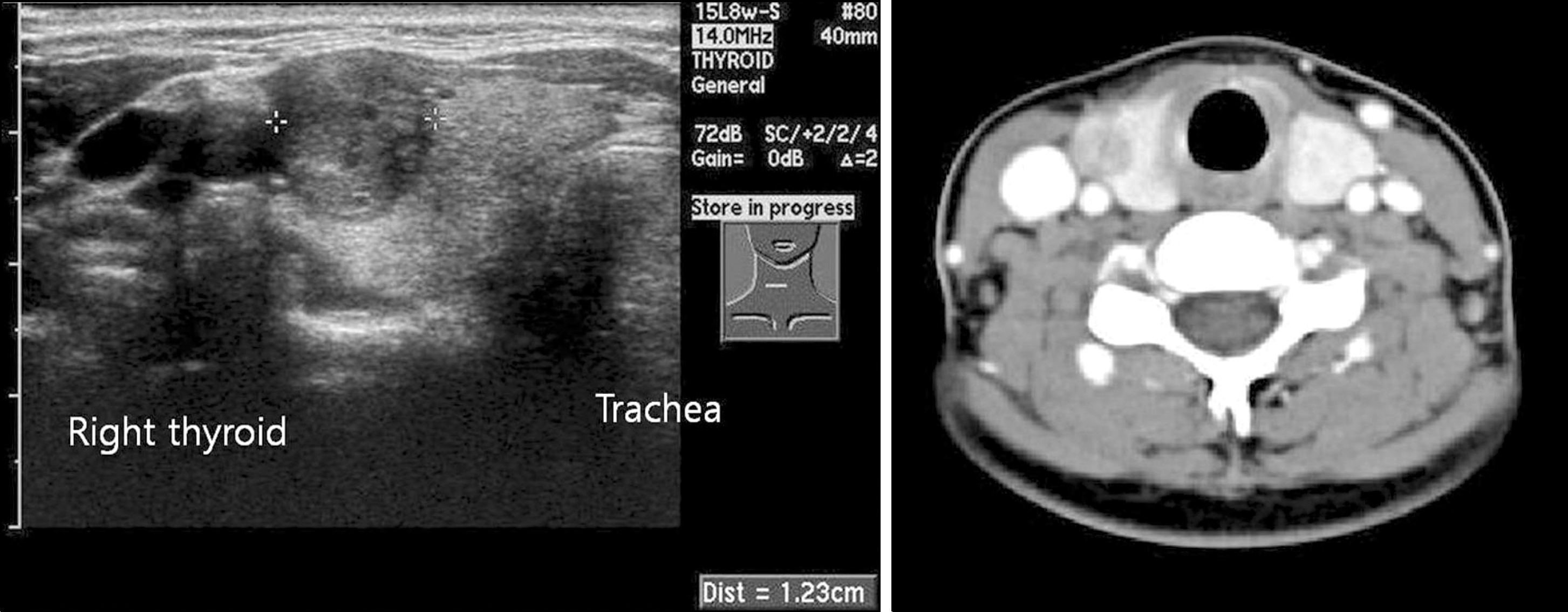 | Fig. 1.Neck ultrasonography and neck computed tomography show a 17×12×13 mm sized ovalshaped, well demarcated solid nodule in the right upper portion of thyroid. |
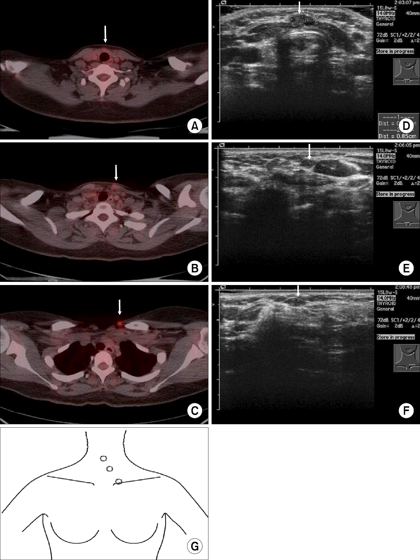
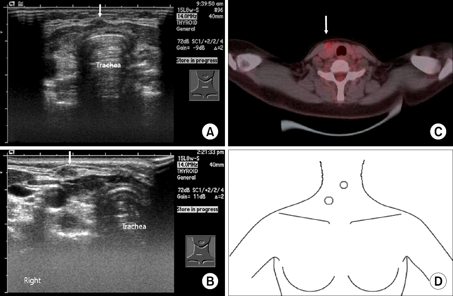 | Fig. 2.The neck ultrasonography shows two nodules in the medial border of right sternocleidomastoid (SCM) muscle, 6 mm sized in the anterior midline and with superior location of another nodule, about 5 mm sized (A, B, D). The PET-CT shows a mildly hypermetabolic nodule at the medial border of right SCM muscle (C). |
 XML Download