Abstract
Hyalinizing trabecular tumor (HTT), a type of thyroid lesion, was first reported by Carney in 1987 and has since been reported continuously. Due to its histological non-specificity, HTT can be misdiagnosed as papillary thyroid cancer or medullary thyroid cancer. For this reason, over treatment might occur; for example, total thyroidectomy and lymphadenectomy. Diagnosis and treatment is a challenge because there is still controversy regarding HTT characters. We report on two cases. One patient was a 48-year-old female and the other was a 46-year-old female. Both patients complained of a thyroid mass and were diagnosed as HTT.
Go to : 
References
1. Akin MR, Nguyen GK. Fine-needle aspiration biopsy cytology of hyalinizing trabecular adenomas of the thyroid. Diagn Cytopathol. 1999; 20:90–4.

2. Lee S, Han BK, Ko EY, Oh YL, Choe JH, Shin JH. The ultrasonography features of hyalinizing trabecular tumor of the thyroid are more consistent with its benign behavior than cytology or frozen section readings. Thyroid. 2011; 21:253–9.

3. Carney JA, Hirokawa M, Lloyd RV, Papotti M, Sebo TJ. Hyalinizing trabecular tumors of the thyroid gland are almost all benign. Am J Surg Pathol. 2008; 32:1877–89.

4. Nga ME, Lim GS, Soh CH, Kumarasinghe MP. HBME-1 and CK19 are highly discriminatory in the cytological diagnosis of papillary thyroid carcinoma. Diagn Cytopathol. 2008; 36:550–6.

5. Carney JA, Ryan J, Goellner JR. Hyalinizing trabecular adenoma of the thyroid gland. Am J Surg Pathol. 1987; 11:583–91.

6. Bishop JA, Ali SZ. Hyalinizing trabecular adenoma of the thyroid gland. Diagn Cytopathol. 2011; 39:306–10.

7. Casey MB, Sebo TJ, Carney JA. Hyalinizing trabecular adenoma of the thyroid gland: cytologic features in 29 cases. Am J Surg Pathol. 2004; 28:859–67.
8. Hirokawa M, Carney JA, Ohtsuki Y. Hyalinizing trabecular adenoma and papillary carcinoma of the thyroid gland express different cytokeratin patterns. Am J Surg Pathol. 2000; 24:877–81.

9. Evenson A, Mowschenson P, Wang H, Connolly J, Mendrinos S, Parangi S, et al. Hyalinizing trabecular adenoma–an uncommon thyroid tumor frequently misdiagnosed as papillary or medullary thyroid carcinoma. Am J Surg. 2007; 193:707–12.

10. Shikama Y, Osawa T, Yagihashi N, Kurotaki H, Yagihashi S. Neuroendocrine differentiation in hyalinizing trabecular tumor of the thyroid. Virchows Arch. 2003; 443:792–6.

11. Gaffney RL, Carney JA, Sebo TJ, Erickson LA, Volante M, Papotti M, et al. Galectin-3 expression in hyalinizing trabecular tumors of the thyroid gland. Am J Surg Pathol. 2003; 27:494–8.

12. Ünlütürk U, Karaveli G, Sak SD, Erdoğan MF. Hyalinizing trabecular tumor in a background of lymphocytic thyroiditis: a challenging neoplasm of the thyroid. Endocr Pract. 2011; 17:e140–3.

13. Hirokawa M, Carney JA. Cell membrane and cytoplasmic staining for MIB-1 in hyalinizing trabecular adenoma of the thyroid gland. Am J Surg Pathol. 2000; 24:575–8.

14. Casey MB, Sebo TJ, Carney JA. Hyalinizing trabecular adenoma of the thyroid gland identification through MIB-1 staining of fine-needle aspiration biopsy smears. Am J Clin Pathol. 2004; 122:506–10.
15. Kim T, Oh YL, Kim KM, Shin JH. Diagnostic dilemmas of hyalinizing trabecular tumours on fine needle aspiration cytology: a study of seven cases with BRAF mutation analysis. Cytopathology. 2011; 22:407–13.

16. Baloch ZW, Puttaswamy K, Brose M, LiVolsi VA. Lack of BRAF mutations in hyalinizing trabecular neoplasm. Cytojournal. 2006; 3:17.
Go to : 
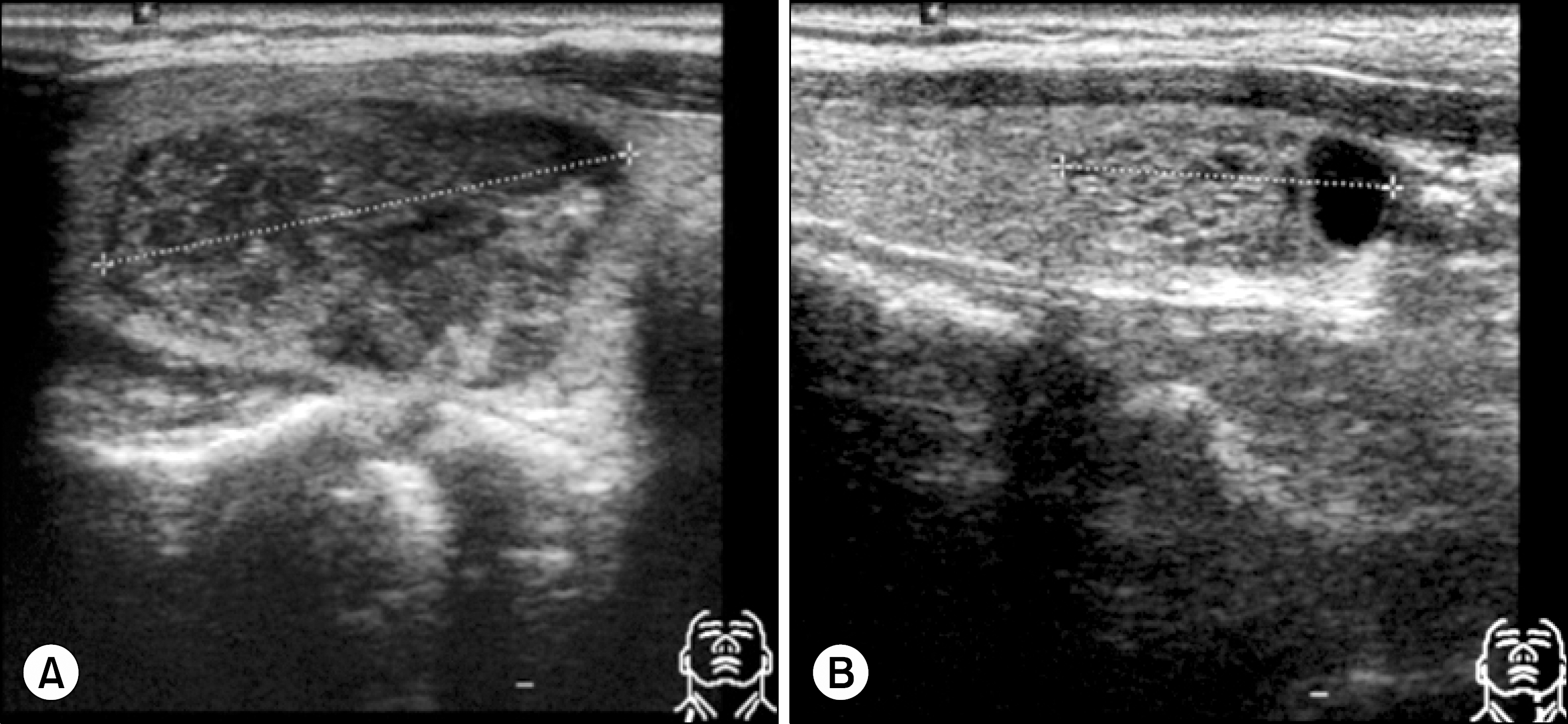
 | Fig. 1.Case 1. Ultrasonography feature of 48-year-old women. Transverse and longitudinal sonogram shows a 2.8 cm solid, cystic and heterogeneous mass in right lobe of thyroid (A). Transverse and longitudinal sonogram show a 1.7 cm solid, cystic and well defined heterogeneous mass in left lobe of thyroid, Fine needle aspiration was done two mass. |
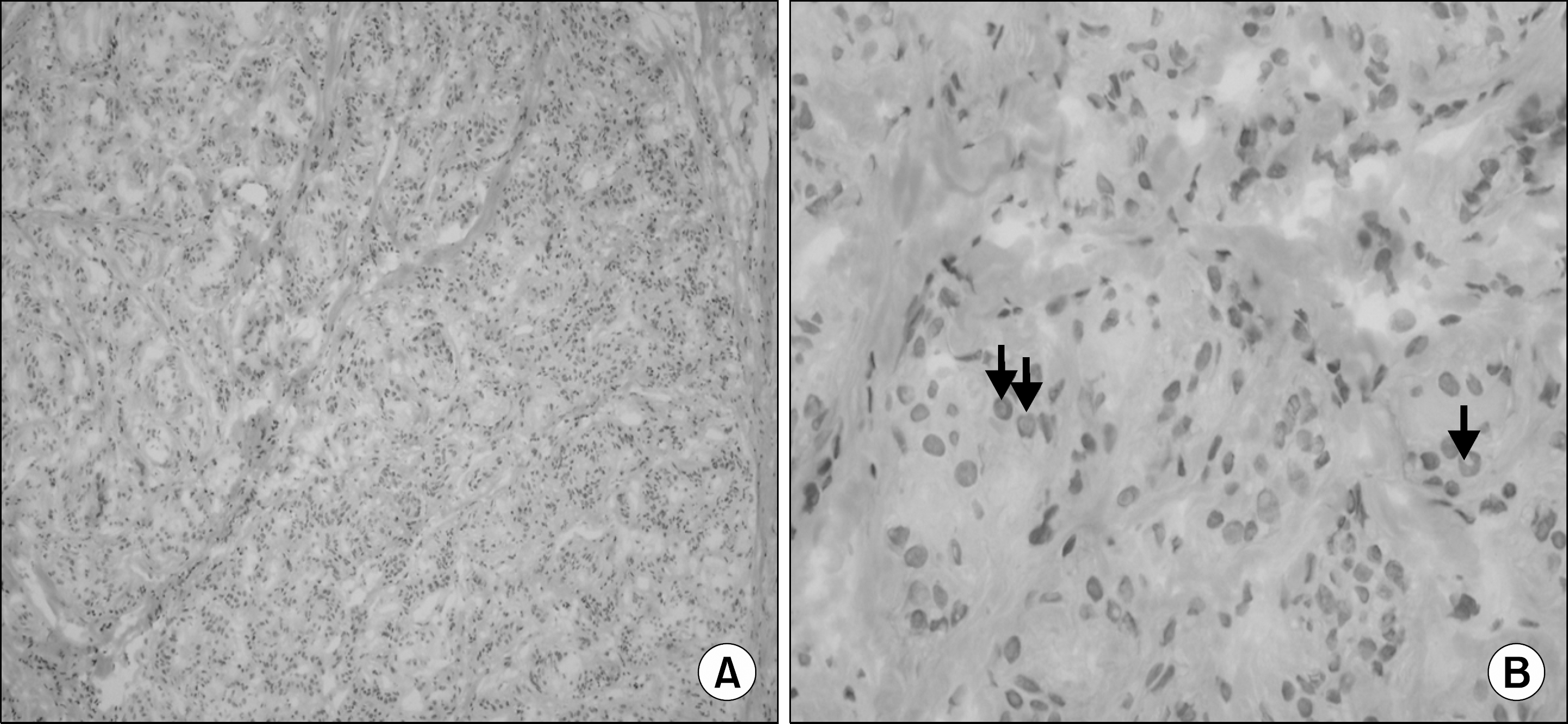
 | Fig. 2.On frozen section show the cluster of tumor cells reveal nuclear overlapping and nuclear inclusion (arrow), which can be misinterpreted as papillary carcinoma (H&E stain, ×100 (A), ×400 (B)). |
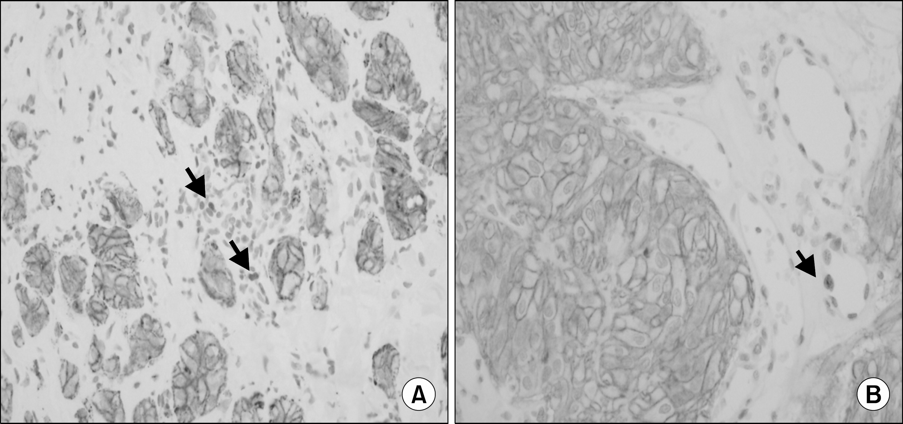
 | Fig. 3.The Ki-67 staining characteristic membranous staining. The endothelial cell or inflammatory cell show usual nuclear staining pattern (arrow). ×400 (A, B). |
 | Fig. 4.Case 2. Ultrasonography feature of 48-year-old women. Transverse and longitudinal sonogram show a 1 cm solid, heterogeneous and intermediate mass, a 0.7 cm irregular hypoechoic mass which suggested malignant lesion in right lobe of thyroid. (A) Transverse and longitudinal sonogram show a 0.9 cm heterogeneous hypoechoic mass in left lobe of thyroid. (B) Fine needle aspiration was done right 0.7 cm, left 0.9 cm mass. |


 PDF
PDF ePub
ePub Citation
 Print
Print


 XML Download