Abstract
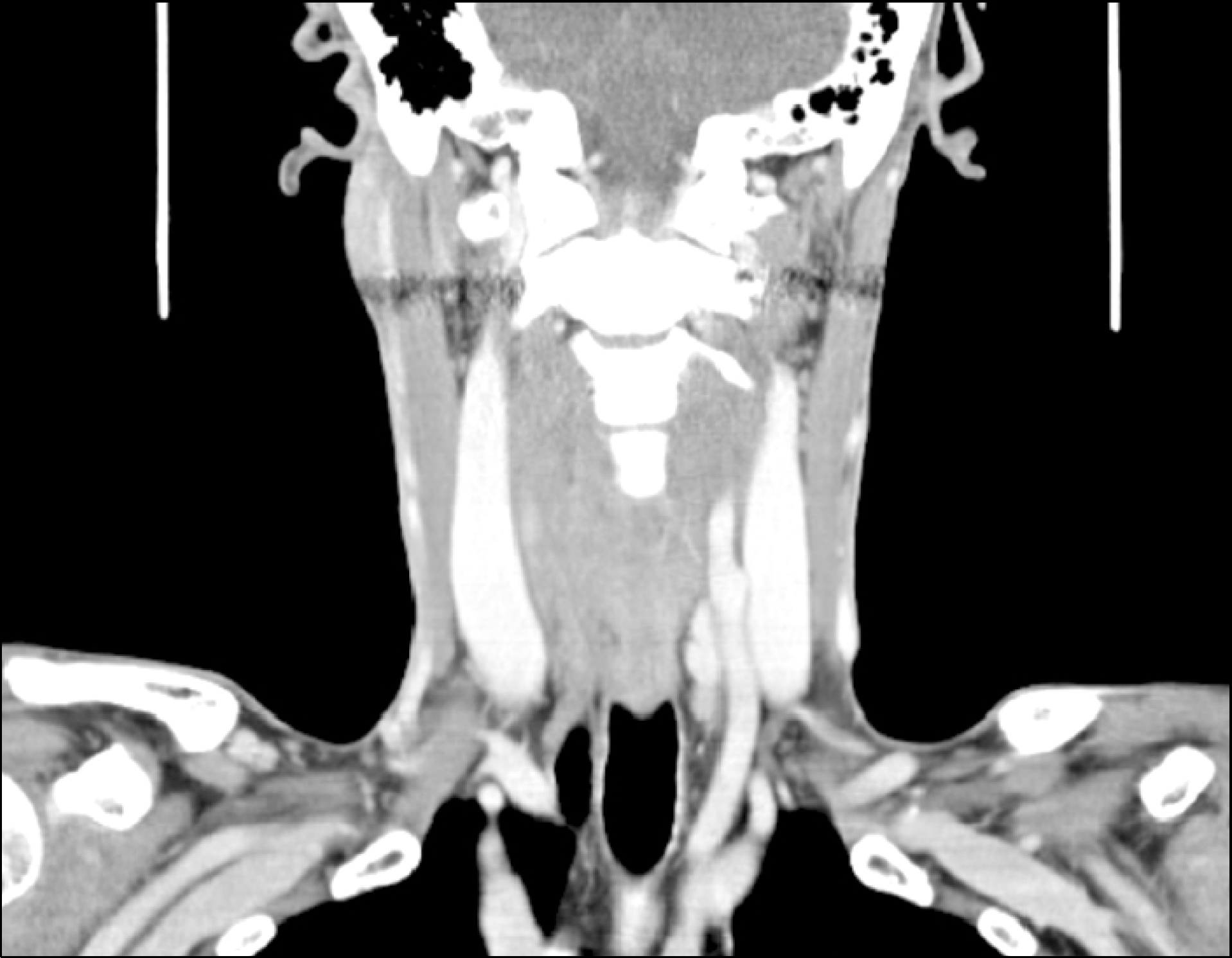
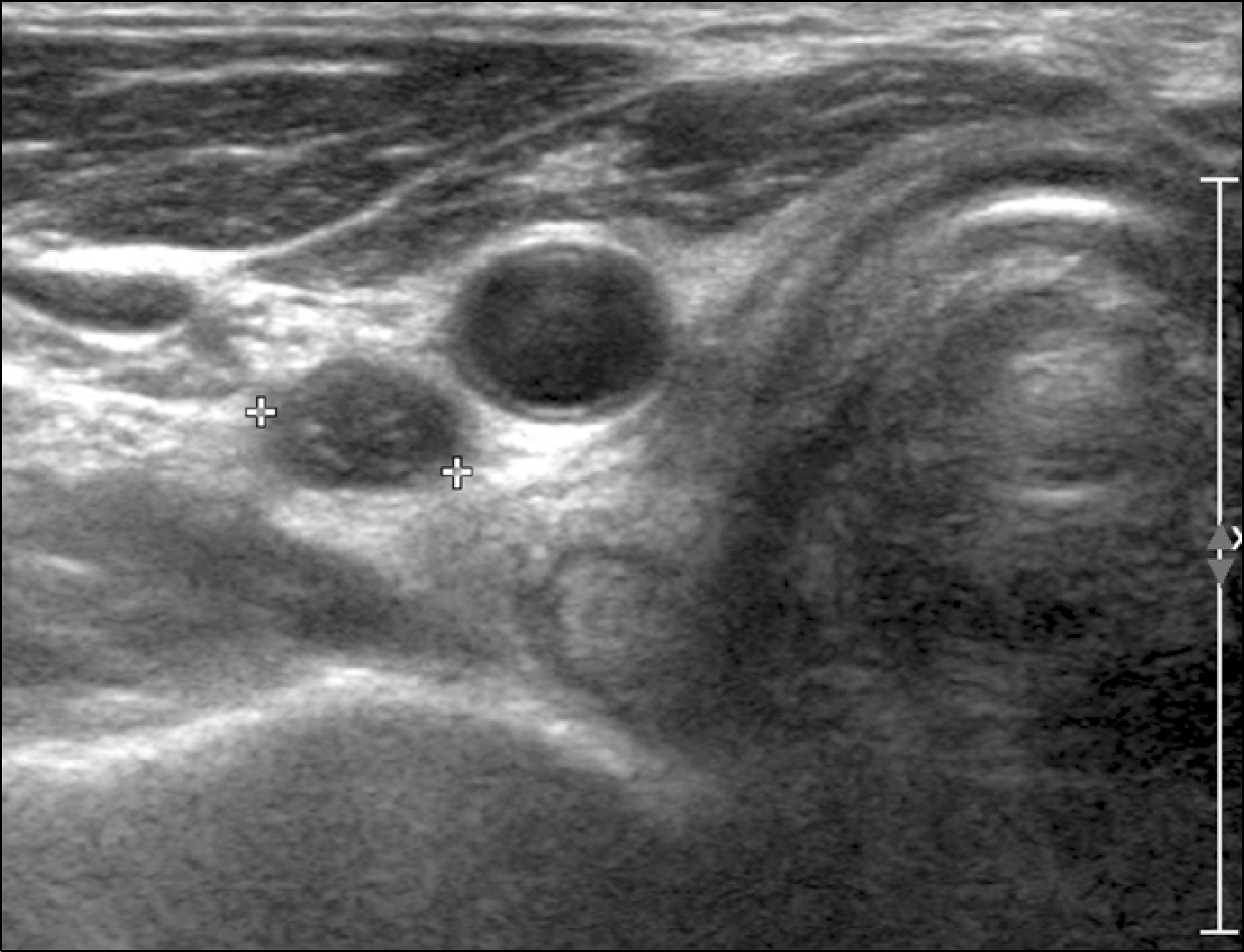
Parathyroid carcinoma is a rare malignancy presenting hyperparathyroidism. At times, diagnosis and localization are difficult. The optimum treatment for parathyroid carcinoma is en bloc resection when malignancy is highly suspicious or diagnosed. However, even after the adequate surgical treatment, persistent or recurrent disease is well encountered. Here we report a case with recurred parathyroid carcinoma presenting discrepancy between image findings and operative findings.
REFERENCES
2.Fujimoto Y., Obara T., Ito Y., Kodama T., Nobori M., Ebihara S. Localization and surgical resection of metastatic parathyroid carcinoma. World J Surg. 1986. 10:539–47.

3.Shaha AR., Shah JP. Parathyroid carcinoma: a diagnostic and therapeutic challenge. Cancer. 1999. 86:378–80.
4.Kirkby-bott J., Lewis P., Harmer CL., Smellie WJ. One stage treatment of parathyroid cancer. Eur J Surg Oncol. 2005. 31:78–83.

5.Sandelin K., Auer G., Bondeson L., Grimelius L., Farnebo LO. Prognostic factors in parathyroid cancer: a review of 95 cases. World J Surg. 1992. 16:724–31.

6.Kebebew E., Arici C., Duh QY., Clark OH. Localization and reoperation results for persistent and recurrent parathyroid carcinoma. Arch Surg. 2001. 136:787–885.

7.Shane E. Clinical review 122: parathyroid carcinoma. J Clin Endocrinol Metab. 2001. 86:485–93.


 PDF
PDF ePub
ePub Citation
 Print
Print


 XML Download