Abstract
Granular cell tumor (GCT) of the thyroid is rare and histogenesis of the carcinoma still remains poorly understood. Here in this study, we report a case of perithyroidal granular cell tumor in a 44-year-old woman, diagnosed as medullary carcinoma upon the interoperative frozen diagnosis. The tumor was comprised of white, solid mass with infiltrating margin in isthmus. Microscopically, the tumor revealed abundant eosinophilic cytoplasm, elongated nucleus and eosinophilic amyloid-like materials. It was composed of diffuse sheets of polygonal cells with abundant eosinophilic cytoplasm and cytologically bland nucleus on permanent section. On immunohistochemical staining, S-100 and CD68 are diffusely positive. Determining the progression and the behavior of the tumor is critical for providing long-term man- agement and preventing aggressive treatment.
REFERENCES
1.Baloch ZW., Martin S., Livolsi VA. Granular cell tumor of the thyroid: a case report. Int J Surg Pathol. 2005. 13:291–4.

2.Compagno J., Hyams VJ., Ste-Marie P. Benign granular cell tumors of the larynx: a review of 36 cases with clinicopathologic data. Ann Otol Rhinol Laryngol. 1975. 84:308–14.

3.Sharon W. Weiss, R. Goldblum J. Benign tumors of peripheral nerves. In: William Schimitt, editors. Soft tissue tumors. 5th ed. Philadelphia: Mosby; p.878–87.
4.Kang H., Hong E., Park M., Lee J. Granular cell tumor of the thyroid. Korean J Pathol. 1998. 32:63–7.
5.Ordonez NG., Mackay B. Granular cell tumor: a review of the pathology and histogenesis. Ultrastruct Pathol. 1999. 23:207–22.
6.Mahoney CP., Patterson SD., Ryan J. Granular cell tumor of the thyroid gland in a girl receiving high-dose estrogen therapy. Pediatr Pathol Lab Med. 1995. 15:791–5.

7.Espinosa-de-Los-Monteros-Franco VA., Martinez-Madrigal F., Ortiz-Hidalgo C. Granular cell tumor (Abrikossoff tumor) of the thyroid gland. Ann Diagn Pathol. 2009. 13:269–71.
9.Milias S., Hytiroglou P., Kourtis D., Papadimitriou CS. Granular cell tumour of the thyroid gland. Histopathology. 2004. 44:190–1.

10.Chang SM., Wei CK., Tseng CE. The cytology of a thyroid granular cell tumor. Endocr Pathol. 2009. 20:137–40.

11.Park J., Kang H., Park M., Cho M., Yoon J., Jaegal Y. A case of granular cell tumor of the thyroid. Korean J Head Neck Oncol. 2006. 22:43–6.
12.Christopher DM. Flecher. Tumors of the thyroid and parathyroid glands. In: Michael Houston, editors. Diagnostic histopathology of tumors. 3rd ed. Philadelphia: Elsevier; p.1026–46.
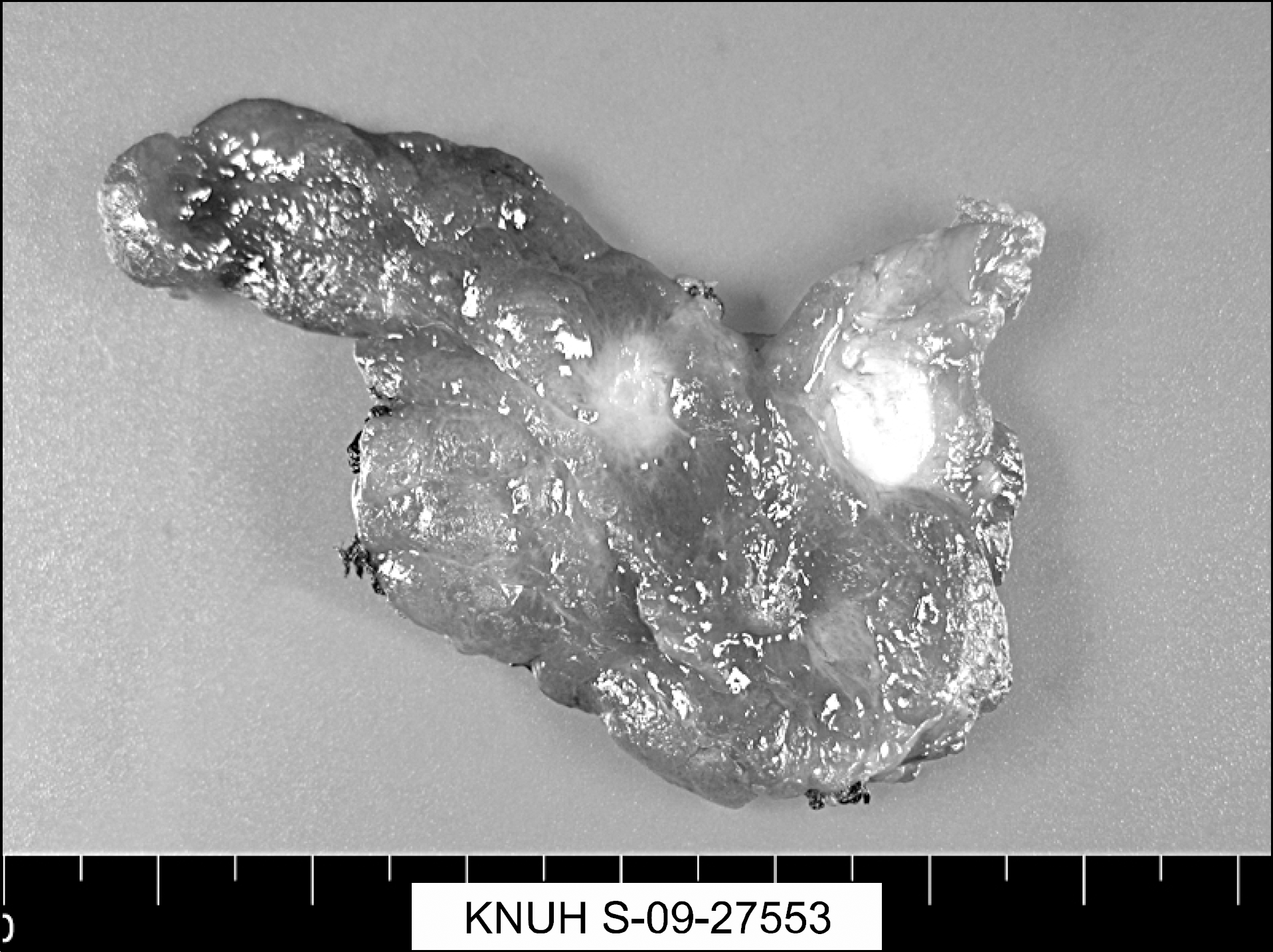
Fig. 1
Gross image of the thyroid gland showing two white solid masses. A 0.7×0.5 cm sized, white and solid nodule with infiltratigns margins located in right thyroid lobe was thyroid papillary carcinoma. The other mass was 0.9×0.7 cm sized, well defined, bright yellow solid tumor with smooth cut surface, located just adjacent to isthmus was GCT.


 PDF
PDF ePub
ePub Citation
 Print
Print


 XML Download