Abstract
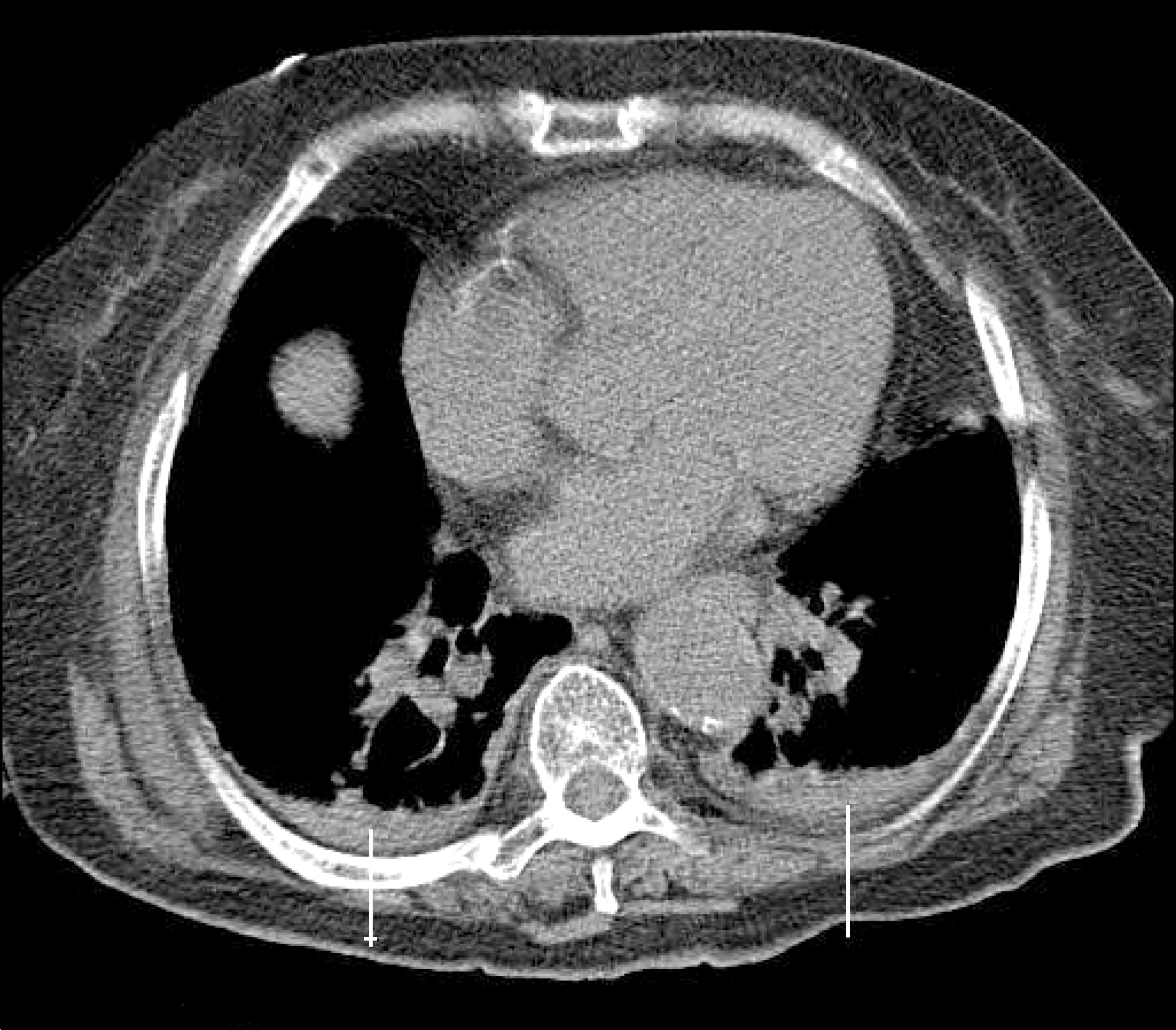
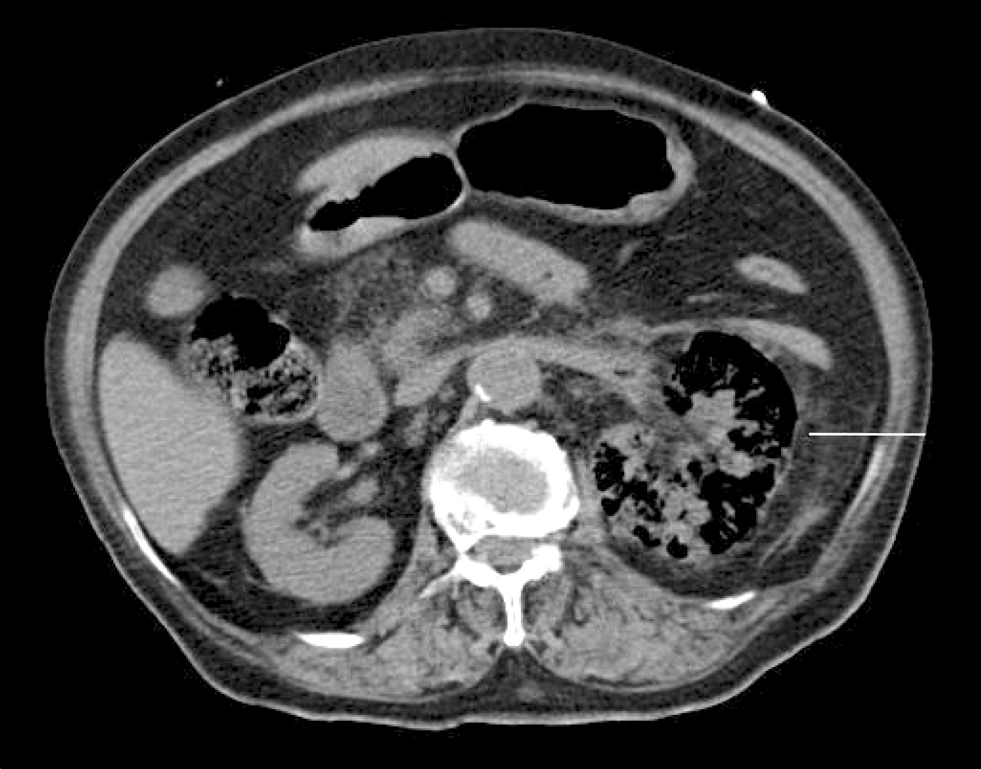
Emphysematous pyelonephritis is an acute gas forming necrotizing infection of the renal parenchyma with high mortality, which shows non-specific clinical findings. Elderly patients with altered mentality and shock require careful monitoring and abdominal computed tomography scanning is thought to be beneficial in prompt detection of infection focus and management. We report on a patient with altered mentality who showed emphysematous pyelonephritis on abdominal computed tomography and provide a review of the literature.
REFERENCES
2. Chen MT, Huang CN, Chou YH, Huang CH, Chiang CP, Liu GC. Percutaneous drainage in the treatment of emphysematous pyelonephritis: 10-year experience. J Urol. 1997; 157:1569–73.

3. Rivers E, Nguyen B, Havstad S, Ressler J, Muzzin A, Knoblich B, et al. Early Goal-Directed Therapy Collaborative Group. Early goal-directed therapy in the treatment of severe sepsis and septic shock. N Engl J Med. 2001; 345:1368–77.

4. McDermid KP, Watterson J, van Eeden SF. Emphysematous pyelonephritis: case report and review of the literature. Diabetes Res Clin Pract. 1999; 44:71–5.

5. Shokeir AA, El-Azab M, Mohsen T, El-Diasty T. Emphysematous pyelonephritis: a 15-year experience with 20 cases. Urology. 1997; 49:343–6.

6. Huang JJ, Tseng CC. Emphysematous pyelonephritis: clinicora-diological classification, management, prognosis, and pathogenesis. Arch Intern Med. 2000; 160:797–805.


 PDF
PDF ePub
ePub Citation
 Print
Print


 XML Download