Abstract
Median raphe cysts of the perineum are rare congenital anomalies of the male genitalia, which form during embryological development, and can be found in the midline from the distal penis to the perineum. However, the incidence of median raphe cyst is likely under-reported and under-recognized. We report on the case of a median raphe cyst extending from the scrotum to the perineum with recurrent infection and purulent discharge in a 28-year-old man, which first developed at the age of 5 years. We believe it is important that urologists recognize median raphe cysts and have knowledge of their management in order to provide appropriate information to patients.
Go to : 
REFERENCES
1. Mermet P. Congenital cysts of the genitoperineal raphe. Revue de Chirurgie. 1895; 15:382–435.
2. Golitz LE, Robin M. Median raphe canals of the penis. Cutis. 1981; 27:170–2.
3. Ravasse P, Petit T, Pasquier CJ. Perineal median raphe canal: a typical image. Urology. 2002; 59:136.

4. Park CO, Chun EY, Lee JH. Median raphe cyst on the scrotum and perineum. J Am Acad Dermatol. 2006; 55(5 Suppl):S114–5.

5. Krauel L, Tarrado X, Garcia-Aparicio L, Lerena J, Sunol M, Rodo J, et al. Median raphe cysts of the perineum in children. Urology. 2008; 71:830–1.

6. Dini M, Baroni G, Colafranceschi M. Median raphe cyst of the penis: a report of two cases with immunohistochemical investigation. Am J Dermatopathol. 2001; 23:320–4.
7. Romani J, Barnadas MA, Miralles J, Curell R, de Moragas JM. Median raphe cyst of the penis with ciliated cells. J Cutan Pathol. 1995; 22:378–81.

8. Otsuka T, Ueda Y, Terauchi M, Kinoshita Y. Median raphe (parameatal) cysts of the penis. J Urol. 1998; 159:1918–20.

9. Lopez-Candel E, Roig Alvaro J, Lopez-Candel J, Fernandez Dozagarat S, Soler J, Hernandez Bermejo JP, et al. Median raphe cysts of the perineum in childhood. An Esp Pediatr. 2000; 52:395–7.
Go to : 
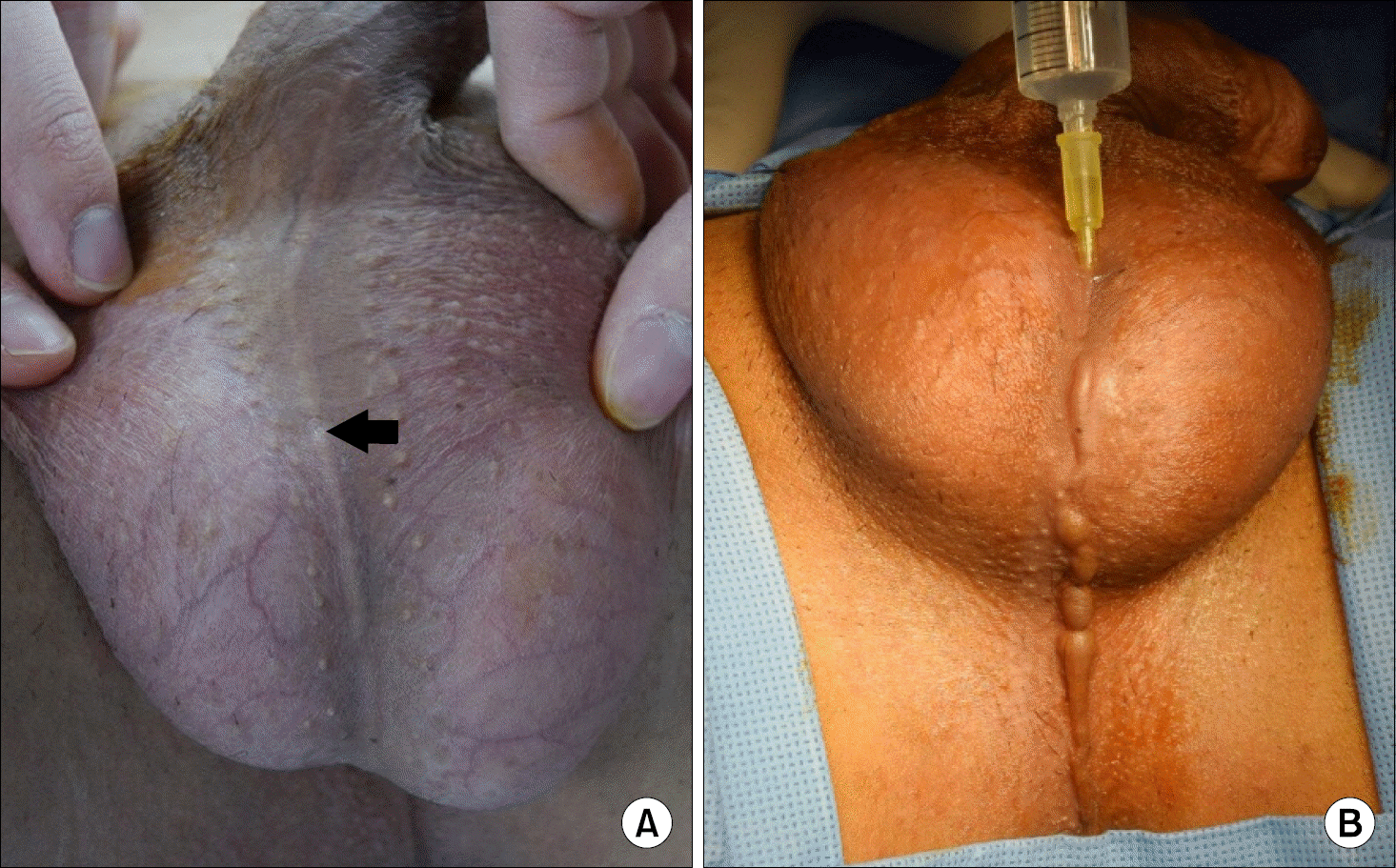
 | Fig. 1.(A) The scrotum and the perineum appear to have an almost normal shape except for the fistula opening (arrow) on the mid-scrotum. (B) After injection of normal saline through the fistula opening, several 5-7 mm canalized cystic lesions are found from the mid portion of the scrotum to the perineum. |


 PDF
PDF ePub
ePub Citation
 Print
Print


 XML Download