Abstract
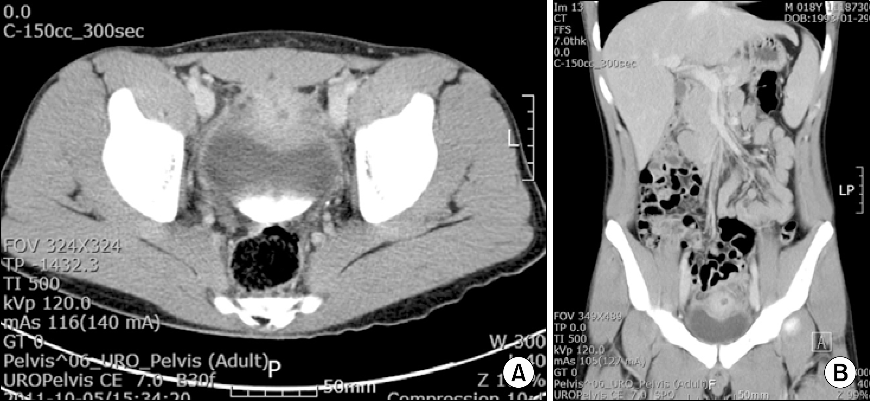
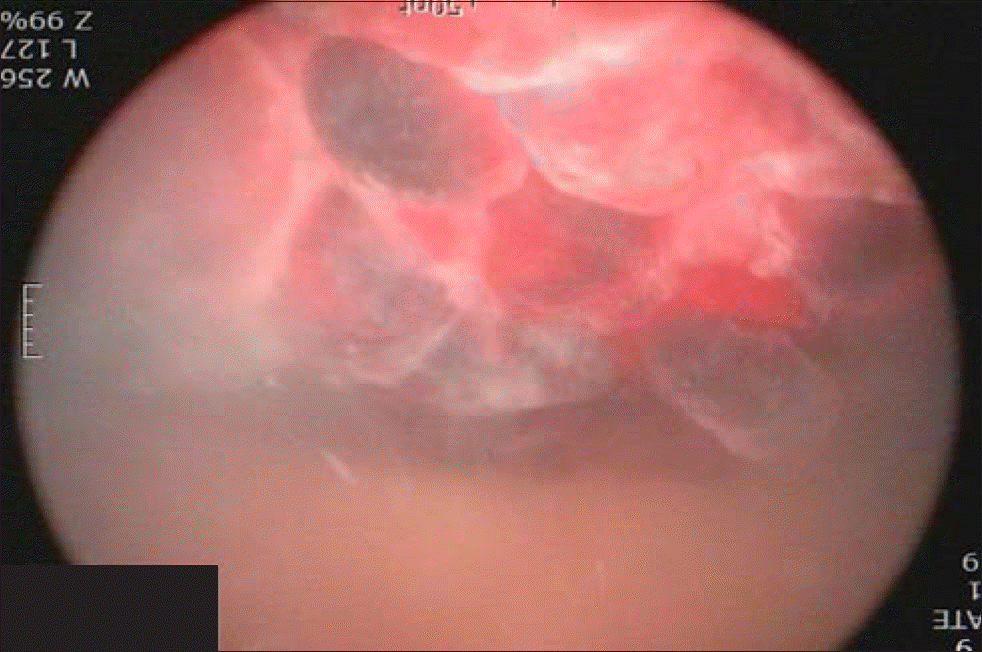
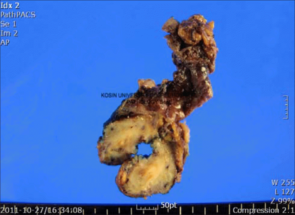
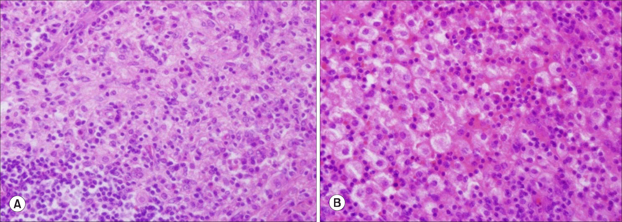
Urachal xanthogranuloma is an extremely rare disease. An 18-year-old male presented with lower abdominal pain, hematuria, and dysuria. An urachal mass with bladder invasion, which was suspected to be an urachal carcinoma or abscess, was observed on computed tomography. Exploratory laparotomy, excision of the urachus, and partial cystectomy was performed by way of a lower midline incision. Histopathologic examination identified the mass as an urachal xanthogranuloma.
REFERENCES
1. Yamamoto T, Mori Y, Katoh Y, Iguchi M, Minamidani K, Sawai Y, et al. A case of urachal xanthogranuloma suspected to be a urachal tumor. Hinyokika Kiyo. 2004; 50:493–5.
2. Carrere W, Gutierrez R, Umbert B, Sole M, Menendez V, Carretero P. Urachal xanthogranulomatous disease. Br J Urol. 1996; 77:612–3.

3. Diaz Candamio MJ, Pombo F, Arnal F, Busto L. Xanthogranulomatous urachitis: CT findings. J Comput Assist Tomogr. 1998; 22:93–5.
4. Kuo TL, Cheng C. Xanthogranulomatous inflammation of urachus mimicking urachal carcinoma. Urology. 2009; 73:443. e13-4.

5. Tian J, Ma JH, Li CL, Xiao ZD. Urachal mass in adults: clinical analysis of 33 cases. Zhonghua Yi Xue Za Zhi. 2008; 88:820–2.
6. Park S, Ji YH, Cheon SH, Kim YM, Moon KH. Urachal xanthogranuloma: laparoscopic excision with minimal incision. Korean J Urol. 2009; 50:714–7.

8. Fornari A, Dambros M, Teloken C, Hartmann AA, Kolling J, Seben R. A case of xanthogranulomatous cystitis. Int Urogy-necol J Pelvic Floor Dysfunct. 2007; 18:1233–5.

9. Kim DY, Kim HS, Kim IK, Moon I, Kim TS, Choi S, et al. Xanthogranulomatous cystitis presenting as a urachal carcinoma. Korean J Urol. 2004; 45:1180–2.
10. Han DH, Choi HJ, Kim JH, Shin JS, Chung KJ, Choi HY, et al. Xanthogranulomatous cystitis. Korean J Urol. 2004; 45:958–61.
Fig. 1.
Ultrasound of bladder shows a protruding cystic mass in bladder dome. Sagittal (A) and transverse (B) plane.

Fig. 2.
Computed tomography of the abdomen shows well enhancing mass in the anterior wall of urinary bladder. Sagittal (A) and transverse (B) plane.


 PDF
PDF ePub
ePub Citation
 Print
Print


 XML Download