

 PDF
PDF ePub
ePub Citation
 Print
Print


Abstract
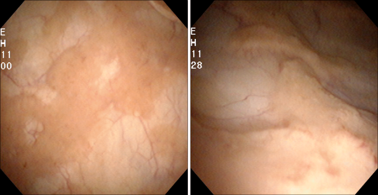
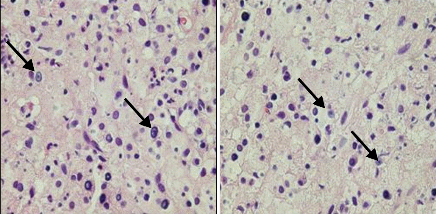
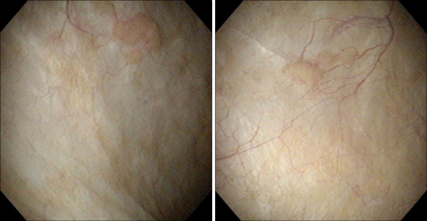
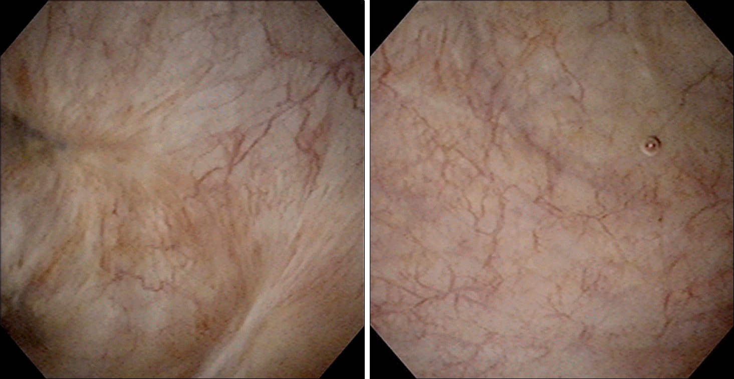
Malakoplakia is a rare chronic granulomatous disease, which was originally described in the urinary bladder, but can involve many other organs and soft tissues. Malakoplakia is often associated with immunosuppression or immunodeficiency and is believed to be caused by an alternation in the bacterial phagocytic system. Histologically, the presence of Michaelis-Gutmann bodies is pathognomonic. We report on a case of malakoplakia of the bladder in a 62-year-old female.
REFERENCES
1. Michaelis L, Gutmann C. Ueber eischlusse in blasentumoren. Z Klin Med. 1902; 47:208.
2. von Hansemann D. Uber malakoplakie der harnblase. Virch Arch Path Anat. 1903; 173:302–8.
3. Stanton MJ, Maxted W. Malacoplakia: a study of the literature and current concepts of pathogenesis, diagnosis and treatment. J Urol. 1981; 125:139–46.

4. Yousef GM, Naghibi B, Hamodat MM. Malakoplakia outside the urinary tract. Arch Pathol Lab Med. 2007; 131:297–300.
5. Lemmens B, Besnier JM, Diot P, Fetissof F, Anthonioz P, Cattier B, et al. Pulmonary malacoplakia and Rhodococcus equi pneumonia in a patient infected with the human immunodeficiency virus. A case report with review of the literature. Rev Mal Respir. 1994; 11:301–3.
6. McClure J. Malakoplakia. J Pathol. 1983; 140:275–330.
7. An T, Ferenczy A, Wilens SL, Melicow MM. Observations on the formation of Michaelis-Gutmann bodies. Hum Pathol. 1974; 5:753–8.
8. van Furth R, van't Wout JW, Wertheimer PA, Zwartendijk J. Ciprofloxacin for treatment of malakoplakia. Lancet. 1992; 339:148–9.
9. Maderazo EG, Berlin BB, Morhardt C. Treatment of malakoplakia with trimethoprim-sulfamethoxazole. Urology. 1979; 13:70–3.
 XML Download