

 PDF
PDF ePub
ePub Citation
 Print
Print


INTRODUCTION
Fish oil consumption has been shown to have a protective role in some chronic degenerative diseases such as cardiovascular disease, autoimmune disorders, type 2 diabetes, rheumatoid arthritis, and cancer [1]. The health beneficial effects of fish oil have been attributed to its high content of the omega-3 (n-3) polyunsaturated fatty acids (PUFAs), such as docosahexaenoic acid (DHA) and eicosapentaenoic acid (EPA) [2]. DHA (all cis-docosa-4,7,10,13,16,19-hexaenoic acid) has 22 carbons with 6 double bonds, and most unsaturated fatty acid commonly found in biological systems. EPA is elongated twice to yield 24:5 n-3 PUFA, followed by desaturation twice to 24:6 n-3 PUFA, which is oxidized to form DHA [3]. Both EPA and DHA produce the resolvins (resolution-phase interaction products) and other endogenous lipid mediators possessing both pro-resolving as well as anti-inflammatory effects (Figure 1). Failure of resolution results in chronic inflammation which contributes to substantial part of colon cancer and other human malignancies. Therefore, both EPA and DHA have attracted much attention due to their chemopreventive and adjuvant chemotherapeutic potential [4].
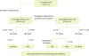 | Figure 1Metabolic formation of EPA and DHA and their conversion to anti-inflammatory and pro-resolving metabolites with chemopreventive and therapeutic potentials.
EPA, eicosapentaenoic acid; DHA, docosahexaenoic acid; n-3, omega-3; ASA, aspirin; COX-2, cyclooxygenase-2; CYP450, cytochrome P450; 18S-HEPE, 18S-hydroxyeicosapentaenoic acid; 18R-HEPE, 18R-hydroxyeicosapentaenoic acid; 18S-RvE1, 18S-resolvin E1; RvE1, resolvins E1; 5-LOX, 5-lipoxygenase; 17S-HDHA, 17S-hydroxy docosahexaenoic acid; 17R-HDHA, 17R-hydroxy docosahexaenoic acid; PD1, protectin D1; RvD1, resolvin D1; 17R-RvD1, 17R-resolvin D1.
|
Colorectal cancer (CRC) is the third most common cause of cancer-associated death in the world [56]. CRC can be caused by familial and hereditary factors as well as environmental lifestyle-related risk factors, such as physical inactivity, obesity, smoking, and alcohol consumption [7]. It has been reported that obese people are at 7% to 60% greater risk of CRC than normal individuals [8]. According to a case-control study conducted in Korea, obesity and hyperglycemia are positively related to advanced colorectal adenoma formation [9].
Both the type and the amount of dietary fats differing in PUFA composition have been implicated in the etiology and pathogenesis of colon cancer. Rather than their absolute intake, a balanced ratio of PUFAs in the diet is essential for normal growth and development [10]. A high fat diet containing saturated fatty acids promotes colonic aberrant crypt foci (ACF) formation, and increases the incidence and the multiplicity of colon tumors [11]. Consistent with this experimental study, epidemiological studies revealed that Greenland Eskimo populations eating their traditional diet rich in n-3 PUFAs compared to reference populations in the West showed a significantly lower incidence of CRC [1213]. In contrast, Western-style diet that contains relatively large proportion of omega-6 (n-6) PUFAs is associated with a high risk for inflammatory bowel disease (IBD) and colon carcinogenesis through enhancement of inflammation [14].
The consumption of n-3 PUFAs has been known to exert protective effects against colon cancer by modulating intracellular signaling processes involved in multistage carcinogenesis [15]. Biochemical mechanisms underlying the multiple health benefits of n-3 PUFAs include suppression of inflammation, inhibition of proliferation, metastasis and angiogenesis of cancer cells, induction of differentiation and apoptosis of cancer cells, modification of the lipid raft, etc. Notably, several studies demonstrated that combination of n-3 PUFAs with anticancer agents potentiated their preventive/therapeutic effects. This review highlights the chemopreventive and therapeutic effects of n-3 PUFAs and their potential as an adjuvant to improve the efficacy of cancer chemotherapeutics.
RATIO OF n-6 TO n-3 PUFAs AS A DETERMINANT OF COLON CANCER RISK
A ratio of n-3 to n-6 PUFAs in the diet has been considered to be a critical determinant of risk for colon cancer. The 2.5:1 ratio of fish oil (n-3 PUFA) to corn oil (n-6 PUFA) elicited a better chemopreventive efficacy as compared to the 1:1 ratio of these fatty acids mix in dihydrochloride-induced colon carcinogenesis by suppressing the cell cycle progression [16]. The fat-1 gene of Caenorhabditis elegans encodes an n-3 fatty-acid desaturase that converts n-6 to n-3 PUFAs [17]. This gene is absent in most mammals including humans. The ratio of n-6/n-3 PUFA in colonic cells of fat-1 transgenic mice was markedly lower compared to wild-type mice [18]. Fat-1 transgenic mice are less susceptible to dextran sulfate sodium (DSS)-induced chronic inflammation with significant decreases in expression of cyclooxygenase-2 (COX-2) and production of prostaglandin E2 (PGE2) and proinflammatory cytokines, such as interleukin (IL)-18, IL-1α, IL-1β, IL-6, and tumor necrosis factor-α (TNF-α) [18]. Likewise, trinitrobenzenesulfonic acid (TNBS)-induced colitis was less severe in fat-1 transgenic mice than the wild-type mice [19]. Moreover, fat-1 transgenic mice exhibited a reduced number of adenocarcinomas and elevated apoptosis compared with wild-type mice [20]. In addition, levels of n-6 PUFA derived eicosanoids such as PGE2, prostaglandin D2 (PGD2), prostaglandin E1 (PGE1) and 12-hydroxyeicosatetraenoic acid (12-HETE) were significantly reduced, whereas that of EPA-derived prostaglandin, prostaglandin E3 (PGE3) was elevated in fat-1 mice [20]. Th1 cells, in part, are responsible for mediating IBD. Fat-1 transgenic mice exhibited a decreased proportion of CD3+ T cells and CD4+ T-helper cells (Th1 and Th2) in lamina propria after 2 weeks of recovery in DSS-induced murine colitis. Above findings suggest that the ratio of n-3/n-6 PUFAs is important factor to determine the cellular generation of pro- and anti-inflammatory eicosanoids.
EFFECTS OF n-3 PUFAs ON LIPID RAFT
Lipid microdomains that include lipid rafts and caveolae are small cholesterol- and sphingolipid-enriched segments present in the membranes of most cells. Perturbations in lipid composition of microdomains alter the function of resident proteins and subsequent downstream signaling. Therefore, lipid rafts are important for cell survival signaling, T-cell activation, and protein and lipid trafficking [21].
Dietary n-3 PUFAs have membrane remodeling properties including alteration of membrane fluidity, phase behavior, permeability, fusion, flip-flop, and resident protein activity [22]. n-3 PUFAs are effectively incorporated into phospholipids of lipid rafts, thereby increasing phospholipid n-3 fatty acyl content and reducing cholesterol and the localization of resident proteins, which contribute to regulation of cell signaling [22]. n-3 PUFAs decreases the localization of H-Ras and eNOS in colonic caveolae, thereby suppressing epidermal growth factor (EGF)-stimulated H-Ras signaling [22]. DHA displaces several raft-associated oncoproteins, including EGF receptor (EGFR), Hsp90, Akt, and Src, from the rafts. Furthermore, DHA reduces total levels and activities of those proteins via multiple processes, including the proteasomal and lysosomal pathways [23]. Effects of DHA on lipid rafts are reversed by cholesterol. Therefore, DHA could exert its anti-cancer effect, at least in part, via modulation of lipid raft-associated signaling events.
P-glycoprotein (Pgp) and multidrug resistance related protein 1 (MRP1) are membrane transporters involved in multidrug resistance of colon cancer cells. The expression of Pgp and MRP1 is increased by large amounts of cholesterol in plasma membrane and detergent resistant membranes [24]. Stability of these proteins is enhanced by lower expression of ubiquitin E3 ligase Trc8. DHA and EPA reduced the amount of Pgp and MRP1 through reactivation of Trc8 E3 ligase which leads to restoration of ubiquitination of 3-hydroxy-3-methylglutaryl-coenzyme A reductase, thereby reducing the cholesterol synthesis and incorporation in detergent resistant membranes [24]. This contributes to decreased transporter activity, restoration of the antitumor effects against chemotherapeutic agents and stimulation of the proper tumor-immune system recognition in response to chemotherapy in multidrug resistant cells [24].
ANTI-INFLAMMATORY EFFECTS OF n-3 PUFAs
Failure of resolution of acute inflammation can result in chronic inflammation. Chronic inflammation-associated disorders, such as Crohn's disease and ulcerative colitis, have been known to increase the risk of developing colon cancer. The protective effect of n-3 PUFAs is mainly attributed to their anti-inflammatory properties. It has been reported that n-3 fatty acids are beneficial in patients suffering from infectious diseases characterized by inflammatory symptoms [25]. According to the population study conducted in Italy, levels of n-3 PUFAs in the serum are correlated with those of anti-inflammatory cytokines [26]. Serum levels of total n-3 PUFAs (DHA, EPA, and α-linolenic acid) were associated with lower production of pro-inflammatory markers (IL-6, IL-1ra, and TNF-α and higher production of anti-inflammatory markers; e.g., soluble IL-6r, IL-10, and transforming growth factor-β [TGF-β]). In contrast, lower n-6 PUFA levels were significantly associated with higher IL-1ra and lower TGF-β production. Moreover, the n-6 to n-3 ratio is inversely correlated with IL-10 [26]. Clinical studies and meta-analysis in surgical patients with colon cancer indicated that supplementing parenteral nutrition regimens with n-3 PUFAs, in particular EPA and DHA, is associated with improved clinical outcomes. Especially, colon cancer patients who consumed various types of n-3 PUFAs showed the relatively low level of inflammation markers (Table 1). Patients consuming n-3 PUFA-enriched emulsions showed statistically and clinically significant reduction in the infection rate and the lengths of stay, both in the intensive care unit and in hospital overall [27]. These patients also showed the reduced markers of inflammation, improved lung gas exchange, liver function, antioxidant status and fatty acid composition of plasma phospholipids, and a trend towards less impairment of kidney function [27].
Table 1
Therapeutic effects of dietary long-chain n-3 PUFAs in colon cancer patients
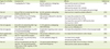
| Type of n-3 PUFAs | Dose/period | Subjects | Effects | Reference |
|---|---|---|---|---|
| Fish oil | 1.2 g/kg/day for 7 days | 42 CRC patients undergoing radical resection | • Reduce the serum IL-6 levels | [73] |
| • Increase the CD4+/CD8+ | ||||
| • Reduce the serum TNF-α levels | ||||
| • Increase the CD3 & CD4 lymphocyte percentage | ||||
| Fish oil | 2.0 g of fish oil containing 600 mg of EPA & DHA for 9 wk | 23 CRC patients undergoing chemotherapy | • Reduce the C-reactive protein/albumin | [74] |
| Oral supplement of n-3 FAs | 2.0 g of EPA & 1.0 g of DHA/day for 7 days before surgery | 148 patients referred for elective CRC surgery | • Increase the production LTB5 | [75] |
| • Reduce the production of LTB4 | ||||
| • Increase the neutrophil 5-HEPE production | ||||
| • Reduce the 5-HETE | ||||
| Fish oil capsule | 2.0 g fish oil containing 600 mg/EPA + DHA/day for 9 wk | 11 CRC patients undergoing chemotherapy | • Increase the body weight | [76] |
| • Reduce the CRP/albumin | ||||
| Fish oil capsule | 2.0 g fish oil containing 1.4 g EPA & 1.0 g DHA/twice/day | 51 patients requiring colon cancer surgery | • Increase the proportion of EPA in the mucosal lipids | [77] |
| Oral supplement of n-3 FAs | 2.0 g of EPA & 1.0 g of DHA/twice/day for 7 days before surgery | 148 patients referred to colon cancer surgery | • Increase the DHA levels in colon tissue | [64] |
| Fish oil | 6.1 g fat with 1.0 g of EPA/twice/day for 12 wk | 10 CRC patients (stage IV) | • Increase body weight | [78] |
| • Enhanced quality of life | ||||
| Fish oil | Fish oil supplements per day (12 mg EPA + 45 mg DHA/capsule) (total dose of 456 mg/day of EPA + DHA) for 2 yr | 104 participants belong to experimental group | • Reduce the ratio of n-6 PUFAs/n-3 PUFAs | [79] |
| • Reduce the colon cancer incidence |
n-3, omega-3; FA, fatty acids; PUFAs, polyunsaturated fatty acids; CRC, colorectal cancer; IL, interleukin; TNF-α, tumor necrosis factor-α; EPA, eicosapentaenoic acid; DHA, docosahexaenoic acid; LTB4, leukotriene B4; LTB5, leukotriene B5; HETE, hydroxyeicosatetraenoic acid; HEPE, hydroxy-eicosapentaenoic acid; CRP, C-reactive protein; n-6, omega-6.
![]()
DHA administration ameliorated experimentally induced colitis in the IL-10−/− mice, as demonstrated by decreased production of proinflammatory cytokines (TNF-α and interferon [IFN]-γ), reduced infiltration of inflammatory cells, and lowered histologic scores of the proximal colon mucosa [28]. Moreover, in the DHA-treated mice, enhanced autophagy was found to be associated with increased expression and restoration of the distribution integrity of microtubule-associated protein 1 light chain 3B (LC3B) in the colon and inhibition of the mechanistic target of rapamycin (mTOR) signaling pathway [28].
Chronic low-grade inflammation in adipose tissue has been recognized as a key step in the development of obesity-associated complications. Obesity is associated with increased all-cause mortality, and cancer accounts for a substantial proportion of obesity-related deaths. Inflammation links obesity and colon cancer. Obese patients with colon cancer show the markedly elevated levels of serum C-reactive protein, which is correlated with poor prognosis [29]. In obesity, accumulation of infiltrating macrophages in adipose tissues and their phenotypic switch to M1-type contribute to chronic inflammation characterized by abnormal secretion of pro-inflammatory adipokines [3031]. In addition, the incidence of obesity and its comorbidities, such as insulin resistance and type II diabetes, are associated with fatty acid composition of commonly consumed human diets. Fish oils reduced the number of adipose macrophages and the expression of plasma macrophage chemoattractant protein 1 (MCP-1) in macrophages and adipocytes in vitro [32].
Beside uncontrolled chronic inflammation, unresolved inflammation leads to colon carcinogenesis. DHA and its metabolites have a preventive potential in the management of human cancer via generation of anti-inflammatory and pro-resolving mediators [33]. For instance, resolvins, protectins, and maresins are endogenously generated from n-3 PUFAs during the spontaneous resolution phase [34]. These pro-resolving bioactive lipids act as “stop-signaling” of the inflammatory response and have potent anti-inflammatory and anti-carcinogenic properties. A deficit in the production of these endogenous anti-inflammatory and pro-resolving signals was demonstrated in adipose tissue from obese individuals [35]. n-3 PUFAs increased production of DHA-derived pro-resolving mediators in women with obesity [36]. In addition, generation of oxylipins (oxygenated metabolites of fatty acids) from α-linolenic acid, linoleic acid, and arachidonic acid by lipoxygenases has been reported to decrease the triglyceride accumulation in lipid droplet and blocked lipid production [37]. The restoration of their levels by either exogenous administration of these mediators or feeding diets enriched with n-3 PUFAs, improves the inflammatory status of adipose tissue and blocks or retards the inflammation-associated carcinogenesis [35]. Elevated production of resolvins and upregulation of the resolvin receptor occurred in parallel with activation of peroxisome proliferator receptor-α. Lipoxins and resolvins are considered as new targets for regulation of inflammation and inflammation-associated cancers [38].
COMBINATION/SYNERGETIC EFFECTS OF n-PUFAs WITH CHEMOTHERAPEUTIC AGENTS
The combination therapy using synthetic and natural compound is considered as one of the most reliable strategies to enhance the efficacy of therapeutic agents. Dietary intake of n-3 PUFAs has been shown to improve the efficacy of chemotherapeutic agents in the in vitro, animal and clinical studies (Table 2). For example, 5-fluorouracil (5-FU) is an anti-metabolite that is used as a chemotherapeutic agent for a wide variety of cancers including CRC [3940]. The combined treatment of the colon cancer cells with fish oil and 5-FU markedly enhanced growth inhibition compared to cells exposure to either substance alone [39]. 5-FU in combination with fish oil synergistically increased the survival rate of animals and inhibited the tumor growth and serum sialic acid levels in a 1,2-dimethylhydrazine dihydrochloride plus DSS-induced mouse colon cancer model [41]. Additionally, fish oil ameliorated hematologic depression and gastrointestinal, hepatic and renal toxicity caused by 5-FU as substantiated by a marked improvement in structural and functional alterations of these organs [41]. Moreover, DHA potentiates the proapoptotic effect of 5-FU through downregulation of antiapoptotic proteins Bcl-2 and Bcl-XL [42]. DHA also markedly induced overexpression of c-Myc which confers a driving force to induce apoptosis and sensitize cancer cells to the therapeutic action of 5-FU [42].
Table 2
Combination/synergistic effects of n-3 PUFAs with chemotherapeutic or chemopreventive agents
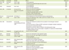
| n-3 PUFAs | Chemicals | Models | Effects | Reference |
|---|---|---|---|---|
| DHA, EPA | Paclitaxel | Caco-2 cells | • Induce apoptosis | [80] |
| DHA | 5-FU | Caco-2 cells | • Inhibit the cell growth through cell cycle arrest | [39] |
| • Induce apoptosis | [42] | |||
| DHA | Celecoxib | HCA-7 cell | • Induce apoptosis | [43] |
| • Reduce the COX-2 expression | ||||
| DHA, EPA | 5-FU & OX | HT-29, HCT-116 | • Reduce the CSC/CSLC population | [44] |
| SCID mice xenografts of CR | • Suppression of tumor growth | |||
| • Increase the phosphorylation of PTEN | ||||
| • Reduction of Akt phosphorylation | ||||
| • Normalization of β-catenin expression | ||||
| Fish oil | 5-FU & OX & IRI | HT-29 (Bax+/+) | • Induce apoptosis via mitochondrial membrane depolarization | [81] |
| DHA | p-XSC | CaCo-2 cells | • Reduce the expression COX-2, iNOS, cyclin D1, β-catenin, NF-κB | [46] |
| • Inhibit the cell growth | ||||
| • Induce apoptosis | ||||
| DHA, EPA | Doxorubicin | HT-29 cells, chemoresistant HT-29-dx cells | • Reduce the cholesterol synthesis & incorporation in the detergent resistant membrane | [24] |
| • Reduced the amount of Pgp and MRP1 contained in detergent resistant membrane | ||||
| • Decreased the transporters activity | ||||
| • Restored the antitumor effects of different chemotherapeutic drugs | ||||
| • Restored a proper tumor-immune system recognition in response to chemotherapy in multidrug resistant tumor | ||||
| Fish oil | Cisplantin | Xenografts with colon cancer cells | • Reduce the tumor weight | [82] |
| DHA | Butyrate | HCT-116 cells | • Reduce the methylation of pro-apoptotic genes | [50] |
| Fish oil | Butyrate | AOM-induced colon cancer model | • Reduce the aberrant crypt height and apoptosis | [52] |
| • Induces apoptosis | ||||
| • Increase the p27 protein levels | ||||
| Fish oil | Olive oil | DSS-induced colitis model | • Suppress the NO synthase expression | [83] |
| • Reduce the colonic TNF-α and LTB4 levels | ||||
| Fish oil | Curcumin | DSS-induced colitis model | • Enhance the resolution of chronic inflammation | [56] |
| • Suppress the NF-κB | ||||
| • Improve the repair of colonic epithelium | ||||
| Fish oil | Quercitrin | DSS-induced colitis model | • Reduce the MPO & AP activities | [61] |
| • Restore the colonic glutathione content | ||||
| • Reduce the colonic insult |
n-3, omega-3; PUFAs, polyunsaturated fatty acids; DHA, docosahexaenoic acid; EPA, eicosapentaenoic acid; 5-FU, 5-fluorouracil; COX-2, cyclooxygenase-2; OX, oxaliplatin; SCID, severe combined immunodeficiency; CR, complete response; CSC, cancer stem cell; CSLC, cancer stem-like cell; PTEN, phosphatase and tensin homolog; IRI, irinotecan; p-XSC, 1,4-phenylenebis(methylene)selenocyanate; iNOS, inducible nitric oxide synthase; NF-κB, nuclear factor-kappa B; Pgp, p-glycoprotein; MRP1, multidrug resistance related protein 1; AOM, azoxymethane; DSS, dextran sulfate sodium; NO, nitric oxide; TNF-α, tumor necrosis factor-α; LTB4, leukotriene B4; MPO, myeloperoxidase; AP, alkaline phosphatase.
![]()
DHA also stimulated the apoptosis and suppressed COX-2 expression and activity in HCA-7 colon cancer cells treated with celecoxib [43]. EPA was found to be as effective as the combinatorial treatment with 5-FU and Oxaliplatin (FuOx). EPA and FuOx reduced the proportion of CD44+/CD166low cells, which are specific surface epitopes of cancer stem/stem-like phenotype, in HT-29 cells [44]. Combined treatment of EPA with FuOx reduced the size of the tumor in HT-29 xenograft mice. EPA in combination with FuOx induced phosphatase and tensin homolog (PTEN) phosphorylation, resulting in the decreased Akt activity. Moreover, combination of EPA and FuOx significantly reduced the levels of inflammatory mediators, leukotriene B4 (LTB4), and PGE2 in the colon of the mouse [44]. 1,4-phenylenebis(methylene)selenocyanate (p-XSC), an organoselenium compound, has been shown to be effective in several experimental cancer models [45]. Exposure to a combination of DHA and p-XSC synergistically reduced β-catenin and inducible nitric oxide synthase (iNOS) expression in comparison with the control CaCo-2 cells [46].
COMBINATION/SYNERGETIC EFFECTS OF n-PUFAs WITH NATURAL CHEMOPREVENTIVE AGENTS
Several studies suggest synergistic effects achieved by combination of n-3 PUFAs and cancer chemopreventive phytochemicals in various experimental models [47]. Consumption of dietary fibers increases levels of butyrate, one of the end-products derived from microbial fermentation, in the lumen of the colon, and this protects against CRCs [48]. Co-treatment of colonocytes with DHA and butyrates synergistically enhanced apoptosis via the Ca2+-mediated intrinsic mitochondrial pathway [49]. In addition, the combination of DHA and butyrates induced apoptosis in HT116 colon cancer cells, in part, by suppressing methylation of promoters present in the proapoptotic genes including Bcl2, Bcl2l11, Cideb, Dapk1, Ltbr, and Tnfrsf25 [50]. Dietary administration of fish oil decreased DNA damage and proliferation in the colonic crypt cells of rat, and increased differentiation of these cells to a greater extent than those achieved with corn oil [51]. When combined with butyrates, fish oil also enhanced apoptosis compared with a corn oil-butyrate diet in azoxymethane (AOM)-induced colon carcinogenesis [52].
Curcumin is a yellow colouring agent present in the dried rhizomes of Curcuma longa (turmeric), which has been used since ancient times for the treatment of various diseases, especially those associated with inflammation [53]. Curcumin modulates multiple molecular pathways involved in the carcinogenic process. These include promoting apoptosis, inhibiting survival signals, scavenging reactive oxidative species (ROS), and reducing the inflammatory cancer microenvironment [54]. Curcumin has also been known to suppress PGE2 formation by blocking the expression of COX-2 and activity and expression of microsomal PGE2 synthase-1 [55]. Fish oil combined with curcumin in the diet promoted repair of colonic epithelium during the chronic inflammation by suppressing the TLR4/COX-2/PGE2 signaling axis. However, fish oil alone or even in combination with curcumin failed to exert protective effect on acute phase inflammation induced by DSS [56]. Signal transducer and activator of transcription 3 (STAT3) has been known to play an important role in development of inflammation-associated carcinogenesis. Fish oil and curcumin treatment also suppressed the DSS-induced proinflammatory gene expression as well as phosphorylation of STAT3, a key event in STAT3 activation [56]. Besides STAT3, NF-κB is also a key transcription factor which is over-activated in many inflammation-associated tumors. Diet containing both fish oil and curcumin reduced the expression of Erc1, an essential regulatory subunit of IκB kinase complex, during the chronic inflammation. Insulin resistance and obesity are associated with increased colon cancer risk and higher reoccurrence rates [57]. The insulin and insulin-like growth factor (IGF) trigger the IGF1-R/Akt/Bcl-xL signaling which accounts for the aggressive phenotype of CRCs [58]. Curcumin and DHA can block insulin-induced colon cancer cell proliferation in vitro via suppression of the mitogen-activated protein kinase kinase (MEK)/extracellular signal-regulated kinase (ERK) pathway [59].
Quercetin (3,3',4',5,7-pentahydroxyflavone), a flavonoid ubiquitous in plant-based diet, has a potential chemopreventive and anticarcinogenic properties [60]. Both colonic myeloperoxidase and alkaline phosphatase activities were significantly reduced by fish oil combined with quercetin in rat colitis induced by DSS compared with untreated colitic rats [61]. Both colonic myeloperoxidase and alkaline phosphatase activities were significantly reduced in rat colitis induced by DSS compared with untreated colitic rats [61]. In addition, rats treated with quercetin plus fish oil exhibited completely restored colonic glutathione content, which was depleted as a consequence of the colitis, compared with colitic rats given fish oil diet alone. When compared with the control colitic group or the rats given fish oil diet without the flavonoid, the combined treatment showed a significantly greater inhibitory effect on colonic NOS and COX-2 expression and markedly reduced the levels of proinflammatory mediators, such as LTB4, TNF-α, and IL-1β [61].
EFFECTS OF n-3 PUFAs ON CACHEXIA
Cachexia, the massive loss of both adipose tissue and skeletal muscle mass, is a multidimensional, multifactorial syndrome and a significant factor which contributes to the poor life quality and high mortality rate of cancer patients [62]. Several inflammatory cytokines including TNF-α, IL-1, IL-6, and IFN-γ have been identified as mediators of the cachectic process [63]. High serum levels of TNF-α, IL-1, and IL-6 have been found in some cancer patients, which correlate with the progression of the tumors [64]. Several clinical studies revealed beneficial effects of fish oil administration in cancer cachexia under radio- and chemotherapy (Table 1). Long-term ingestion (6 months) with EPA, DHA, and α-linolenic acid induced a significant decrease of IL-1, IL-6, TNF-α, and IFN-α levels in the serum of patients with CRC [65]. However, the effects of n-3 PUFAs on these cytokines were attenuated at three months after cessation of essential fatty acid intake. EPA suppressed catabolism through inhibition of adenosine triphosphate (ATP)-dependent proteolytic pathways. EPA inhibits the activity of proteasomes by repressing the activity of 20S proteasome α-subunits and p42 regulator in cachectic mice, which is correlated with enhanced expression of the skeletal muscle protein, myosin [66]. EPA inhibited the protein degradation in the skeletal muscle of cachectic animals through suppression of the rise in muscle PGE2 in response to a tumor-produced proteolytic factor [67]. In addition, EPA showed the anti-lipolytic effect by inhibiting adenylcyclase activity and consequently the cyclic adenosine monophosphate (AMP) formation in adipocytes [67]. Fish oil administration suppressed the loss of body weight in the mice bearing cachexia-induced colon adenocarcinoma [68]. In addition, cancer patients who consumed n-3 PUFAs as a supplement showed the weight gain and an increase in the lean body mass than the individuals without n-3 PUFA supplementation, which was associated with enhancement of plasma EPA levels [6970].
CONCLUSION
n-3 PUFAs, especially the long-chain PUFAs (EPA and DHA) abundant in fish oil, prevent colon carcinogenesis through multiple mechanisms (Figure 2). Dietary intake of n-3 fatty acids also improves efficacy of chemotherapeutic agents in the clinical and preclinical studies through suppression of inflammation and induction of cancer cell apoptosis. Moreover, supplementation of n-3 PUFAs appears to offer health benefits for colon cancer patients, such as amelioration of the symptoms of cachexia, weight gain and increased lean body mass, thereby conferring enhanced quality of life. Therefore, n-3 PUFAs may be of use as an adjuvant in the therapy of colon cancer.
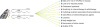 | Figure 2Chemotherapeutic effects of n-3 PUFAs on colon carcinogenesis.
n-3, omega-3; PUFAs, polyunsaturated fatty acids; EPA, eicosapentaenoic acid; DHA, docosahexaenoic acid; n-6, omega-6.
|
Although n-3 PUFAs would improve health, they could form secondary lipid oxidation products, especially in the presence of heme iron or pro-oxidants. It has been suggested that fish oil-rich diet with heme iron induce a 130-fold increase in the levels of urinary malondialdehyde, a representative lipid peroxidation product [71]. Fecal extracts from rats fed a diet containing heme iron and n-3 PUFAs were more toxic to epithelial colon cells than those from rats given diets containing ferric citrate or on a diet containing heme iron alone [71]. In addition, rats subjected to bile duct ligation and given fish oil supplement showed a high level of TGF-β1 in the liver and an impaired liver function. Therefore, fish oil supplementation to subjects with biliary atresia might be potential hazard and should be used with caution [72].
 XML Download