Journal List > Clin Nutr Res > v.5(2) > 1059824

Han, Lee, and Bu: The Level of Serum Cholesterol is Negatively Associated with Lean Body Mass in Korean non-Diabetic Cancer Patients

Abstract

Due to poor nutrition and abnormal energy metabolism, cancer patients typically experience the loss of muscle mass. Although the diabetic conditions or dyslipidemia have been reported as a causal link of cancer but the consequence of such conditions in relation to gain or loss of skeletal muscle mass in cancer patients has not been well documented. The purpose of this study was to investigate the relationship of lean body mass and systemic parameters related to lipid metabolism in non-diabetic cancer patients using data from the Korean National Health and Nutrition Examination Survey (KNHANES) 2008-2011. As results the level of serum total cholesterol (total-C) was negatively associated with both total lean body mass and appendicular lean body mass in cancer patients after adjustment for sex, physical activity, energy intake and comorbidity. The associations between consumption of dietary factors (energy, carbohydrate, protein and fat) and lean body mass were disappeared after adjusting comorbidities of cancer patients. Multivariate-adjusted linear regression analysis by quartiles of serum total-C showed that higher quartile group of total-C had significantly lower percent of lean body mass than reference group in cancer patients. The data indicate that serum lipid status can be the potential estimate of loss of skeletal muscle mass in cancer patients and be referenced in nutrition care of cancer patients under the onset of cachexia or parenteral/enteral nutrition. This data need to be confirmed with large pool of subjects and should be specified by stage of cancer or the site of cancer in future studies.

Introduction

Many studies have reported that cholesterol, triglycerides, and lipoproteins are involved in cancer incidence and severity. Moses et al. [1] reported that increased level of low density lipoprotein (LDL) was associated with a higher risk of prostate cancer in African American men. Tada et al. [2] found that high levels of serum triglycerides were positively correlated to cancer incidence in Japanese hypertriglyceridemia patients. Also increased levels of fasting glucose and c-peptide were observed with elevated body weight and increased plasma cholesterol in breast cancer patients at stage I-II [3]. Also a recent report from meta-analysis suggested that total serum cholesterol and high density lipoprotein cholesterol (HDL-C) were inversely associated with breast cancer risk [4].
Such dysregulated lipid status changes patterns of energy utilization, affects immune and inflammatory pathways, interacts with catecholamines and leads an alteration of body composition including fat and muscle amount [567]. Hence many cancer patients who sustain altered energy homeostasis (e.g. insulin resistance) in conjunction with uncontrolled blood lipid finally experience the accelerated loss of skeletal muscle mass [89101112]. For example Prado et al. [11] reported that decreased muscle mass and muscle strength (i.e. handgrip strength) were observed in male cancer patients with advanced lung or colon cancer. Gale et al. [12] suggested that less grip strength due to low muscle mass was associated with increased mortality from cancer in the analysis of the data from UK Department of Health and Social Security survey. Cancer patients with cachexia also showed catabolic conditions including elevated carbohydrate utilization, protein degradation and increased fat lipolysis, and these conditions led weight loss and were associated with increased morbidity and mortality of cancer patients [1314].
However it has been well suggested that elevated glucose level or longer duration of diabetes is associated with declines in skeletal muscle mass even in non-cancer subjects [8910]. Additionally metabolic changes observed in cancer patients are more complicate than those in patients only suffering from diabetic conditions [11131415]. Therefore, in cancer patients, diabetes or insulin resistance will not be the only factor affecting loss of skeletal muscle mass. Beside glycemic status, dyslipidemia and related complications observed in cancer patients were mostly explained in terms of decreased fat mass [131415] but their relationship with decreased skeletal muscle mass in cancer patients has not been well documented. Also muscle mass could be influenced by dietary factors or health conditions affecting lipid status specific to cancer hence the associations among altered metabolic conditions and muscle mass which differ from non-cancer subjects need to be explored in cancer patients. Based on such research gaps this study was conducted to investigate the relationship of serum lipids with relative lean body mass in cancer patients without a diagnosed diabetes by utilizing the Korean National Health and Nutrition Examination Survey (KNHANES) data from 2008-2011.

Materials and Methods

Study participants

KNHANES used a stratified multistage probability design to provide nationally representative estimates of Korean population [1617]. The data from KNHANES 2008-2011 containing the assessment of dual-energy X-ray absorptiometry (DXA) in participants were used. Of 28071 potential participants aged ≥ 30 years, 230 participants who were diagnosed cancer (stomach, colorectal, liver, lung, breast, ovary) were included in the study. Participants with known history of diabetes and/or use of insulin therapy were excluded and further participants who had incomplete lab data were excluded. Finally the data of 179 subjects were analyzed for the present study. Control subjects were also used for differentiating the health characteristics of cancer patients from subjects without cancer before exploring the possible variables associated with lean body mass in cancer patients. Total of 19172 participants who were older than 30 years, has no history of diabetes or insulin use but never been diagnosed cancer were used as control subjects only for comparing general health or diet characteristic of cancer patients to that of non-cancer subjects. Since the number of cancer subjects from the data of this complex sampling design was not large enough to be matched by the data from non-cancer subjects the comparison between cancer patients and control group was performed on the raw data without stratification or adjustment. KNHANES was approved by the Institutional Review Board of the Korea Center for Disease Control and Prevention (IRB No. 2008-04EXP-01-C, 2009-01CON-03-2C, 2010-02CON-21, 2011-02CON-06-C).

Assessment of lean body mass

The whole body DXA examination in KNHANES was acquired according to the manufacture's instruction on a Discovery-W fan-beam densitometer (Hologic Inc., Bedford, MA, USA). All subjects were asked to take off all accessories which could interfere with the DXA examination. The coefficient of variation (CV) of bone mineral density (BMD) measurement of lumbar spine (L1-L4), femoral neck and whole femur were less than 1.9%, 2.5% and 1.8%, respectively [16]. Additional information for the precision of the DXA instrument used or calibration in the KNHANES study was reported previously [161718]. Total lean body mass was obtained from all anatomical regions of skeletal muscle. Appendicular lean body mass was obtained from combined lean body masses in the right and left arms and legs [16]. All obtained values of lean body mass (kg) were divided by total body weight (kg) and multiplied by 100% to provide standardized estimates of percent lean body mass which could be better account for lean mass relative to body size as reported previously [19].

Measurement of covariates

Height was measured by a balanced beam scale (Seca 225, Seca, Germany) and body weight was measured using a digital electronic scale (GL-6000-20, G-tech, Korea). Body mass index (BMI) was calculated as the weight (kg) to the square of height (m2). Blood samples were obtained from a cephalic vein of the study subjects. Fasting plasma glucose, hemoglobin A1c (HbA1c), blood urea nitrogen (BUN), total cholesterol (total-C), triglyceride and HDL-C levels were measured using a Hitachi 7600 chemistry analyzer (Hitachi, Tokyo, Japan).
Participants were asked if they did physical activity designed for enhancement of muscle strength such as push-ups, situps, lifting weights or the horizontal bar in the last week. Status of smoking was categorized either a smoker: a person who had smoked more than five packs of cigarettes in the past, or a non-smoker: a person who had no history of smoking. Status of alcohol consumption was defined based on whether a person currently drinks alcohol more than one time per month (drinkers) or not (non-drinkers).
Nutrient intakes such as carbohydrate, lipid and proteins were assessed with a 24-hour dietary recall questionnaire administered by a trained survey researcher. The results from a 24-hour recall were calculated using the Food Composition tables developed by the National Rural Resources Development Institute (7th revision) [20].
History of comorbidities including arthritis, cardiovascular disease (CVD), asthma, hypertension or chronic obstructive pulmonary disease (COPD) was self-reported by study subjects.

Statistical analysis

All statistical analyses were conducted using SAS software version 9.3 (SAS Institute Inc., Cary, NC, USA). Values of demographic and health-related variables were expressed as mean and standard error for continuous variables or, number of frequency and percentages for categorical variables. For the comparison with non-cancer healthy subjects the t-test was used for continuous variables and the chi-square test for categorical variables. Pearson's correlation test was used to explore simple correlation of percent lean body mass and health related continuous variables. The 'domain' statement was used to provide less-biased estimates for subgroup analysis in the survey data. Before selecting variables for linear regression model collinearity was checked for each independent variable related to lean body mass. Linear regression models were created and tested to characterize the association of percent lean body mass and variables related to energy metabolism in cancer patients. Key covariates accounted in regression models were as follows; Model I: adjusted for age, sex, physical activity, smoking, height, serum total-C, serum total triglyceride and fasting glucose, Model II: Model I + energy intake and Model III: Model II + comorbidities which are the presence of CVD (n = 22), arthritis (n = 3), COPD (n = 15), asthma (n = 4) or hypertension (n = 50). These covariates were chosen as potential confounders of the association between hyperglycemia/hypercholesterolemia and muscle mass. Since lean body mass was initially standardized to body weight, the height was adjusted for the analysis. With these characterized models further analyses were performed to estimate beta coefficient, 95% CIs for percent lean body mass by quartiles of serum total-C, taking the lowest quartiles group as the reference group, in male and female subjects separately. Quartiles of serum total-C were categorized for male as follows: 1st quartile (Q1): total-C < 161, 2nd quartile (Q2): 161 ≤ total-C < 180, 3rd quartile (Q3): 180 ≤ total-C < 211, 4th quartile (Q4): total-C ≥ 211, for female as follows: Q1 total-C < 145, Q2:145 ≤ total-C < 182.5, Q3:182.5 ≤ total-C < 208, Q4:total-C ≥ 208, and for whole subjects (both male and female) as follows: Q1:total-C < 156, Q2:156 ≤ total-C < 180, Q3:180 ≤ total-C < 209, Q4:total-C ≥ 209. A p value of less than 0.05 was considered statistically significant.

Results

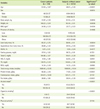
Initially data of skeletal muscle mass and related outcomes were compared between cancer patients and non-diabetic subjects without cancer (Table1). The mean age of cancer patients was higher than that of healthy non-cancer subjects (p < 0.0001). The body weight and BMI were not different betwe en two groups but the lean body mass (p = 0.0004) and appendicular lean body mass (p < 0.0001) were significantly low in cancer patients. The level of HbA1c, fasting glucose, total-C, HDL-C, TG and BUN was not different between cancer patients and non-cancer subjects. Also overall nutrient intake except for carbohydrate consumption was lower in cancer patients compared to non-cancer subjects. The number of subjects who performed physical activity designed for muscle enhancement was not different between two groups. The frequency ratio of the presence of alcohol drinking and the history of cigarette smoking in cancer patients were less than non-cancer subjects (p < 0.0001 for both parameters).
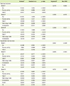
Table 2 shows results from simple correlation test between lean body mass and variables which could affect skeletal muscle before any adjustment was made for the weighted data. The simple correlation analysis revealed that the height, the level of BUN, the intake of energy and nutrients (protein, carbohydrate, fat) were positively correlated with percent lean body mass in both cancer patients and non-cancer subjects. The lean body mass and the level of total-C was negatively correlated both in cancer patients (r = -0.2765, p < 0.0001) and in non-cancer subjects (r = -0.1710, p < 0.0001). Correlation between lean body mass and age was positive in cancer patients (p = 0.0002) while negative correlation was observed in non-cancer subjects.
Three linear regression models for cancer patients were developed using the variables which showed relatively high level of correlation coefficient with lean body mass in order to explore the main predictor of lean body mass in cancer patients (Table 3). First linear model (Model I) demonstrated that sex, physical activity and total-C were significantly associated with lean body mass. However weak or no association was observed between lean body mass and fasting glucose (p = 0.0948) or serum triglycerides (p = 0.2692). After nutrient intakes (energy, carbohydrate, protein and fat intake) were adjusted (Model II) the energy and carbohydrate intake were significantly associated with lean body mass while the direction of these associations were opposite each other. The association of lean body mass with sex, physical activity, total-C, total energy intake and carbohydrate intake was still significant after accounting for diabetes-related comorbidities (Model III). When the use of cholesterol lowering drugs (daily use) was incorporated in the model the association between lean body mass and total-C or other variables were not affected. (data not shown). However the effect of total energy intake and carbohydrate on the lean body mass was disappeared after adjusting for HDL-C (data not shown). Also same regression models were tested for appendicular lean body mass (Table 3) and the association between total-C and appendicular lean body mass was same as seen in the models for total lean body mass with higher R2 value for all three regression models (R2 = 0.6521, 0.6676, and 0.6712 for Model I, II, and III, respectively).
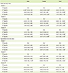
Since sex, physical activity and the total-C were significantly associated with percent lean body mass at the total body including appendicular sites in all linear regression models, the relationship was further analyzed within same sex by quartiles of total-C (Table 4). Male cancer patients were getting lower lean body mass compared to reference (the lowest quartile of total-C) by increased quartiles of total-C in all three regression models but the trend was not significant. In female cancer patients only the highest quartile (total-C ≥ 208 mg/dL) of total-C had significantly lower percent lean body mass compared to reference in models adjusted for physical activity (Model I), the nutrient intake (Model II) and in fully adjusted model (Model III). For whole subjects including both male and female all three higher quartiles of total-C versus reference were also associated with significantly lower lean body mass in all three tested regression models. When these regression models were tested (Table 4) for appendicular lean body mass male cancer patients showed significantly lower lean body mass compared to reference in the highest quartile (total-C ≥ 211 mg/dL) of total-C (p < 0.0001). In whole subjects only the highest quartile (total-C ≥ 209 mg/dL) of total-C had significantly lower appendicular lean body mass compared to reference in all three regression models (p < 0.05).

Discussion

The loss of muscle mass is associated with decreased muscle strength, consequently, and negatively impacting functional muscle capacity, and leads increased mortality [122122]. Also subjects with sarcopenia have higher risk of infection than older patients with a normal skeletal muscle mass [2324]. Cancer patients usually experience progressive weight loss accompanied with reduction of lean body mass which is responsible for a poor quality of life and poor response to chemotherapy with a shorter survival time than in cancer patients without weight loss [2425]. In this study percent lean body mass was relatively low in cancer patients compared to non-cancer subjects without significant difference in BMI or body weights indicating the change of body composition toward a decrease of skeletal muscle mass in cancer patients. Also the results showed that sex and physical activity were associated with lean body mass in cancer patients and were consistent with well-established findings from previous studies which supported the difference of skeletal muscle mass by sex and the response of skeletal muscle to exercise or the level of physical activity in normal healthy subjects [2627]. On the other hand, the muscle loss by aging has been well evidenced by past studies [232427] but aging induced muscle loss was not observed in cancer patients of this study and rather positive correlation was observed between muscle mass and aging. These results implicate that decreased muscle mass in cancer patients is likely due to metabolic alteration specific to cancer at the age of their illness and is different than gradual downfall of muscle mass by aging process.
Of many other parameters which were found to be correlated with skeletal muscle mass in this study total-C was an only serum factor showing consistently negative association with lean body mass in cancer patients. Serum cholesterol has been reported to be responsible for incidence or progression of cardiovascular diseases and metabolic disease such as cancer [282930]. Studies reported that men with low serum cholesterol had a lower risk of prostate cancer [28] and the long-term use of cholesterol lowering drugs attenuated the progression of prostate cancer [29]. Also the low level of serum total-C was positively associated with the incidence of high grade prostate cancer and the risk was higher in subjects with higher BMI [30]. In regards to the role of cholesterol on muscle mass, so far, there have been no human studies which specify the role of cholesterol in muscle mass of cancer patients. However, numerous experimental studies suggest that the increase of serum total-C is attributed in reduced lipoprotein lipase (LPL) activity in cancer [313233] which mediates decreased cellular uptake of fatty acids by hydrolysis of lipoproteins and is associated with hypertriglyceridemia, and hyperlipidemia accompanied with weight loss [34]. Also lower levels of serum TG, glycerol and free fatty acids have been observed in cancer patients with cachectic conditions [3334] though the LPL activity was not determined in these studies. In our current study two higher quartiles of serum total-C were negatively associated with lean body mass in the regression models with quartile level of total-C for estimation of skeletal muscle mass of cancer patients. This result suggests that uncontrolled catabolic conditions of cancer patients underlie both decreased lean body mass but increased serum level of cholesterol.
In this study the pattern of relationship between lean body mass and quartile of total-C was different between male and female cancer patients. Though the results were not significant the trend that proportional decline in lean body mass by the level of serum total-C was more noticeable in male subjects than female subjects who showed dramatic downfall of muscle mass only in bearing with highest level of total-C (more than 208 mg/dL). In appendicular lean body mass, however, a subtle change of muscle mass was observed in male subjects and whole subjects in the other two quartile levels of total-C which considered normal range of serum total-C value. There is no obvious reason to explain differences between male and female but also it's difficult to judge whether such difference is reliable in limited number of subjects within each quartile group. Nevertheless the differences in the amount and distribution of skeletal muscle between men and women could be a part of this result. For example skeletal muscle in lower limb took part greater proportion of appendicular lean body mass in male than in female and then muscle strength (i.e., handgrip strength) was associated with less amount of appendicular lean body mass in women [35]. Furthermore, since sex-related differences exist in the rate of weight loss [2627], the response of skeletal muscle to substances from blood circulation (e.g., steroid hormone, systemic lipid or cytokines) or nutrient changes might be different between male and female.
In this study energy or nutrient consumption was associated with muscle mass but when other disease factors were adjusted the effect of dietary factors were disappeared. Since 24h recall data from this cross-sectional KNHANES did not precisely reflect nutrient consumption, further incorporation of data from food frequency questionnaire (FFQ) will give more reliable information about the nutritional status of the subjects. However, results from regression analyses in this study are supported by previous findings that the dietary factor is not the only factor accounting for muscle loss [363738]. There are numerous evidences that, rather than change of nutrient intake, more of systemic inflammation induced by tumor cell derived cytokines disturb metabolic balance under cachexia [363738]. In addition, studies with cancer patients suffering from muscle loss reported that metabolic alterations including elevated carbohydrate utilization and decreased fat stores in cancer patients were mainly due to increased lipolysis rather than reduced lipid biogenesis [3233]. Also if nutrient supply is low then this would be represented by blood parameters but, in this study, the level of blood glucose or serum triglycerides was not different between cancer patients and non-cancer subjects and their associations with lean body mass in regression models were not significant. Furthermore, incorporation of the parameter which indicate the use of lipids lowering drugs did not affect the power of the model (R2 value in model III, 0.5964 vs. 0.5967) and did not change the value or significance of the beta coefficient for selected variables in the model (data not shown). Taken together these data indicate that loss of skeletal muscle mass in cancer patient is related to the dysregulated metabolic changes more likely than supply aspect.
The study also has limitations. First, we cannot conclude that all cancer subjects in this KNHANES study were experiencing muscle loss but also cannot generalize the finding of this study as the general phenomenon of cancer patients. However, many cancer patients (more than 50%) experience muscle wasting [353940] and patients with weight loss, even at 15% loss, are related to the reduced survival time of cancer patients due to the loss of respiratory function or circulation capacity [40]. Second, the number of cancer subjects in KNHANES data was small and the data were analyzed from mixed subjects who suffered from various types of cancer so the findings from such small and mixed pool may hardly reflect common features of cancer patients. In spite of weak regression coefficients the association between total-C and total lean body mass was consistent throughout all possible regression models in this study. Hence, the data from this study could provide applicable foundation for future study of metabolic characteristics and skeletal muscle or muscle function only specific to cancer patients. Concerning several beneficial roles of skeletal muscle in energy metabolism, further identification of mechanisms and mediators of skeletal muscle loss under cancer illness would help developing early nutrition interventions to prevent further muscle wasting and to improve survival in cancer patients. Last, a type of cancer, stage of cancer should be accounted for the future analysis.

Conclusion

This study was performed to explore the independent relationship of serum lipids with relative lean body mass in nondiabetic cancer patients by utilizing KNHANES data from 2008-2011. In Korean cancer patients higher level of serum total-C was negatively associated with total and appendicular lean body mass (p < 0.0001) after adjusting key covariates (sex, age, physical activity, comorbidity, and energy intake etc.) while the correlation or the association of the other blood parameter (fasting glucose, HbA1c, BUN, and TG) with lean body mass were not statistically significant. However the effect of total-C was consistently significant in various linear regression models for estimation of skeletal muscle mass in cancer patients while the effects of nutrient consumption on lean body mass were disappeared after adjusting comorbidities of cancer patients. When linear regression models were tested with a quartile of total-C the higher quartile of total-C had lower lean body mass than reference quartile within male and female group of cancer patients and in whole subjects. These data suggest that the level of cholesterol can be the potential indicator of the loss of skeletal muscle in cancer patients and the data can be referred to a nutrition care of cancer patients with cachexia or parenteral/enteral nutrition. Causality of the findings in this study needs to be investigated in longitudinal study with large pool of subjects and the type or stage of cancer should be specified in future study.

Figures and Tables

Table 1

Baseline characteristics of study subjects

cnr-5-126-i001
Cancer patients
(n = 179)
Subjects without cancer
(n = 14172)
Significance**
(p value)
Age 53.59 ± 1.05* 48.61 ± 0.20 < 0.0001
Sex 0.0016
 Male 66 (33.7) 6064 (49.3)
 Female 113 (66.3) 8108 (50.7)
Body weight, kg 61.87 ± 0.96 63.54 ± 0.12 0.0850
Height, cm 160.23 ± 0.84 163.16 ± 0.10 0.0006
BMI, kg/m2 24.06 ± 0.33 23.77 ± 0.03 0.4034
Presence of obesity 0.4868
 Lean 6 (4.52) 517(3.44)
 Normal 108 (58.27) 9133 (63.70)
 Obese 65 (37.21) 4522 (32.86)
Lean body mass, % 65.37 ± 0.75 68.05 ± 0.11 0.0004
Appendicular lean body mass, % 26.68 ± 0.42 28.54 ± 0.06 < 0.0001
HbA1c, % 5.91 ± 0.13 5.94 ± 0.04 0.4577
Fasting glucose, mg/dL 97.43 ± 1.23 95.72 ± 0.19 0.1701
Total-C, mg/dL 184.86 ± 3.67 191.01 ± 0.39 0.0951
HDL-C, mg/dL 51.16 ± 1.38 52.00 ± 0.15 0.8101
TG, mg/dL 138.76 ± 9.30 140.65 ± 1.30 0.8398
BUN, mg/dL 14.34 ± 0.49 14.23 ± 0.05 0.8186
Total energy intake, kcal/day 1680.46 ± 62.83 1988.85 ± 10.54 < 0.0001
Protein intake, g/day 58.28 ± 2.46 71.00 ± 0.50 < 0.0001
Carbohydrate intake, g/day 304.01 ± 12.08 323.21 ± 1.72 0.1137
Fat intake, g/day 28.69 ± 1.80 38.04 ± 0.39 < 0.0001
Alcohol intake§ < 0.0001
 Yes 79 (47.7) 10123 (75.7)
 No 100 (52.3) 4049 (24.3)
Cigarette smoking < 0.0001
 Yes 6 (4.7) 2947 (25.4)
 No 173 (95.3) 11225 (74.6)
Physical activity 0.1114
 Yes 40 (21.8) 3571 (27.8)
 No 139 (78.2) 10601 (72.2)

HbA1c: hemoglobin A1c, Total-C: total cholesterol, HDL-C: high density lipoprotein cholesterol, TG: triglyceride, BUN: blood urinary nitrogen.

*Mean (S.E); N (%); Lean BMI < 18.5, Normal: 18.5 ≤ BMI < 25, Obese: 25 ≤ BMI; §Alcohol drink: drink alcohol more than one time per month (Yes) or not (No); Cigarette smoking: smoked more than five packs of cigarettes in the past (Yes) or no history of smoking (No); Physical activity: Any physical activity designed for muscle mass enhancement last week; **For the comparison with non-cancer healthy subjects the t-test was used for continuous variables and the chi-square test for categorical variables.

Table 2

Correlation of percent lean body mass and factors related to energy metabolism in cancer patients, KNHANES 2008-2011

cnr-5-126-i002
Variables Cancer patients (n = 179) Subjects without cancer (n = 14172)
Coefficient* p-value Coefficient p-value
Age 0.1111 0.0412 -0.0489 < 0.0001
Body weight -0.2043 0.0008 -0.0010 0.8818
Height 0.4831 < 0.0001 0.4878 < 0.0001
BMI -0.5663 < 0.0001 -0.3983 < 0.0001
HbA1c 0.1117 0.3718 -0.0401 0.0115
Fasting glucose -0.0082 0.9060 -0.0224 0.0018
Total-C -0.2765 < 0.0001 -0.1710 < 0.0001
HDL-C -0.1056 0.1692 -0.0120 0.1167
TG -0.0611 0.3771 0.0069 0.3364
BUN 0.1410 0.0407 0.0811 < 0.0001
Total energy intake 0.2236 0.0004 0.3119 < 0.0001
Protein intake 0.1873 0.0033 0.2453 < 0.0001
Carbohydrate intake 0.1924 0.0025 0.2537 < 0.0001
Fat intake 0.0760 0.2367 0.1974 < 0.0001

BMI: body mass index, HbA1c: hemoglobin A1c, Total-C: total cholesterol, HDL-C: high density lipoprotein cholesterol, TG: triglyceride, BUN: blood urinary nitrogen.

*Pearson's correlation test was used to explore simple correlation of percent lean body mass and health related continuous variables.

Table 3

Linear regression model for the association of serum total cholesterol with percent lean body mass in cancer patients

cnr-5-126-i003
Estimate* Standard error p-value Adjusted R2 Root MSE
Total lean body mass
 Model I 0.5778 4.8983
  Sex -11.4129 1.4145 < 0.0001
  Physical activity 2.2150 0.8955 0.0137
  Total-C -0.0417 0.0110 0.0002
 Model II 0.5920 4.8783
  Sex -12.1336 1.4318 < 0.0001
  Physical activity 2.0460 1.0137 0.0441
  Total-C -0.0428 0.0107 < 0.0001
  Total energy intake -0.0082 0.0035 0.0203
  Carbohydrate 0.0351 0.0137 0.0104
 Model III§ 0.5964 4.8531
  Sex -11.7404 1.5568 < 0.0001
  Physical activity 2.2875 1.0568 0.0309
  Total-C -0.0440 0.0106 < 0.0001
  Total energy intake -0.0080 0.0035 0.0195
  Carbohydrate 0.0334 0.0137 0.0132
Appendicular lean body mass
 Model I 0.6521 2.4289
  Sex -6.4288 0.7843 < 0.0001
  Physical activity 1.1929 0.4836 0.0139
  Total-C -0.0147 0.0058 0.0117
 Model II 0.6676 2.3903
  Sex -6.5307 0.8177 < 0.0001
  Physical activity 0.7318 0.5245 0.1636
  Total-C -0.0151 0.0056 0.0077
  Total energy intake -0.0056 0.0017 0.0011
  Carbohydrate 0.0230 0.0066 0.0005
  Protein 0.0313 0.0147 0.0341
  Fat 0.0397 0.0190 0.0372
 Model III 0.6712 2.3780
  Sex -6.3680 0.8823 < 0.0001
  Physical activity 0.8211 0.5337 0.1245
  Total-C -0.0145 0.0053 0.0066
  Total energy intake -0.0055 0.0016 0.0008
  Carbohydrate 0.0227 0.0063 0.0004
  Protein 0.0307 0.0147 0.0377
  Fat 0.0404 0.0188 0.0319

*Beta coefficient after adjustment has been made in each regression model; Model I: adjusted for age, sex, physical activity, smoking, height, serum total cholesterol (total-C), serum triglyceride and fasting glucose; Model II: Model I + energy intake (total energy intake, total carbohydrate intake, total fat intake, total protein intake); §Model III: Model II+ comorbidities (the presence of either CVD, arthritis, COPD, asthma or hypertension).

Table 4

The association of quartiles of total cholesterol with lower lean body mass in cancer patients

cnr-5-126-i004
Male Female Total
Total lean body mass
 Model I
  1st Quartile Ref Ref Ref
  2nd Quartile +3.40* (-0.39, 7.19) -0.46 (-0.88, -0.04) -0.26 (-1.30, 0.78)
  3rd Quartile -0.65 (-5.45, 4.15) -0.67 (-1.61, 0.28) -1.20 (-1.79, -0.60)
  4th Quartile -2.60 (-6.38, 1.18) -3.04 (-3.49, -2.59) -3.15 (-3.58, -2.73)
 Model II
  1st Quartile Ref Ref Ref
  2nd Quartile +3.00 (-1.19, 7.20) -0.43 (-0.89, 0.03) -0.98 (-1.91, -0.05)
  3rd Quartile -0.05 (-5.27, 5.18) -0.23 (-1.10, 0.63) -1.13 (-1.63, -0.64)
  4th Quartile -1.84 (-5.77, 2.08) -3.08 (-3.41, -2.74) -3.14 (-3.44, -2.84)
 Model III§
  1st Quartile Ref Ref Ref
  2nd Quartile +3.14 (-1.43, 7.70) -0.23 (-0.58, 0.13) -0.85 (-1.78, 0.07)
  3rd Quartile +1.38 (-3.61, 6.37) -0.20 (-1.00, 0.59) -1.07 (-1.56, -0.58)
  4th Quartile -1.21 (-5.38, 2.96) -3.37 (-3.75, -2.99) -3.23 (-3.62, -2.83)
Appendicular lean body mass
 Model I
  1st Quartile Ref Ref Ref
  2nd Quartile -0.05 (-0.27, 0.18) +2.37 (0.03, 4.70) -0.28 (-1.95, 1.38 )
  3rd Quartile -0.26 (-0.80, 0.28) +0.29 (-2.67, 3.25) -0.37 (-2.09, 1.34)
  4th Quartile -1.47 (-1.70, -1.24) -0.07 (-2.72, 2.58) -1.81 (-3.23, -0.38)
 Model II
  1st Quartile Ref Ref Ref
  2nd Quartile -0.09 (-0.32, 0.14) +2.67 (0.34, 5.00) -0.21 (-2.04, 1.62)
  3rd Quartile -0.05 (-0.51, 0.42) +0.85 (-1.94, 3.64) -0.16 (-1.99, 1.67)
  4th Quartile -1.48 (-1.65, -1.31) +0.66 (-1.79, 3.11) -1.49 (-2.93, -0.05)
 Model III§
  1st Quartile Ref Ref Ref
  2nd Quartile +0.04 (-0.15, 0.23) +2.67 (1.01, 4.33) +0.19 (-1.37, 1.74)
  3rd Quartile +0.03 (-0.43, 0.50) +1.86 (-0.43, 4.16) +0.05 (-1.71, 1.80)
  4th Quartile -1.54 (-1.71, -1.37) +1.20 (-0.72, 3.12) -1.42 (-2.75, -0.10)

*Beta coefficient with 95% confidence intervals was shown; Model I: adjusted for age, gender, physical activity, smoking, height, serum total cholesterol (total-C), serum triglyceride and fasting glucose; Model II: Model I + energy intake (total energy intake, total carbohydrate intake, total fat intake, total protein intake); §Model III: Model II + comorbidities (the presence of either CVD, arthritis, COPD, asthma or hypertension), For male: 1st quartile: total-C < 161, 2nd quartile: 161 ≤ total-C < 180, 3rd quartile: 180 ≤ total-C < 211, 4th quartile total-C ≥ 211, For female: 1st quartile: total-C < 145, 2nd quartile: 145 ≤ total-C < 182.5, 3rd quartile: 182.5≤ total-C < 208, 4th quartile total-C ≥ 208, For total subjects: 1st quartile: total-C< 156, 2nd quartile: 156 ≤ total-C < 180, 3rd quartile: 180 ≤ total-C < 209, 4th quartile total-C ≥ 209; p < 0.05; p < 0.001 compared to reference group of total-C within same sex or total subjects.

Acknowledgement

This research was supported by grants from the National Research Foundation of Korea (NRF) funded by the Ministry of Education, Science and Technology (2012R1A1A1019253).

Notes

Conflict of interest This manuscript has no conflict of interest with third parties.

References

1. Moses KA, Abd TT, Goodman M, Hsiao W, Hall JA, Marshall FF, Petros JA, Issa MM. Increased low density lipoprotein and increased likelihood of positive prostate biopsy in black americans. J Urol. 2009; 182:2219–2225.
crossref
2. Tada H, Kawashiri MA, Nakahashi T, Yagi K, Chujo D, Ohbatake A, Mori Y, Mori S, Kometani M, Fujii H, Nohara A, Inazu A, Mabuchi H, Yamagishi M, Hayashi K. Clinical characteristics of Japanese patients with severe hypertriglyceridemia. J Clin Lipidol. 2015; 9:519–524.
crossref
3. Bell KE, Di Sebastiano KM, Vance V, Hanning R, Mitchell A, Quadrilatero J, Russell C, Dubin JA, Bahl M, Califaretti N, Campbell C, Mourtzakis M. A comprehensive metabolic evaluation reveals impaired glucose metabolism and dyslipidemia in breast cancer patients early in the disease trajectory. Clin Nutr. 2014; 33:550–557.
crossref
4. Touvier M, Fassier P, His M, Norat T, Chan DS, Blacher J, Hercberg S, Galan P, Druesne-Pecollo N, Latino-Martel P. Cholesterol and breast cancer risk: a systematic review and meta-analysis of prospective studies. Br J Nutr. 2015; 114:347–357.
crossref
5. Jacob S, Machann J, Rett K, Brechtel K, Volk A, Renn W, Maerker E, Matthaei S, Schick F, Claussen CD, Häring HU. Association of increased intramyocellular lipid content with insulin resistance in lean nondiabetic offspring of type 2 diabetic subjects. Diabetes. 1999; 48:1113–1119.
crossref
6. Itani SI, Ruderman NB, Schmieder F, Boden G. Lipid-induced insulin resistance in human muscle is associated with changes in diacylglycerol, protein kinase C, and IkappaB-alpha. Diabetes. 2002; 51:2005–2011.
crossref
7. De Pergola G, Giorgino F, Benigno R, Guida P, Giorgino R. Independent influence of insulin, catecholamines, and thyroid hormones on metabolic syndrome. Obesity (Silver Spring). 2008; 16:2405–2411.
crossref
8. Kalyani RR, Tra Y, Yeh HC, Egan JM, Ferrucci L, Brancati FL. Quadriceps strength, quadriceps power, and gait speed in older U.S. adults with diabetes mellitus: results from the National Health and Nutrition Examination Survey, 1999-2002. J Am Geriatr Soc. 2013; 61:769–775.
crossref
9. Lee CG, Boyko EJ, Strotmeyer ES, Lewis CE, Cawthon PM, Hoffman AR, Everson-Rose SA, Barrett-Connor E, Orwoll ES. Osteoporotic Fractures in Men Study Research Group. Association between insulin resistance and lean mass loss and fat mass gain in older men without diabetes mellitus. J Am Geriatr Soc. 2011; 59:1217–1224.
crossref
10. Lee CG, Boyko EJ, Barrett-Connor E, Miljkovic I, Hoffman AR, Everson-Rose SA, Lewis CE, Cawthon PM, Strotmeyer ES, Orwoll ES. Osteoporotic Fractures in Men (MrOS) Study Research Group. Insulin sensitizers may attenuate lean mass loss in older men with diabetes. Diabetes Care. 2011; 34:2381–2386.
crossref
11. Prado CM, Lieffers JR, Bowthorpe L, Baracos VE, Mourtzakis M, McCargar LJ. Sarcopenia and physical function in overweight patients with advanced cancer. Can J Diet Pract Res. 2013; 74:69–74.
crossref
12. Gale CR, Martyn CN, Cooper C, Sayer AA. Grip strength, body composition, and mortality. Int J Epidemiol. 2007; 36:228–235.
crossref
13. Tsoli M, Swarbrick MM, Robertson GR. Lipolytic and thermogenic depletion of adipose tissue in cancer cachexia. Semin Cell Dev Biol. 2015; Forthcoming.
crossref
14. Fearon K, Arends J, Baracos V. Understanding the mechanisms and treatment options in cancer cachexia. Nat Rev Clin Oncol. 2013; 10:90–99.
crossref
15. Arner P, Langin D. Lipolysis in lipid turnover, cancer cachexia, and obesity-induced insulin resistance. Trends Endocrinol Metab. 2014; 25:255–262.
crossref
16. Korea Centers for Disease Control and Prevention. Guidelines for utilization of raw data from the fifth Korea National Health and Nutrition Examination Survey (2010-2012). Cheongwon: Korea Centers for Disease Control and Prevention;2013.
17. Kweon S, Kim Y, Jang MJ, Kim Y, Kim K, Choi S, Chun C, Khang YH, Oh K. Data resource profile: the Korea National Health and Nutrition Examination Survey (KNHANES). Int J Epidemiol. 2014; 43:69–77.
crossref
18. Joo NS, Dawson-Hughes B, Kim YS, Oh K, Yeum KJ. Impact of calcium and vitamin D insufficiencies on serum parathyroid hormone and bone mineral density: analysis of the fourth and fifth Korea National Health and Nutrition Examination Survey (KNHANES IV-3, 2009 and KNHANES V-1, 2010). J Bone Miner Res. 2013; 28:764–770.
crossref
19. Janssen I, Heymsfield SB, Ross R. Low relative skeletal muscle mass (sarcopenia) in older persons is associated with functional impairment and physical disability. J Am Geriatr Soc. 2002; 50:889–896.
crossref
20. Rural Development Administration (KR). Food composition table. 7th ed. Suwon: Rural Development Administration;2006.
21. Bouchard DR, Dionne IJ, Brochu M. Sarcopenic/obesity and physical capacity in older men and women: data from the Nutrition as a Determinant of Successful Aging (NuAge)-the Quebec longitudinal Study. Obesity (Silver Spring). 2009; 17:2082–2088.
crossref
22. Cruz-Jentoft AJ, Baeyens JP, Bauer JM, Boirie Y, Cederholm T, Landi F, Martin FC, Michel JP, Rolland Y, Schneider SM, Topinková E, Vandewoude M, Zamboni M. European Working Group on Sarcopenia in Older People. Sarcopenia: European consensus on definition and diagnosis: report of the European Working Group on Sarcopenia in Older People. Age Ageing. 2010; 39:412–423.
crossref
23. Newman AB, Kupelian V, Visser M, Simonsick EM, Goodpaster BH, Kritchevsky SB, Tylavsky FA, Rubin SM, Harris TB. Strength, but not muscle mass, is associated with mortality in the health, aging and body composition study cohort. J Gerontol A Biol Sci Med Sci. 2006; 61:72–77.
crossref
24. Cosquéric G, Sebag A, Ducolombier C, Thomas C, Piette F, Weill-Engerer S. Sarcopenia is predictive of nosocomial infection in care of the elderly. Br J Nutr. 2006; 96:895–901.
crossref
25. Dodson S, Baracos VE, Jatoi A, Evans WJ, Cella D, Dalton JT, Steiner MS. Muscle wasting in cancer cachexia: clinical implications, diagnosis, and emerging treatment strategies. Annu Rev Med. 2011; 62:265–279.
crossref
26. Denison HJ, Cooper C, Sayer AA, Robinson SM. Prevention and optimal management of sarcopenia: a review of combined exercise and nutrition interventions to improve muscle outcomes in older people. Clin Interv Aging. 2015; 10:859–869.
27. Waters DL, Baumgartner RN, Garry PJ, Vellas B. Advantages of dietary, exercise-related, and therapeutic interventions to prevent and treat sarcopenia in adult patients: an update. Clin Interv Aging. 2010; 5:259–270.
crossref
28. Platz EA, Till C, Goodman PJ, Parnes HL, Figg WD, Albanes D, Neuhouser ML, Klein EA, Thompson IM Jr, Kristal AR. Men with low serum cholesterol have a lower risk of high-grade prostate cancer in the placebo arm of the prostate cancer prevention trial. Cancer Epidemiol Biomarkers Prev. 2009; 18:2807–2813.
crossref
29. Solomon KR, Freeman MR. Do the cholesterol-lowering properties of statins affect cancer risk? Trends Endocrinol Metab. 2008; 19:113–121.
crossref
30. Mondul AM, Clipp SL, Helzlsouer KJ, Platz EA. Association between plasma total cholesterol concentration and incident prostate cancer in the CLUE II cohort. Cancer Causes Control. 2010; 21:61–68.
crossref
31. López-Soriano J, Argilés JM, López-Soriano FJ. Sequential changes in lipoprotein lipase activity and lipaemia induced by the Yoshida AH-130 ascites hepatoma in rats. Cancer Lett. 1997; 116:159–165.
crossref
32. Notarnicola M, Miccolis A, Tutino V, Lorusso D, Caruso MG. Low levels of lipogenic enzymes in peritumoral adipose tissue of colorectal cancer patients. Lipids. 2012; 47:59–63.
crossref
33. Rydén M, Agustsson T, Laurencikiene J, Britton T, Sjölin E, Isaksson B, Permert J, Arner P. Lipolysis--not inflammation, cell death, or lipogenesis--is involved in adipose tissue loss in cancer cachexia. Cancer. 2008; 113:1695–1704.
crossref
34. Dahlman I, Mejhert N, Linder K, Agustsson T, Mutch DM, Kulyte A, Isaksson B, Permert J, Petrovic N, Nedergaard J, Sjölin E, Brodin D, Clement K, Dahlman-Wright K, Rydén M, Arner P. Adipose tissue pathways involved in weight loss of cancer cachexia. Br J Cancer. 2010; 102:1541–1548.
crossref
35. Barbat-Artigas S, Plouffe S, Pion CH, Aubertin-Leheudre M. Toward a sex-specific relationship between muscle strength and appendicular lean body mass index? J Cachexia Sarcopenia Muscle. 2013; 4:137–144.
crossref
36. Tsoli M, Robertson G. Cancer cachexia: malignant inflammation, tumorkines, and metabolic mayhem. Trends Endocrinol Metab. 2013; 24:174–183.
crossref
37. Wigmore SJ, Plester CE, Richardson RA, Fearon KC. Changes in nutritional status associated with unresectable pancreatic cancer. Br J Cancer. 1997; 75:106–109.
crossref
38. Wigmore SJ, Ross JA, Falconer JS, Plester CE, Tisdale MJ, Carter DC, Fearon KC. The effect of polyunsaturated fatty acids on the progress of cachexia in patients with pancreatic cancer. Nutrition. 1996; 12:S27–S30.
crossref
39. Esper DH, Harb WA. The cancer cachexia syndrome: a review of metabolic and clinical manifestations. Nutr Clin Pract. 2005; 20:369–376.
crossref
40. Dewys WD, Begg C, Lavin PT, Band PR, Bennett JM, Bertino JR, Cohen MH, Douglass HO Jr, Engstrom PF, Ezdinli EZ, Horton J, Johnson GJ, Moertel CG, Oken MM, Perlia C, Rosenbaum C, Silverstein MN, Skeel RT, Sponzo RW, Tormey DC. Eastern Cooperative Oncology Group. Prognostic effect of weight loss prior to chemotherapy in cancer patients. Am J Med. 1980; 69:491–497.
crossref
TOOLS
ORCID iDs

Ji Eun Han
https://orcid.org/http://orcid.org/0000-0002-8902-7140

Jun Yeup Lee
https://orcid.org/http://orcid.org/0000-0002-8998-2930

So Young Bu
https://orcid.org/http://orcid.org/0000-0001-9801-5435

Similar articles