

 PDF
PDF ePub
ePub Citation
 Print
Print


INTRODUCTION
In 2009, approximately 33.3 million individuals were living with human immunodeficiency virus (HIV) and approximately 1.8 million deaths (worldwide) were attributed to Acquired Immune Deficiency Syndrome (AIDS) [1]. In 1998, highly active antiretroviral therapy (HAART) was introduced. Since then, there has been a substantial decrease in HIV/AIDS-associated mortality [2]. HAART combines different drugs with varying mechanisms of action that target specific stages of the HIV-virus life cycle [2]. HAART effectively decreases viral load and has had a significant impact on the life expectancy of individuals living with HIV/AIDS. This change in clinical course was accompanied by an increased frequency of non-AIDS defining malignancies (non-ADMs), such as glioblastoma multiforme, hereinafter referred to as glioblastoma.
Glioblastoma is the most common malignant form of adult primary brain tumor and may arise in patients with HIV/AIDS, increasing an already elevated mortality risk [3]. Negative prognostic factors for patients with glioblastoma include age over 60 years, male sex, and prior low-grade astrocytoma or a history of genetic disorders, such as neurofibromatosis [4]. Median survival time without treatment is approximately 3 months, and with maximal treatment, may be up to 15 months [5]. Despite a multi-modal approach, combining surgery, chemotherapy, and radiation, the prognosis remains grim [6]. The purpose of this study was to determine the clinical outcomes glioblastoma in HIV positive patients and to discuss the molecular pathogenetic mechanisms underlying the development of glioblastoma in individuals living with HIV/AIDS.
MATERIALS AND METHODS
Between January and February 2010, a comprehensive PubMed search was performed by the first author (WC) using a strategic combination of search terms including “HIV glioma” AND “glioblastoma,” and “AIDS glioma” AND “glioblastoma.” Case reports and series describing HIV-positive patients with glioblastoma (histologically-proven World Health Organization grade IV astrocytoma) and reporting on HAART treatment status, clinical follow-up, and overall survival (OS), were included for the purposes of quantitative synthesis. Patients without clinical follow-up data or OS were excluded. No age or language restrictions were enforced. Remaining articles were assessed for data extraction eligibility. References of included articles were reviewed; studies and texts discussing the molecular pathogenetic mechanisms underlying tumorigenesis in the setting of HIV/AIDS and those examining the association between glioblastoma and HIV/AIDS are summarized in the Discussion.
Statistical analysis
Student's t test was used to compare the average survival of HIV-positive glioblastoma patients not treated and treated with HAART. Log-rank test was performed to compare the survival curves of the two patient groups. Patients who did not receive treatment for their glioblastoma was excluded from survival analysis. All tests were two sided with a two-tailed p-value less than or equal to 0.05 deemed statistically significant. Continuous variables are presented as means with corresponding standard deviations, and ranges when applicable.
RESULTS
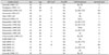
From our search, 13 studies (from 1987 to 2009), reporting on 21 cases of HIV-associated glioblastoma, were identified. Of the 21 cases, 4 cases (from three studies) were deficient in outcomes data and were excluded [789]. A total of 17 patients met our inclusion criteria. Of these patients, 14 (82.4%) were male and 3 (17.6%) were female, with a mean age of 39.5±9.2 years (range, 19–60 years). Patients were HIV-positive for an average of 4.4 years (range, 0–11 years) prior to diagnosis of glioblastoma. At the time of glioblastoma diagnosis, average CD4 count was 358.9±193.4 cells/mm3 (range, 80–610 cells/mm3). The cases reviewed are summarized in Table 1.
HAART and survival
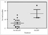
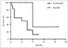
Mean survival for all patients was 7.7±6.8 months (range, 0–26 months). Mean survival was 16.67±8.1 months for patients treated with HAART (n=3) and 5.75±4.8 months (p=0.006) for patients who did not receive HAART (n=14) (Fig. 1). Patients had been treated with HAART for an average of 4.5 years (range, 3–6 years). There was a trend towards increased median survival with HAART treatment (12.0 vs 7.5 months, p=0.10) (Fig.2). Age and CD4 count at initial diagnosis of glioblastoma were not correlated with survival and were not prognostic indicators based on our analysis. Tumor progression (as opposed to HIV-associated complications) dictated patient survival and was the cause of death in all patients included.
Glioblastoma treatment
Of the 17 cases, two cases reported no glioblastoma treatment and five cases reported biopsy as the only treatment. Subtotal resection was the initial treatment in 7 cases, with 5 of those resections followed by adjuvant radiotherapy and chemotherapy. Eight patients received radiation therapy, and the mean total dose of those reported was 57.5 Gy (range, 55–60 Gy). One patient received only 12 Gy (of the intended 60 Gy) before death from disease progression. Radiation therapy was well tolerated and no unanticipated toxicity was reported in any of the cases. Six patients received adjuvant chemotherapy. Three patients received and tolerated lomustine (CCNU) and retinoic acid chemotherapy. One patient received hydroxyurea and intratumoral trisaziridylthiophosphoric acid (Thio-Thepa) and adjuvant radiotherapy and chemotherapy [procarbazine, lomustine, and vincristine (PCV-3)]. However, the chemotherapy dose was reduced because of myelosuppression. Two cases received temozolomide and the treatment was well tolerated. A slight drop in lymphocyte count below normal was noted in both cases, but eventually returned to baseline levels [11].
DISCUSSION
Neurological complications are frequent in HIV/AIDS patients. Roughly 40 to 60% of patients with AIDS will develop neurological sequelae at some stage of the disease [1020]. Additionally, an autopsy review found that up to 80% of AIDS patients had evidence of neurological pathology [2122]. Neurotrophic opportunistic infections like toxoplasmosis and cryptococcal meningitis can develop in the immunocompromised state. NeuroAIDS is a syndrome that manifests as dementia, sensory neuropathy, myelopathy, seizures, and aseptic meningitis. NeuroAIDS arises from HIV-associated neuronal damage resulting from the secretion of viral proteins and inflammatory [host] responses to these proteins [23].
Advancements in HIV management have led to a reduction in the incidence of opportunistic infections and have improved survival. Unfortunately, this has led to an increased incidence of non-ADMs, such as glioblastoma. A retrospective cohort study of HIV patients, by Hajjar et al. [24], reported a 5.4-fold increase in frequency of non-ADMs in patients with HIV compared to the non-HIV population. Blumenthal et al. [18] studied 1,384 HIV patients and found 6 patients with gliomas (0.43%), which translates to a 45-fold increase in non-ADMs compared to the general population. Moulignier et al. [13] retrospectively reviewed 70 HIV patients with intracerebral lesions undergoing stereotactic biopsy and reported a glioma frequency of 5.7%. Similarly, Tacconi et al. [25] reported a glioma frequency of 6.2% in their AIDS patients. Given the increased incidence of non-ADMs in HIV-positive patients, it is important to include glioblastoma in the differential diagnosis of intracranial lesions, especially in patients presenting with neurological symptoms.
The mean age for patients reviewed in our study is 39.5±9.5 years (range, 19–60 years). This is significantly younger than the mean age for diagnosis of non-HIV glioblastoma (61.3 years), reported by Ohgaki et al. [26] in 2004. This age discrepancy suggests a potential oncogenetic effect of HIV and/or suppressed antitumor responses in the immunocompromised state. Of the 17 cases reviewed, 4 patients had a presumptive diagnosis of toxoplasmosis, and were treated empirically with antitoxoplasmosis therapy [10111213]. Regimen failure was followed by stereotactic biopsy. Of note, one patient had a presumed diagnosis of lymphoma prior to being diagnosed with glioblastoma on pathology, which highlights the need for histological diagnosis in these patients as the treatment differs significantly between the two [15]. Most of the patients did not have end-stage HIV, and the CD4 counts in the cases reported were not substantially low. In our study, the average CD4 count at diagnosis of glioblastoma was 358.9±193.4 cells/mm3 (n=9). In all cases reviewed, tumor progression (rather than AIDS-associated complications) dictated patient survival. Thus, early and accurate diagnosis is imperative, as these individuals would gain similar benefits from aggressive treatment as patients with non-HIV glioblastoma [1819]. While there has been some concern for toxicity and increased risk for opportunistic infections from radiotherapy and chemotherapy in HIV patients, we found no evidence to suggest that the treatment strategy for HIV-associated glioblastoma should be any different than non-HIV glioblastoma, with the addition of HAART.
HIV and glioblastoma
As a neurotrophic virus [1227], HIV mainly targets microglia and macrophages within the central nervous system (CNS). HIV has been found in glial cells in vitro [132829]. A number of studies have identified selective expression of viral proteins within astrocytes of HIV patients. Astrocytes can serve as an important HIV reservoir and act as potential mediators of HIV-induced neuronal damage [30]. HIV infection of microglia depends on CD4 and the chemokine co-receptors (e.g., CCR5 and CCR3), whereas HIV infection of glial cells is CD4-independent [30]. The mechanism of HIV infection is thought to include the human mannose receptor [30]. Interestingly, HIV is unable to exert its typical cytopathic effects in astrocytes [1331]. Astrocytes cannot sustain HIV gene expression and replication due to restrictions on the HIV viral life cycle [32]. These restrictions of viral replication include inadequate expression and activity of viral Rev protein in astrocytes [32]. The Rev protein regulates nuclear export [3334], translation, and packaging of viral ribonucleic acid (RNA) [35]. However, given its resistance to HIV-mediated cytotoxicity and persistence of the virus in the host genome, astrocytes have the potential to transform during HIV infection [13], with activation of oncogenes or inactivation of tumor suppressors [36].
Numerous studies have recognized the transforming potential of HIV regulator genes. The transactivator gene (Tat) is a critical regulator of HIV replication and can up-regulate HIV gene expression by modifying transcriptional and post-transcriptional processes [37]. Vogel et al. [38] found that expression of HIV Tat in transgenic mice induces dermal lesions resembling Kaposi's sarcoma. Another study found that the Tat gene could directly transform human keratinocytes in culture and contribute to epidermal hyperplasia, which is associated with development of squamous and basal cell carcinomas in AIDS patients [37].
Other investigators have demonstrated neuron-specific transforming potential of Nef, another regulatory HIV protein [394041]. Nef expression alters the growth and morphology of astrocytes to resemble that of neoplastic transformation in vitro [3942]. Murine neural stem cells expressing HIV Nef exhibited alterations associated with cell transformation, including morphological changes, increased motility, loss of contact and anchorage growth inhibition, and increased cell proliferation. Mice undergoing intracranial injection with Nef-expressing neural stem cells formed tumors, while those injected with Nef-defective stem cells did not. Human astrocytoma cells expressing Nef injected into mice led to a greater incidence of tumor formation than injection with Nef-mutated astrocytoma cells. Interestingly, these tumors resembled glioblastomas [39]. Thus, Nef can induce tumor formation in neural stem cells and increase the malignancy of low-grade astrocytomas [39]. While the mechanisms underlying tumorigenesis are unclear, the in vivo data shows the transforming potential of Nef and its abundant expression in HIV-infected astrocytes, which suggests that Nef is important in the development of glial tumors in HIV-positive patients [1139]. HIV infection also induces the secretion of a number of cytokines including interleukin -1, -6, -8 and tumor necrosis factor-α that may facilitate glioma development [132943].
The immunocompromised state of HIV/AIDS patients may create an environment permissive to the development of neoplasms, as is the case for primary cerebral lymphoma and Kaposi sarcoma [14]. Several lines of evidence support a complex interplay between the immune system, specifically immunosurveillance within the CNS, and glioma pathogenesis. A number of large epidemiological studies have found that patients with a history of allergic disease have a reduced risk for developing gliomas, suggesting that a heightened immune status may be associated with a more robust intracranial defense against certain neoplasms [4445]. Immunosuppression resulting from treatment with immunosuppressive drugs has been strongly associated with increased frequency of intracranial gliomas in organ transplant recipients [4647]. In a study of 6,700 transplant recipients, Schiff et al. [48] reported 6 cases of gliomas consisting of 5 glioblastomas and 1 oligodendroglioma. Another series of 1,597 renal transplant recipients found 3 glioblastomas among 106 tumors [49]. Consistent with these studies, HIV mediated immunosuppression is associated with an increased frequency of gliomas.
In addition to HIV's primary cytotoxic effects on helper T cells, HIV-associated immunodeficiency can be mediated by the overexpression of transforming growth factor beta (TGF-β) by infected macrophages in the brains of HIV-infected patients. TGF-β2 is a cytokine with immunosuppressive effects and is detected in the brains of HIV-infected patients, but not in uninfected individuals [1350]. Similarly, TGF-β2 is overexpressed in many astrocytomas and in glioblastoma, but not in normal brain [13].
Immuno-glioma interface
The immuno-glioma interface represents the distinct immune environment of the CNS and the local immunosuppression induced by glioma-secreted factors. Tumors found in the CNS do not elicit the same type and degree of immunological responses as tumors found peripherally. This phenomenon is attributed to the classical understanding that the brain is strictly immune privileged [5152]. However, this concept of "immune privilege" has evolved to mean a modified immune status, rather than a state devoid of immune reactivity [5354]. The differences between systemic and CNS immunological surveillance are not qualitative, but rather quantitative [55]. Very low lymphocyte levels and low antibody diffusion normally characterize the anti-inflammatory CNS environment. During pathological states, the normally absent dendritic cells (DCs) can be recruited to inflammatory foci. Microglia that do not normally express detectable levels of major histocompatibility complex (MHC) class I and II molecules, can be induced to express MHC class II after exposure to stimulatory signals and brain trauma [56]. Expression of MHC antigens, normally absent in neurons and astrocytes in the CNS, is upregulated at sites of CNS inflammation and neoplasia [51].
Although studies examining the immunogenicity of glioma-specific antigens are scarce, evidence suggests that gliomas can elicit an immune response [57]. For example, tumor associated antigens gp100 and (melanoma-associated antigen 1) MAGE-1 are expressed in glioblastoma cell lines and it is thought that cognate cytotoxic T cells (CTLs) could recognize glioblastoma cells in an antigen-specific MHC class I manner [57]. Moreover, treatment with interferon-gamma has demonstrated greater CTL recognition through induction of MHC class I expression in glioblastoma cells [5758].
EGFRvIII, a sequence variant of the epidermal growth factor receptor, is found in 20–25% of glioblastoma patients [5759]. Wu et al. [59] found EGFRvIII peptide-pulsed DCs can induce autologous EGFRvIII-specific CTLs in vitro, and these CTLs are able to mediate cytotoxic effects on glioma expressing EGFRvIII. Although the brain does not exhibit a traditional lymphatic drainage system, studies of multiple sclerosis patients and experimental autoimmune encephalomyelitis (EAE) animal models have demonstrated that intracranial antigens can drain to cervical lymph nodes, suggesting a potential line of communication between the CNS and the peripheral lymphatic system that may be important in initiating an intracranial tumor response [60]. Activated microglia expressing MHC class I and II are candidate antigen presenting cells (APCs) within the CNS [61]. Recent studies using animal and EAE models have suggested that DCs may be important APCs involved in intracranial immunity. Further studies are necessary to elucidate the role of DC in antigen presentation in the setting of glioblastoma [576263].
Several studies utilizing autoimmune disease models have demonstrated that lymphocytes can cross the blood-brain barrier (BBB) [6465]. Antigen activation can mediate specific T cell migration into brain parenchyma [5166]. Calzascia et al. [67] demonstrated that brain tissue-specific CTLs, cross-primed within the cervical lymph nodes, can be imprinted through APCs presenting brain tumor antigens with a CNS-homing phenotype [57]. Additionally, glioma-induced angiogenesis can compromise the integrity of the BBB. The more porous blood-tumor barrier exhibits a number of alterations at the cellular level, including hyperdilated or absent tight junctions, downregulation of critical tight junction molecules, and loss of the basement membrane [5768].
Although human gliomas have been demonstrated to elicit a spontaneous antigen-mediated immune response, mainly comprised of CD8+ T cells, this is insufficient to control tumor growth [69]. Glioma-induced immunomodulation may prevent effective local and systemic response, and likely contribute to malignant progression and treatment resistance [70]. The immunosuppression in the glioma microenvironment is attributed to a number of factors including low MHC class I expression and expression of human leukocyte antigen (HLA) type G. HLA-G, is a non-classical MHC I molecule that serves as a potent immunosuppressor by decreasing glioma susceptibility to an antigen-specific anti-tumor response through mechanisms that include inhibition of tumor specific effector cell priming and T cell proliferation/cytotoxicity [55]. Additionally, secretion of tumor factors by glioma cells contributes to immune paralysis within the glioma microenvironment through disrupting T cell receptor signaling and APC function [51].
As previously mentioned, TGF-β2 is one of many cytokines over-expressed in glioblastomas. TGF-β2 is a multifunctional molecule normally involved in regulating cell growth and motility, and has been implicated in glioma angiogenesis, immunosuppresssion, and immune escape. In vivo data has demonstrated that TGF-β2 can down-regulate MHC II expression, inhibit natural killer lymphocyte activity, and hamper T-cell-mediated anti-tumor activity [7172]. Thus, an intracranial antitumor response must overcome several challenges to be effective. Barriers include insufficient extravasation of activated T cells across the BBB, lack of antigens that allow proper discrimination between malignant and normal cells, insufficient glioma MHC expression, and local immunosuppression within the tumor microenvironment [5356].
A number of studies have examined the relationship between the presence of tumor infiltrating lymphocytes (TILs) and tumor progression, and have demonstrated the presence of an immunosurveillance process in humans. These studies have begun to unravel the extent of the immune system's role in intracranial tumor pathogenesis. While several investigators have examined TILs, namely in melanoma, ovarian cancer, and colorectal cancer, the significance of TILs in glioblastoma progression is less clear. Brooks et al. reviewed biopsies of 149 patients with gliomas, of which 45% exhibited lymphocyte infiltration. Infiltration within the perivascular spaces was correlated with a statistically significant increase in survival (range, 2–4 months) compared to patients whose gliomas did not exhibit lymphocyte cuffing. Palma et al. [73] published a similar study of 200 non-HIV glioblastoma patients and found that patients with resection specimens exhibiting definite lymphocyte infiltration (23/200 patients) were associated with significantly longer survival compared to those exhibiting slight or absent TILs (p<0.01). Similarly, in another study of 199 glioblastomas, Böker et al. [74] noted that 72.6% of samples possessed histological evidence of TILs, which correlated with prolonged survival (8.1 months) versus those without TILs (5.6 months). Other studies assessing TILs and tumor progression have reported conflicting results. Schiffer et al. [75] found no correlation between the presence of lymphocyte infiltration and clinical outcome in their study of 324 malignant gliomas. In a review of 68 grade III/IV astrocytomas, Rossi et al. [76] found no correlation between the presence of CD4+ and CD8+ T cells, or macrophages and increased survival. Furthermore, in a study of 342 grade III/IV astrocytomas, Safdari et al. [77] reported a negative correlation and found that patients with TILs had a shorter average survival than those lacking infiltration. Recently, Yang et al. [64] found that the extent of CD8+ infiltrate in non-HIV glioblastomas is positively correlated with survival. Patients with long term survival (>403 days) were more likely to have intermediate or extensive CD8+ infiltration than short-term survivors (<93 days) (p<0.06). These studies have provided mounting evidence for the critical role of the immune system in the clinical progression of glioblastoma.
HAART and HIV
HAART has been a treatment standard for HIV patients. Clinical outcomes of HAART have been dramatic and include, but are not limited to, prolonged disease-free survival, HIV replication suppression, immunologic repletion, and reduction in hospitalization rates [78]. The effect of HAART on the immune system is not completely understood [79]. Although recovery of the immune system can occur in most individuals when viral replication is suppressed [79], time to and extent of immune recovery is uncertain [80]. Additionally, not only are CD4+ T cell numbers depleted during HIV infection, but immune cell functions are impaired and are not fully recovered under HAART. For example, plasmacytoid DCs and natural killer cell activity levels remain incompletely reconstituted after a year of effective therapy [81].
Despite nearly 15 years of experience with antiretroviral therapy for the treatment of HIV-positive individuals, consensus on the optimal time to initiate HAART has yet to be reached [82]. Aggressive early treatment in the course of infection was initially purported to present significant side effect risks and development of drug resistance [83]. Plettenburg et al. [82] recently identified that initiation of HAART in patients with higher CD4 counts correlated with improved outcomes. Aggressive and early treatment, even in asymptomatic HIV-positive patients, is recommended by most clinicians [84]. A recent analysis by Leone et al. [85], found that even treatment experienced HIV-positive individuals with over a decade of HAART therapy were at greater risk of death compared to the uninfected population. However, the leading cause of death was discovered to be malignancies (rather than AIDS-associated complications). While a direct effect of HIV on malignancy development has not been defined, there is an association between malignancy development and immune suppression. Leone et al. [85] reported significantly lower CD4 counts in patients presenting with aids-defining malignancies (ADMs) versus patients that did not.
In addition to HIV-associated illnesses, complications secondary to HAART can arise. A complication that has been recognized is immune reconstitution inflammatory syndrome (IRIS). As the immune system recovers due to HAART initiation, dormant infections can awaken. This pathological inflammatory response to previously treated or subclinical infections can result in aseptic meningitis or necrotizing lymphadenitis. It is recommended that HAART-naïve patients who initiate antiretroviral drug treatment and have a rapid decline in HIV RNA level should be monitored for the development of IRIS [86]. Other complications include reverse transcriptase inhibitor-associated peripheral neuropathy, pancreatitis, lipoatrophy, and hypersensitivity reactions [87].
HAART plays a role in restoring immune function. A rapid increase in CD4+ memory cells is followed by a more gradual increase in T-cells [88]. Immune reconstitution following HAART is found at all stages of disease and is thought to be responsible for the decrease in ADMs [88]. It is believed that CD4+ T cells are released from lymphoid tissues (in which they were sequestered) as viral replication is suppressed in lymphoid tissue following HAART treatment [89]. Additionally, the introduction of HAART leads to a decrease in level of immune activation as evidenced by reduction of expression of HLA-DR and CD38 activation markers on CD4+ and CD8+ T cells. Despite partial immune restoration, the increase in CD4+ T cells may not be adequate [89]. Immunotherapy has been suggested as a beneficial adjunct to HAART, but further studies are required to establish its role (if any) in HIV-associated glioblastoma [90].
HAART and glioblastoma
Neurological complications associated with HIV infection are common [91]. The BBB can prevent permeation of antiviral drugs, resulting in chronic intracerebral HIV infection. Once HIV enters the brain, it invades microglia and macrophages in the CNS, leading to chronic infectious diseases such as HIV-associated dementia (HAD) [91]. Although HIV enters the nervous system early after infection, damage to the CNS almost never occurs until systemic immunosuppression is established. Before the introduction of HAART, the incidence of HAD in AIDS patients was about 10%. After the introduction of HAART in 1996, the incidence of HAD decreased to approximately 50%. Maso et al. [92] conducted a record-linkage study to analyze the changing incidence of cancer in persons with HIV/AIDS (from years 1986 to 2005) using the year 1997 (the introduction of HAART) as the point of comparison to determine if cancer risk had changed in the HAART era. CNS cancer slightly decreased from 3.5 (95% CI: 1.5–7.0) to 3.2 (95% CI: 1.4–6.3) standardized incidence ratios (SIR) (from 1986–1996 and 1997–2004, respectively) after the introduction of HAART. Shiels et al. [93] found a decrease of 1.9 (0.66, 5.7) to 1.0 (0.63, 1.7) SIR of brain cancer in a meta-analysis of forty-two studies comparing the pre-HAART and HAART era. A case report by Aboulafia et al. [94] described a patient with primary CNS lymphoma that remained in complete remission with just HAART, nullifying the need for palliative whole-brain radiotherapy and the use of corticosteroids. Bayraktar et al. [95] also confirmed these findings in a retrospective study from 1999-2008, reporting that OS was better in HIV-associated lymphoma patients that received HAART.
Protease inhibitors may have a potential therapeutic effect against glioblastoma [11]. Ritonavir blocks the ubiquitin/proteasome pathway and has been demonstrated to exhibit anti-tumor activity through blocking cell cycle progression and inducing secondary apoptosis [96979899]. The ubiquitin/proteasome pathway regulates protein degradation, cell cycle progression, and cell survival, and is often disrupted in glioblastoma [96100101102]. Laurent et al. [96] demonstrated that ritonavir has cytotoxic effects on glioblastoma-derived cell lines in vitro. However, ritonavir was not found to control tumor proliferation, which was potentially due to sub-therapeutic levels of ritonavir in the cerebrospinal fluid. Neldinavir (another HIV protease inhibitor) down-regulates vascular endothelial growth factor (VEGF) and hypoxia-inducible factor (HIF) by disrupting the phosphatidylinositol 3-kinase (PI3K)/protein kinase B (AKT) pathway. The PI3K/AKT pathway is commonly activated in many cancers and regulates the expression of VEGF and HIF-1a among other cell processes [102103104]. Neldinavir treatment decreases angiogenesis and increases radiosensitivity in glioblastoma cells from mice bearing xenographed tumors [105]. The specific therapeutic role of protease inhibitors, as part of HAART regimens, has yet to be defined. As in the treatment of HIV, it is likely that the combination of drugs, rather than a single drug, is responsible for the immunomodulatory effects.
In conclusion, our analysis of 17 cases of HIV-associated glioblastoma revealed a male predilection, and a younger mean age at glioblastoma diagnosis in HIV-positive patients (when compared to HIV-negative individuals with glioblastoma). Age and CD4 count at initial diagnosis of glioblastoma were not correlated with survival and were not prognostic indicators (unlike non-HIV glioblastoma). Tumor progression (rather than AIDS-related complications) dictated survival. Treatment trends for HIV-associated glioblastoma are similar to non-HIV glioblastoma, with temozolomide being well tolerated in this specific patient population. Our data suggests that HAART is associated with improved survival in patients with HIV-associated glioblastoma, although the precise mechanisms underlying this improvement remain unclear. Future studies should further elucidate the potential protective mechanisms of HAART against the development of non-ADMs and the molecular underpinnings of HIV-associated glioblastoma.
 XML Download