

 PDF
PDF ePub
ePub Citation
 Print
Print


INTRODUCTION
Cystic sellar masses are relatively common findings on imaging studies. They are found in as many as 33% of general biopsies, including pituitary adenomas, Rathke cleft cysts (RCCs), craniopharyngiomas and arachnoid cysts. Symptoms related to tumor pressure include headache, visual disturbance and pituitary hormone deficiency [1].
Common indications for operation of sellar masses are as follows: impairment of anterior pituitary function, extrasellar extension, rapid growth and symptomatic impairment of vision [2]. Hemorrhage or necrosis in existing pituitary tumors can cause precipitous visual loss associated a condition called, pituitary apoplexy, which present headache, adrenal insufficiency, cranial neuropathy. This apoplexy is a surgical indication in some cases [3]. Some reports suggest that surgery may be the best option for tumors larger than 1 cm in diameter due to its further expansion at some unpredictable rate of growth [24].
Surgery is a highly effective option for most cases. However, complications that arise in some cases merit a close examination. The rates of surgical complications are as follows: 1% for carotid artery injury, 1% for stroke, 1% for hematoma, 3% for cerebrospinal fluid leakage, and 2% for meningitis. Moreover, the rate of permanent diabetes insipidus was 2 to 5%. Other complications, which occur at a rate of 1 to 7%, include nasal congestion, anosmia and facial pain and septal perforation [5678].
To date, there have been many reports regarding the conservative management of cystic sellar mass, but there have only been limited reports regarding the spontaneous involution of cystic sellar mass without surgery. We reported 14 patients with spontaneous involution of RCCs in our hospital.
MATERIALS AND METHODS
Patients
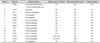
All patients who have visited our institution between 2005 and 2015 were retrospectively studied. The inclusion criteria were as follows: 1) pituitary cyst in the initial MRI, 2) non-surgical treatment, 3) decreased cyst volume in repeated MRI, and 4) no visual symptoms. In decreased cyst volume, a meaningful change in the volume was defined as a change of more than 25%. Clinical symptoms, hormone study, and MRI were evaluated for all patients. A total of 14 patients were identified to satisfy the inclusion criteria. The median age was 35 years (range, 5–67) (Table 1), and 8 patients were male. The initial MRI showed all 14 patients with RCCs. The mean follow up was period 17.3 months (range, 5.7–42.8). The study approved by the Institutional Review Board at SNUBH (B-1603-340-108).
Clinical data
Clinical data included age at diagnosis, sex, initial symptom, symptom duration, sudden headache, nausea and vomiting. If patients showed symptoms, we evaluated the change of symptoms after conservative management. All patients were checked for electrolytes (including serum sodium and potassium levels) at the initial admission day for hyponatremia.
Patients were evaluated for visual acuity, visual field defect and pituitary hormone levels if they presented clinical or laboratory suspicion of abnormality. Repeated evaluations of visual acuity, visual field and pituitary function were performed if there was a change in clinical status. All ophthalmologic evaluations were reviewed by 2 independent ophthalmologists. All patients were closely followed through the clinical and laboratory evaluations.
Patients were scheduled to visit the outpatient clinic for 3, 6, 12, 24, and 60 months after the baseline study. A follow-up included clinical review, MRI and visual testing if there was a change in symptoms.
MRI protocol
All patients had at least two MRI scans, with a mean interval of 17.3 month between the first and the last MRI (5.7 to 42.8 month). All MRI images were reviewed by 2 independent neuroradiologists. Maximum tumor dimensions were measured in orthogonal planes from the coronal (height and width) and sagittal (antero-posterior length), and were compared between the interval scans. The tumor volume was assessed as the volume of a rotating ellipsoid with the following formula: 3.14/6 (vertical diameter×anteroposterior diameter×transversal diameter). All MRI images were reviewed for T1, T2, T1 enhancement signal intensity and homogeneity. Cavernous sinus invasion, internal carotid artery encasement, optic chiasm compression and bleeding were also evaluated.
Hormonal evaluation
Patients were treated by a steroid (rapison) or levothyroxine (synthyroid) in the event adrenal insufficiency or hypothyroidism existed. Hormone study included the measurement of adrenocorticotrophic hormone, free T4, thyroid stimulating hormone, cortisol, human growth hormone, prolactin, luteinizing hormone, follicle stimulating hormone, estradiol, testosterone and insulin-like growth factor-1. They were repeated on an outpatient basis through hormone measurements by an endocrinologist.
RESULTS
Patient cases
The initial MRI showed all 14 patients with RCCs (Table 1). Eight patients were presented with sudden onset of headache, without any visual acuity or field defect. Hormone replacement therapy was given to 5 out of 8 patients. Four patients showed signs of adrenal insufficiency (hyponatremia with severe headache) and 1 patient showed partial hypopituitarism in combined pituitary stimulation test.
Two patients were presented with pituitary hormonal deficiency as the first symptom: a 17-year-old man with delayed secondary sexual character, and a 5-year-old child with delayed growth. The former patient showed under-developed penis and pubic hair. RCC was suspected, and the volume was 1.57 cm3, without optic compression. He had no visual symptoms and underwent hormone replacement therapy without surgery. The latter patient had already been treated for growth delay in another hospital. Again, RCC was suspected and the volume was shown to be 0.05 cm3.
Cyst change
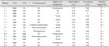
The mean initial tumor size was 1.29 cm3 (range, 0.05 to 3.23) (Table 2). After 17.3 months (range, 5.7 to 42.8), the mean tumor size was decreased to 0.23 cm3 (range, 0 to 0.68). There was a spontaneous decrease in tumor volume of 78% (range, 34 to 99).
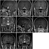
Twenty seven years-old female presented with headache (Fig. 1). Initial cystic mass volume was 3.23 cm3 (Fig. 1A-D). The signal of cyst was hyperintense on T1 (Fig. 1C) and T2 (Fig. 1D)-weighted images. And the cyst showed faint enhancement on T1 enhancement images (Fig. 1A, B). After 7 month of conservative care, mass volume decreased to 0.41 cm3 (Fig. 1E-H). It showed decrease in mass volume of 87%.
The initial MRI showed that the tumor was in contact and in compression with the optic chiasm in 7 patients and 3 patients, respectively. All 14 patients had no visual symptoms.
Symptom change
Eight patients were presented with sudden onset of headache. Headache was improved after conservative treatment in all patients.
Five patients were initially treated with hormone replacement due to hormonal abnormality. After the follow-up period, hormone abnormality was recovered in 3 patients without the need for long-term medication.
DISCUSSION
We have retrospectively studied 14 patients with spontaneous involution of cystic sellar masses. Only 21 other cases with spontaneous involution of RCCs have previously been reported [91011121314].
RCC is derived from the dorsal diverticulum of the stomatodeum. The anterior wall of the developing Rathke pouch forms the pars tuberalis and pars distalis. The residual lumen of the pouch reduces to form a narrow cleft. Failure to regress can result in an expanding and symptomatic RCC [131516].
The overall incidence of RCC regression has not been well quantified. Amhaz et al. [9] reported that overall incidence of cyst regression in 29 conservatively managed patients was 31%. But follow-up was not uniform in length and referral bias may prevent extrapolation of this incidence to RCCs in the general population.
The mechanism regarding the involution of cystic masses involves the repeated cyst rupture or leakage due to sustained elevated intracranial or intrasellar pressure [112]. Imbalances between the secretion and absorption of cystic fluids may lead to a change in the size of the cyst [110].
Hemorrhagic and necrotic degeneration of pituitary tumor was found in as many as 22% of tumors [17]. Hemorrhage in RCCs can occur and present similar characteristics to pituitary apoplexy. The changes may attribute to decreasing tumor size.
RCC might be regrowth after regression [9]. Igarashi et al. [10] reported decrease in RCC size in 4 patients; in 3 of these patients the cysts became symptomatic and required surgery. In our study, there was no case of recurrence in follow-up period.
Of the incidentally detected pituitary masses in other series, 20% became symptomatic during the course of 4 years [18]. Some reports suggested that relatively large masses should be surgically treated because such lesions have a risk for symptomatic growth with or without intratumoral bleeding. Our study included 12 out of 14 patients with a maximal tumor diameter of greater than 1 cm. And 10 patients, the tumor was shown, via the initial MRI, to be in contact with the optic chiasm.
In our study, all 8 patients with a sudden onset of headache showed improvement after the conservative treatment. Amhaz et al. [9] reported 9 patients of involution of RCCs and 5 (71%) out of 7 patients who were presented with headaches experienced improvement as the cyst regressed. This suggests that headaches may be associated with intracystic hemorrhage. Other possible causes of cyst-related headaches may be local inflammation, increased intrasellar pressure, aseptic meningitis and hypocortisolism [9].
No specific similarity was shown in the initial MRI with spontaneous involution of the cystic sellar masses.
The study has some limitations worth mentioning. First, we are not certain whether the cyst involution is a permanent phenomenon. It is possible for the cysts to regrow again: thus extended follow-ups are necessary. Second, despite the diagnosis of suspected pituitary cyst on imaging studies, we did not confirm such suspicion with a histopathological study. Third, the sample size of 14 cases is a limiting factor.
In conclusion, the spontaneous involution of RCCs is not well quantified before. Their incidence has not been well demonstrated, but this phenomenon might be underreported. Con-servative management can be a treatment option in some RCCs without visual symptoms, even in those that are large in size and in contact with the optic nerve via imaging study.
 XML Download