Abstract
We present a case of a subdural osteoma. A 29-year-old female presented with a 3-year history of headaches. Computed tomography scan revealed a homogeneous high-density lesion isolated from the inner table of the frontal bone (a lucent dural line) in the right frontal convexity. Magnetic resonance imaging revealed an extra-axial lesion with a broad base without dural tail sign and punctate enhancement pattern characteristic of abundant adipose tissue. Upon surgical excision, we found a hard bony mass clearly demarcated from the dura. The mass displayed characteristics of an osteoma upon histological examination. The symptom was relieved after operation.
An osteoma is a benign tumor of membranous bone consisting of dense compact bone without soft tissue elements. In the head region, osteomas are commonly found in the frontal and ethmoid sinuses and usually arise from the inner table of the cranium [1]. Intracranial osteomas are very rare. In most cases, they are characterized by an absence of connections to the dura or the skull, with tumor growth along the cerebral cortex. It is difficult to distinguish intracranial osteomas from dural metaplastic ossification commonly observed in elderly patients or calcified non-enhanced meningioma, by computerized tomography (CT) or magnetic resonance (MR) imaging. It is therefore important to develop better diagnostic criteria to distinguish these neoplasms. To the best of our knowledge, about 10 cases of intracranial osteoma have been reported to date [1234567891011]. The majority of patients presented with headaches localized to the site of the lesion, which typically subsided after complete resection of the osteoma. Here, we describe the clinical, radiologic and pathologic findings of subdural osteoma.
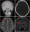
A 29-year-old woman presented with an approximately 3-year history of headaches over the right frontal area. The headache was localized to the region, and had not controlled by medication for the period. She had no history of systemic disease, meningitis, or head injury. Physical and neurological findings were normal. Plain skull radiography showed the presence of an ovoid radiopaque lesion in the right frontal area (Fig. 1A). CT showed a homogeneous high-density lesion isolated from the inner table of the frontal bone (Fig. 1B). MR imaging revealed an extraaxial mass with low-signal intensity in T2-weighted images (Fig. 1C) and a broad base neoplasm was observed by gadolinium enhancement (Fig. 1D). The preoperative diagnosis was an ossified meningioma. At first, the authors did not consider surgical tumorectomy, because the patient was young with benign small tumor, and the lesion being relatively smaller than the symptoms made. But, the headache has aggravated for three years, and the authors thought the headache has provoked by the dural irritation from the localized tumor. A frontoparietal craniotomy was performed under general anesthesia. There were no abnormal findings such as adhesion with or invasion into the bone flap. The dura mater was easy to open along the margin of the lesion, because the mass was located underneath it. After making an incision to expose the dura, we identified a bony, hard mass that was well demarcated from the brain parenchyma. There was no adhesion to the dura and the lesion was excised without difficulty by dissecting it away from the arachnoid membrane. Pathology examination showed fatty marrow within the mature trabecular bone, as is characteristic of osteoma (Fig. 2). Following removal of the mass, the patient has not experienced any headaches in the right frontal area.
Osteomas are benign neoplasms of uncertain etiology consisting of mature, normal osseous tissue. They are commonly found on the long bones of the extremities, in the sinuses of the facial bones, the skull, and the mandible [2]. Intracranial osteomas are uncommon and are usually observed in the inner or outer table of the skull or the inner layer of the dura mater [3]. These neoplasms are often confused with metaplastic dural ossifications or meningiomas. Intracranial intraparenchymal osteomas displaying no attachment to the inner table of the skull or dura are very rare, with about 10 reports described in the literature over the past 20 years [1234567891011]. In these cases, most of the patients were young women (Table 1). It is thus likely that intracranial osteomas are congenital lesions rather than acquired lesions. In the previous nine case reports, the only one case showed epilepsy for chief complaint. The others all described headache for main symptom, and it is interesting that all above headache were localized to the tumor location. An intracranial osteoma is a benign, slowly progressing lesion, which starts with a wide base and grows inward as an expanding mass with a well-demarcated border. As a result, osteomas cause pressure related symptoms by compressing or displacing the underlying brain parenchyma and dura mater. Lee and Lui [4] suggested that headache in subdural osteoma might also be caused by irritation or compression of the adjacent dural membrane. Of the 10 reported cases, there was a single case of intracallosal osteoma protruding into the lateral ventricle. This patient's chief complaint was of Jacksonian seizures, not headache [5].
Notably, osteoma can be confused with meningiomas or meningeal ossification of dura in elderly patients. As we demonstrate, CT examination in the subdural osteoma is a very useful diagnostic tool in differentiating these from other lesions. The lucent dural line seen at the bone window represents the dura between the skull and the osteoma, and can be used to differentiate subdural osteoma from meningeal ossification or osteoma of the inner table (Fig. 1B). Differential diagnosis from meningioma through MR imaging is difficult, but there are some findings specific to osteoma. First, there is no definite dural tail sign visible in meningioma. This is in contrast to osteomas, which are clearly demarcated from the dura. Second, grossly high signal change in T2-weighted images can be found in osteomas due to abundant adipose tissue. It is also found in hematoxylin and eosin staining that the intertrabecular marrow spaces are occupied by abundant adipose tissues (Fig. 2B). A punctuate enhancement pattern is also observed due to the presence of bone marrow with substantial adipose tissue (Fig. 1C, D).
The pathogenesis of intracranial intraparenchymal osteoma is unclear. According to Haddad et al. [6], old hematomas, calcified tuberculomas, or calcified abscesses can be confused with intracranial intraparenchymal osteomas. As a result, half of the 22 reported cases analyzed were misdiagnosed as intraparenchymal osteoma. Akiyama et al. [7] postulated that the primitive mesenchymal cells from the connective tissue might have migrated into the subarachnoid space along the intracerebral blood vessels. In support of this hypothesis, we observed many endothelial cells that adhere to osteoid layers with a medullary component of fibrofatty marrow in the case presented here.
In summary, subdural osteoma shows characteristic CT findings of a lucent dural line, and MR findings of punctate enhancement pattern. It is a rare case, but if the patient complain headache from dural compression or irritation, the surgical tumorectomy can be considered.
Figures and Tables
 | Fig. 1A: Plain radiography showed a dense calcified mass in the right frontal area. B: CT scan of the bone window showed an intraparenchymal calcified lesion separated from bone. The arrows indicate a typical lucent dural line manifest in intracranial intraparenchymal osteoma. C: Magnetic resonance T2-weighted image. The arrow denotes cerebrospinal fluid under subarachnoid space. D: Gadolinium enhanced T1-weighted image showed the typical punctate enhancement pattern characteristic of abundant adipose tissue in osteoma. It is a consideration in differential diagnosis with meningioma. |
 | Fig. 2Pathological findings. A: Microscopically lamellated bony trabeculae are lined by osteoblasts (H&E staining, ×10). B: The intertrabecular marrow spaces are occupied by abundant adipose cells and loose fibrovascular tissues (H&E staining, ×100). Endothelial cells (arrows) adhere to the osteoid layers. H&E, hematoxylin and eosin. |
Table 1
Reported intracranial osteomas

| Year | Authors | Sex | Age | Symptom | Location of tumor | Outcome |
|---|---|---|---|---|---|---|
| 1983 | Vakaet et al. [5] | F | 16 | Seizure | Intracallosal | Died |
| 1995 | Choudhury et al. [10] | F | 20 | Rt. frontotemporal headache | Rt. frontal | Recovered |
| 1997 | Lee and Lui [4] | F | 28 | Lt. frontal headache | Lt. frontal | Recovered |
| 1998 | Aoki et al. [9] | F | 51 | Headache | Rt. frontal | Not available |
| 1998 | Kim et al. [2] | F | 55 | Lt. temporo-parietal headache | Lt. parietal | Recovered |
| 2002 | Cheon et al. [1] | F | 43 | Lt. frontal headache | Lt. frontal | Recovered |
| 2003 | Pau et al. [8] | F | 33 | Headache | Convexity | Recovered |
| 2005 | Akiyama et al. [7] | M | 24 | Headache | Rt. frontal | Recovered |
| 2007 | Jung et al. [3] | M | 60 | Headache | Rt. frontal | Not available |
| - | Present case | F | 29 | Rt. frontal headache | Rt. frontal | Recovered |
References
1. Cheon JE, Kim JE, Yang HJ. CT and pathologic findings of a case of subdural osteoma. Korean J Radiol. 2002; 3:211–213.

2. Kim JK, Lee KJ, Cho JK, et al. Intracranial intraparenchymal ostemoa. J Korean Neurosurg Soc. 1998; 27:1450–1454.
3. Jung TY, Jung S, Jin SG, Jin YH, Kim IY, Kang SS. Solitary intracranial subdural osteoma: intraoperative findings and primary anastomosis of an involved cortical vein. J Clin Neurosci. 2007; 14:468–470.

5. Vakaet A, De Reuck J, Thiery E, vander Eecken H. Intracerebral osteoma: a clinicopathologic and neuropsychologic case study. Childs Brain. 1983; 10:281–285.

6. Haddad FS, Haddad GF, Zaatari G. Cranial osteomas: their classification and management. Report on a giant osteoma and review of the literature. Surg Neurol. 1997; 48:143–147.

7. Akiyama M, Tanaka T, Hasegawa Y, Chiba S, Abe T. Multiple intracranial subarachnoid osteomas. Acta Neurochir (Wien). 2005; 147:1085–1089. discussion 1089.

8. Pau A, Chiaramonte G, Ghio G, Pisani R. Solitary intracranial subdural osteoma: case report and review of the literature. Tumori. 2003; 89:96–98.

10. Choudhury AR, Haleem A, Tjan GT. Solitary intradural intracranial osteoma. Br J Neurosurg. 1995; 9:557–559.

11. Constantinidis J. [Intrathalamic osteoma]. Psychiatr Neurol (Basel). 1967; 154:366–372.


 PDF
PDF ePub
ePub Citation
 Print
Print


 XML Download