

 PDF
PDF ePub
ePub Citation
 Print
Print


INTRODUCTION
Meningiomas are most common benign intracranial tumors and make up 13-26% of all primary intracranial tumors. In most cases meningiomas present with symptoms such as headache, dizziness, seizures or the gradual progression of neurological deficits [1]. Presentation of intracranial hemorrhage is rare in meningiomas, approximately 1.3 or up to 2.4% may bleed according to the reported cases [2]. Subdural hemorrhages are even more uncommon, with 17.5% to 25% of intracranial hemorrhage associated with meningioma in the literature [3,4]. A low incidence of this condition not only makes the diagnosis of meningioma challenging but it also makes, the mechanism of hemorrhage harder to understand.
We present a case of 61-year-old woman with a benign meningioma that presented as subdural hemorrhage. In addition, we discuss related points by reviewing the literature of previous studies.
CASE REPORT
A 61-year-old woman was admitted to National Cancer Center with a persistent headache. Her headache started suddenly about one week before being admitted to the hospital, after a cough. Right frontal area headache was the only symptom, her vital signs were normal and the neurologic exam showed no abnormalities. She wasn't on any medications and had a past history of ovarian cancer (stage IIIc, histologic type: adenocarcinoma, mixed endometrioid/mucinous) that was diagnosed 9 years ago. She underwent an operation and multiple cycles of adjuvant chemotherapy for 2 years from diagnosis. During her follow-up, recurrence was found in the pelvic lymph nodes 2 years ago and she received radiotherapy for 3 months. Since then there was no evidence of disease and she was having regular check-ups on outpatient clinic basis.
A computed tomography scan showed a lesion in the right frontal area with a 5 cm sized conglomerated high density lesion and a cresenteric low density lesion in the ipsilateral subdural area (Fig. 1A). Magnetic resonance imaging revealed a lobulated, contoured, well-defined dural based mass with a central portion of iso-to-high signal intensity in T1-weighted image and low signal intensity in T2-weighted image suggesting acute- to subacute intratumoral hemorrhage (Fig. 1B, C). The cresenteric subdural lesion appeared as a low signal intensity in T1-weighted image and high signal intensity in T2-weighted image which is consistent with a liquefied subdural hemorrhage. On gadolinium enhancement, the mass showed strong homogenous enhancement except for the central portion representing hemorrhage (Fig. 1D). Our first impression from these findings was acute an intratumoral hemorrhage and subacute stage subdural hemorrhage in a dural metastatic tumor due to her past history and presentation of hemorrhages which occurs more frequently in malignant or metastatic tumors than in benign tumors [5]. Meningioma was also the differential diagnosis because of the tumor location and homogenous enhancement. We planned a curative resection of the tumor and adjuvant therapy was undetermined until we received pathology results.
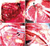
An elective operation was planned and performed without any complications. Bicoronal incision was done and an enlarg-ed middle meningeal artery heading towards the tumor was made visible by opening the frontal lobectomy bone flap. Dura mater was incised and the subdural hemorrhage was evacuated (Fig. 2A). The subdural hemorrhage was composed of mixed components ranging from a liquefied hemorrhage to a dense acute hematoma (Fig. 2B). The surface of the brain contacting tumor was intact and not been invaded by the tumor. At the deepest portion of tumor, a capsule attached to pial surface and cortical vein was observed (Fig. 2C). Intraoperative findings suggested that this cortical vein adhered to the tumor arachnoid capsule might be a focus of bleeding in this case. A frozen biopsy of the main mass turned out to be a meningioma but the histologic subtype was uncertain. The pathologist reported tumor involvement of the pia attached to cortical vein. Thus, the cortical vein and underlying pia mater was removed with negative margins. The subdural hemorrhage had a membranous structure and the frozen biopsy suggested tumor involvement (Fig. 2D). Therefore, the membranes surrounding the hemorrhage were removed totally and all resection margins were tumor-free, indicating Simpson grade I operation. She experienced an immediate postoperative partial seizure which was controlled with 'add-on' anti-epileptics to preventive one. She recovered without any neurological deficit. Final pathology results revealed a conventional meningioma of fibrous type (World Health Organization grade I) with intratumoral hemorrhage revealed among the thin-walled dilated capillaries (Fig. 3). She was discharged without further event and is now being observed on an outpatient basis.
DISCUSSION
Intracranial hemorrhage associated with brain tumors occurs in 1.7-9.6% of all cases and makes 0.9-10.2% of all intracranial hemorrhages [2]. Hemorrhage is more frequently associated with gliomas and certain metastatic tumors than with benign neoplasms. This may be explained by the characteristic difference of tumors. Rapid growth, immature friable blood vessels and tumor necrosis are contributing factors of tumor bleeding [3]. Meningiomas are the most common benign intracranial tumors, making up 13-26% of all primary intracranial tumors. Wakai et al. [2] reported the incidence of meningioma with the sign of hemorrhage in pathologic study as 1.3%, but clinically symptomatic hemorrhages are supposed to be much more uncommon [1,3]. Two large study of meningiomas by Cushing et al. and Hoessly et al. looked at 313 and 280 cases, respectively, and none of the cases showed clinically significant hemorrhage at presentation [6,7].
Martínez-Lage et al. [4] studied 57 cases of meningioma with hemorrhagic onset and 10 of those cases (18%) presented as subdural hemorrhage. Another large review investigating the association of hemorrhage with meningiomas was done by Helle and Conley [3]. From this article, there are 44 cases of meningiomas with hemorrhage. The most commonly hemorrhage associated with meningiomas was subarachnoid hemorrhage, which was present in 24 cases. Eleven cases (25%) of subdural hemorrhage were reported and among those, 4 cases (9%) had a solitary subdural component and 3 cases (7%) were combined with intratumoral components. In our case, the meningioma with an intratumoral component was seen within mixed types of subdural hemorrhage.
The mechanism of hemorrhage in meningiomas is uncertain and may vary according to histologic type, location and the type of hemorrhage. Predisposing factors such as trauma, hypertension or other stressful conditions are commonly described in several articles [1,3,4]. There are many suggested mechanisms, such as vessels in the tumor rupturing because they developed abnormally with weak, thin walls. Other mechanisms include intratumoral necrosis, infarction due to rapid growth of the meningioma, enlargement and a tortuous change of feeding arteries causing them vulnerable to blood pressure fluctuations. Also, expansion of the meningioma stretches the bridging, subdural veins and consequently makes the vessels friable and vulnerable to minor trauma [3,8,9].
In this case, the patient had the characteristic cortical vein attachment with the tumor capsule that was observed during surgery. The pathology revealed intratumoral vessels with thin walls that were dilated. Intratumoral necrosis and infarction were not observed in this case. Based on the gross and pathologic findings, the rupture of thin intratumoral vessels and the stretching of the subdural vein attached to the tumor caused the intratumoral hemorrhage and subdural hemorrhage, respectively. The cause may have been a minor traumatic event in this case it was likely an aggressive cough. The clinical presentation of a headache developing suddenly right after a cough supports our hypothesis.
In conclusion, the clinical presentation of subdural hemorrhage in benign meningioma is rare but should be considered when a bleeding event occurs in mass with benign appearance.


 XML Download