

 PDF
PDF ePub
ePub Citation
 Print
Print


INTRODUCTION
Radiation therapy has an important role in postoperative treatment of neoplasms originating from the central nervous system, but may induce secondary malignancies such as sarcomas, gliomas, and meningiomas [123]. The risk of development of a secondary osteosarcoma due to irradiation is estimated at 0.01% to 0.03% among all irradiative patients [45]. Historically, Cahan et al. [6] suggested the criteria for sarcoma arising in an irradiated bone fulfill several factors that include histologic or radiologic evidence of nonmalignant features in the initial bone condition, development of sarcoma in an irradiated area, at least 5 year period between radiotherapy and the arising of sarcoma, and histologic confirmation of the sarcoma. Later, Arlen et al. [7] modified the Cahan's criteria to include malignancies lacking osteoblastic activity, and reduction of the latency period from radiotherapy and arising of sarcoma from irradiated bone to 3-4 years. The prognosis of radiation-induced osteosarcomas is known to be poor, because of their aggressive nature invasive locally and intractability to multiple treatment strategies like as surgical resection, chemotherapy, and other [8].
We report a case of radiation-induced osteosarcoma that developed in the skull after 7 years of craniospinal radiotherapy for a pineoblastoma.
CASE REPORT
The patient was 34-year-old female with headache, and who had been diagnosed with a pineoblastoma on February, 2005. Craniotomy and tumor removal was done, and the tumor was subtotal resected. A third ventriculostomy was simultaneously provided with tumorectomy. After surgery, craniospinal radiotherapy (total 5,400 cGy, daily tumor dose 180 cGy, 30 fractions for brain, total 3,600 cGy, daily tumor dose 150 cGy, 24 fractions for thoracic and lumbar spine) for the residual tumor and prevention of recurrence was performed, from 4th March to 29th May, 2005 (Fig. 1). After that, patient underwent four cycles of etoposide plus cisplatin regimen from 25th March to 6th September, 2005. During adjuvant chemotherapy, out patient department follow-up was continued. There was no evidence of disease at the last brain MRI follow-up, and thereafter no problem with daily life has been observed.
She had no specific symptoms, but recognized a palpable mass on her right forehead. The mass was firm, non-tender, nonpitting, and not discolored. Considering her history of a brain tumor a brain MRI was performed, which revealed a large heterogenous enhancing mass that seemed to originate from the left frontal skull with epidural and subcutaneous invasion. We decided on surgery, and the second operation was performed on 13th December, 2012. The pathologist confirmed that the tumor tissue was an osteosarcoma, osteoblastic type (Fig. 2). The immunochemical study showed that the positive rate of Ki-67 was 40%. In the operating field, there was no evidence of invasion to the brain parenchyma. The tumor was totally resected grossly. There were no distant metastases to other internal organs or skeletal system as confirmed by abdomino-pelvic CT scan and whole-body bone scan.
Adjuvant chemotherapy was provided again with high-dose intravenous methotrexate-doxorubicin-cisplatin regimen as recommended by our oncologist, but follow-up of brain MRI showed progression of tumor in the previous operative field which was the left frontal skull. She had to receive another operation for the recurred lesion on 3rd January, 2013. When we tried to close her skin wound, there was some trouble because of the large skin defect, so we had received help from a plastic surgeon for a skin graft using an occipital scalp.
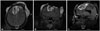
After that, wound dehiscence occurred, and full-thickness skin graft of the head and neck was done by a plastic surgeon on12th February, 2013. After skin graft operation, various different chemotherapy trial regimens were attempted, but despite the efforts of the oncologists, the brain MRI showed consistent disease progression (Fig. 3). At the last tumor conference, we decided to stop the chemotherapy because antitumoral agents could not control the tumor.
Her general condition became worse, including confused mentality. Because of the long period of bed-ridden state, recurrent pneumonia developed, leading refractory pleural effusion. We attempted administration of antibiotics and insertion of percutaneous drainage of the pleural effusion, but those were not effective at all. As a result, she expired at 23rd November, 2013.
DISCUSSION
As mentioned before, Cahan et al. [6] described the following four criteria for radiation-induced osteosarcomas in 1948: 1) microscopic or radiologic evidence of a nonmalignant initial bone condition; 2) sarcoma originating in an area within the previous radiotherapy; 3) a relatively long, asymptomatic latent period following irradiation before the clinical appearance of a bone sarcoma; and 4) histological confirmation of sarcomas. This case we mentioned here is applicable to these criteria.
Cases of radiation-induced sarcoma have previously been reported in the literature. The incidence of sarcoma resulting from irradiation has been estimated to be 0.03% to 0.3% [459].
Radiation-induced tumors are related to cumulative dose of radiation exposure, and there is some difference as to which typevof secondary tumors, for example, which benign tumors, such as meningiomas, tend to occur after lower-dose irradiation (less than 15 Gy), and malignant tumors, such as glioma or sarcoma, which tend to occur after higher-dose radiotherapy (15-56 Gy) [10].
Larger radiation dosages and higher degrees of malignancy may be related to shorter latency before the development of post-irradiative brain tumors [111213]. The mean latency of meningiomas with high-dose radiation is 18.4 years, compared with 36.8 years for low doses [14]. The mean latency for both gliomas and osteosarcomas has similarly been estimated to be about 9.1 years [15].
Osteosarcoma is not a common complication of radiotherapy for brain tumors that usually develop several years after doses exceeding 30 Gy [7]. Radiation-induced osteosarcoma accounts for 3.1-5.5% of all osteosarcomas, and the risk of occurring an osteosarcoma after irradiation is estimated as around 0.01-0.03% of all irradiated patients [1216]. Most radiation-induced osteosarcomas of the skull occur in or around the facial bone or paranasal sinus after radiotherapy for retinoblastoma [671718]. A genetic predisposition to develop a second neoplasm after radiotherapy may exist in patients with hereditary retinoblastoma [17]. There are also several reports of development for radiation-induced osteosarcoma after radiotherapy for pituitary adenoma [161920] or craniopharyngioma [21]. We had attempted to find any cases of secondary osteosarcoma after irradiation for a pineoblastoma such as our case, but none could be located. It seems to be a very uncommon case.
In our case, radiation-induced osteosarcoma arose 7 years after radiotherapy (54 Gy). This dosage level is similar to those reported in the literature, but latency seems to be shorter than estimated duration before.
As known before, the prognosis of radiation-induced osteosarcoma is poorer than that of primary osteosarcoma because of its aggressive clinical behavior such as the high rate of local recurrence and rapid progression [6712161921]. Surgical removal with chemotherapy has been effective in only a several reported cases [121622]. In these patients, chemotherapeutic regimens combining cisplatin, doxorubicin, and ifosfamide have been used. Effective treatment of osteosarcoma requires resection with tumor-free margins, but such a strategy is often disturbed by irregular, widespread invasion [12]. Although additional radiotherapy may be administered in some cases, the risk for radiation damage need to be carefully considered.
Our patient died because of rapid progression of radiation-induced osteosarcoma in spite of surgical resection and chemotherapy. Radiotherapy is an essential treatment option after surgical resection of neoplasms arising from the central nervous system, but prognosis of radiation-induced osteosarcomas are very poor, so cautious attention and consideration of the which treatment strategy is the best option for long-term survival of patients with irradiation is necessary.

 XML Download