

 PDF
PDF ePub
ePub Citation
 Print
Print


INTRODUCTION
Solitary fibrous tumor (SFT) is a rare collagen-rich, spindle-cell, mesenchymal-origin neoplasm. Klempere and Rabin first reported pleural SFT in 1931. Several cases have been reported at pleural and extrapleural sites, including the pericardium, peritoneum, lung, liver, upper respiratory tract, mediastinum, thyroid gland, parotid glands, and central nervous system (CNS). Extrapleural SFT, especially CNS SFT, is very rare. CNS SFT has been reported at the cerebellopontine angle, spinal dura, parasagittal region, meninges, and the intraventricular region [123]. SFT is thought to arise from the fibroblast and needs to be differentiated from some tumors, like fibrous meningioma and hemangiopericytoma, and from myxoid variants like myxochordoid meningioma and myxoid peripheral nerve sheath tumor [3456]. Reported herein is a case of CNS SFT in a 63-year-old female patient.
CASE REPORT
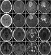
A 63-year-old female patient visited hospital with headache and intermittent confused mentality for 1 month. She had a history of coronary artery occlusive disease. In the neurologic examination, the patient was found to have had disorientation to time, without other neurological abnormalities. The brain magnetic resonance imaging (MRI) showed an 8.0×5.2×6.4 cm ovoid mass in the right frontal convexity, with peritumoral edema. The tumor showed intermediate-low signal intensity in the T1-weighted image (T1WI), and slightly increased signal intensity in T2-weighted image (T2WI). The upper and medial portions of the mass showed heterogenous and relatively low signal intensity in T2WI, and suggested a fibrotic mass. The mass showed strong enhancement in the gadolinium-enhanced T1 image. The initial impression was meningioma (Fig. 1A-D).
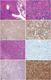
Gross total resection was done. The tumor was a well-encapsulated and whitish solid, round, and firm mass. In the histologic examination, the tumor showed mixed hypercellular and hypocellular areas, with multifocal intervening collagen laid down, and scattered vessels. The tumor cells were short and spindle-shaped and showed diffuse, strong immunoreactivity for CD34. The mitosis was less than 1/10 high power field (HPF), with an about 1.60% Ki-67 labelling index, and there was no evidence of necrosis. Sparse reticulin fibers were observed among the tumor cells on special stain. With these results, hemangiopericytoma was ruled out, and a diagnosis of SFT was made (Fig. 2A-D).
After the surgery, the patient was symptom-free, and the MRI showed no recurrence at 6 months, 1 year, and 36 months after the surgery. During the follow-up visit, at 55 months after the surgery, the patient revisited the hospital due to confusion for a few weeks. Brain MRI was again conducted, and the tumor was shown to have recurred at the primary site and at an adjacent area (Fig. 1E-H). Reoperation was thus done, and the tumor was grossly and totally removed. The pathologic findings were relatively the same as the previous ones, but the tumor was more cellular, with increased mitosis (about 2/10 HPF) and Ki-67 labelling index (12.85%). There was no evidence of necrosis. Still, the tumor cells showed diffuse strong immunoreactivity for CD34, and sparse reticulin fiber (Fig. 2E-H). After the operation, the patient recovered and became symptom-free again. Brain MRI was again conducted 10 months later; three small (5×5×5 mm) recurring nodules were found in the right frontal base, and the patient did not show any symptom (Fig. 1I-L). As the affected area was too small, it was difficult to perform surgery again therein; as such, radiation therapy was planned. The radiation dose totaled 5,940 cGy and was fractionated 33 times. Three months after the radiation therapy, follow-up brain MRI was done, and it was found that the nodules in the right frontal base had become smaller (Fig. 1M-P). Outpatient follow-up brain MRI will again be conducted.
DISCUSSION
SFT is an uncommon tumor that mostly occurs in the visceral pleura. SFT has been reported to occur outside the thoracic cavity, including the pericardium, peritoneum, lung, liver, upper respiratory tract, mediastinum, thyroid glands, and parotid glands. It has also been reported to occur in the CNS, including the cerebellopontine angle, spinal dura, parasagittal region, meninges, and the intraventricular region. It is th-ought that SFT is a mesenchymal-origin tumor and should thus be differentiated from fibrous meningioma, fibrosarcoma, meningeal sarcoma, meningeal myofibroblastoma, schwannoma, neurofibroma, and gastrointestinal stromal tumor [1234].
Patients may present several non-specific symptoms related with increased intracranial pressure or with the location of the tumor, such as headache, dizziness, gait disturbance, hemiparesis or hemiplegia, hearing loss, and mental change [7].
Immunohistochemically, although it is still being debated on, SFT has fibroblastic or myofibroblastic differentiation, which means that it has a mesenchymal fibroblast origin rather than a meningothelial origin. In an immunochemical study, CD34 was found to be strongly expressed in most SFT cases; Bcl-2 in 80-100% of SFT cases; and CD99 in 75-100% of SFT cases. Generally, its diagnosis is confirmed through positive staining for CD34 and negative staining for S-100 [2389], as distinguished from that of meningioma (usually negative for CD34 and S-100) and neurinoma (usually negative for CD34 and positive for S-100), and it should have a differential diagnosis with hemangiopericytoma. Most hemangiopericytoma cases, in contrast to SFT cases, are hypercellular and are oval to round rather than spindle-shaped, and immunohistochemically, they are reactive with CD34 and Bcl-2 only in a few cases [456].
Radiologically, it is difficult to differentially diagnose SFT through image study. SFT has variable signal intensities in MRI. T1WI and T2WI show hypointensity and/or hyperintensity with enhancement diffusely or heterogeneously [17810].
The authors' first impression was meningioma as the mass was seen as a dural-based enhanced mass with heterogenous and relatively low signal intensity in T2WI, and a fibrotic mass was suggested, but the final diagnosis was changed to SFT. As such, there is a need to consider a differential-diagnosis fibrotic mass, especially meningioma, hemanigompericytoma, and SFT.
The treatment of choice is complete resection. Even though SFT is considered benign, it should be followed up regularly due to a possible recurrence [4911]. Due to the rarity of the tumor, its adjuvant therapy and prognosis have not been well studied, and the indications of adjuvant therapy are uncertain [212]. Adjuvant radiotherapy was not done in the case reported herein, but several reports recommend it [13]. As such, close follow-up without radiation therapy was decided. As the tumor recurred at the primary site, however, radiation therapy was eventually done.
In conclusion, SFT is a rare mesenchymal-origin neoplasm especially occurring in the CNS. Image study and immunohistochemistry are crucial for its correct diagnosis. Surgery is the treatment of choice, and some reports recommend adjuvant radiotherapy. SFT is generally considered benign, but recurrences have been reported. Thus, long-term follow-up is necessary.
 XML Download