

 PDF
PDF ePub
ePub Citation
 Print
Print


INTRODUCTION
Intracranial ventricular ependymomas are relatively rare adult brain tumors that occur more frequently in the supratentorial region of the brain [1]. These lesions are derived from the ependymal and subependymal cells that line the choroid plexus and the ventricles [2]. Additionally, ependymomas can be located in the brain parenchyma outside the ventricular system [3]. These tumors are termed extraventricular ependymomas and are extremely rare. Only a few cases of supratentorial extraventricular anaplastic ependymoma have been reported in the literature [4]. We report the case of a patient with a supratentorial extraventricular anaplastic ependymoma that presented with repeated intratumoral hemorrhage.
CASE REPORT
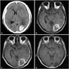
A 23-year-old male presented with a persistent moderate headache and blurred vision on the right side following the sudden onset of a severe headache 2 weeks previously. Computed tomography (CT) revealed a 4 cm-diameter hyperdense intracerebral hematoma in the left occipital lobe and a hypodense, thin chronic subdural hemorrhage along the left cerebral convexity (Fig. 1A). Magnetic resonance imaging (MRI) showed no enhanced lesions besides the hemorrhage (Fig. 1B, C), and cerebral angiography revealed no abnormal findings. Left occipital craniotomy and hematoma removal were performed for exploration. Intraoperative findings revealed the presence of mixed hematomas (liquid and dense hematomas) and small-sized abnormal vessels. Biopsy specimens of both hematomas and vessels were obtained. A histopathological examination revealed no tumor cells besides the hematomas and abnormal vessels. The patient underwent annual follow-up MRI evaluations, and no new lesions were detected for over 4 years (Fig. 1D).
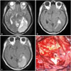
Five years later, a MRI revealed that a 7.5×4×5 cm-sized mass in the same location as the previous hematoma. The mass was visualized as mixed intensity on T1- and T2-weighted MR images and as a region of strong enhancement after intravenous gadolinium injection. There was no calcification and little cerebral edema was observed surrounding the mass (Fig. 2A-C). Based on these findings, a brain tumor with intratumoral hemorrhage was suspected. The patient underwent a left occipital craniotomy and gross total resection of the tumor. Intraoperative findings confirmed that the tumor showed no continuity with the ventricular system (Fig. 2D). Histopathological examination revealed clear cells with a sheet-like, papillary, or columnar to tubular arrangement. In addition, perivascular pseudorosettes were observed in the tumor. Immunohistochemical staining was focally positive for glial fibrillary acidic protein (GFAP) and epithelial membrane antigen, suggestive an ependymoma. Moreover, the tumor cells had diffuse nuclear pleomorphism, high cellularity, hemorrhage, necrosis, and a relatively high Ki-67 index (approximately 10%) (Fig. 3). Taken together, these factors confirmed a diagnosis of anaplastic ependymoma. Whole-spine MRI was performed for cerebrospinal fluid (CSF) dissemination workup, and there was no evidence of dissemination. Adjuvant local field radiotherapy (5,940 cGy in 180 cGy daily fractions) was administered to the left occipital lobe.
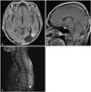
Four years later, the patient was re-admitted to the hospital because of tumor recurrence at the cervicomedullary junction and dissemination at the intradural space of the eighth to ninth thoracic levels, without recurrence at the primary site (Fig. 4). He underwent a midline suboccipital craniotomy and hemilaminectomy at T8-9. Histopathological examination confirmed recurrence and dissemination of the tumor. Adjuvant spine local radiotherapy (4,500 cGy in 180 cGy daily fractions) was then administered.
DISCUSSION
Intracranial ependymomas are common in children, accounting for 10% of pediatric brain tumors [1]. However, they are relatively rare in adults, representing only 2-6% of all intracranial tumors [3]. Although they most commonly develop in the fourth ventricle, followed by the lateral and third ventricles, supratentorial ependymomas occur more frequently in adults.
Ependymomas can occur at any site in the central nervous system, but supratentorial ependymomas outside of the ventricular system are quite uncommon. This type of ependymoma variant is termed "supratentorial extraventricular ependymoma" [5], but the pathophysiology of this type of tumor is not well established. Vernet et al. [6] hypothesized that the pathogenesis of supratentorial extraventricular ependymomas could be described as follows: 1) tumors that develop from intraparenchymal or subarachnoid ependymal cysts that result from disorders of migration from the germinal matrix, 2) tumors that represent primitive neuroectodermal tumors that have differentiated extensively along the ependymal lineage, and 3) tumors that might be the result of neoplastic growth within an ectopic ependymal cell and that are the consequence of a migration error [6].
Histologically, anaplastic ependymomas [World Health Organization (WHO) grade III] are defined by the presence of 2 or more of the following characteristics: 4 mitoses per 10 high-power fields, hypercellularity, endothelial proliferation, and necrosis. On immunohistochemical staining, the phenotypic profiles of anaplastic ependymomas resemble those of ependymoma (WHO grade II), but GFAP expression may be reduced. In the present case, these criteria were fulfilled, and the lesion was confirmed as an anaplastic ependymoma. Supratentorial extraventricular anaplastic ependymomas are extremely rare, and only 10 cases, including the present case, have been reported in the literature (Table 1) [3].
Anaplastic ependymomas are occasionally accompanied by intratumoral hemorrhage. Intracranial tumors that cause hemorrhage are usually high-grade tumors, and hemorrhaging is caused by their extensive and abnormal vascularization [7]. Ernestus et al. [8] reported that the factor that most commonly predisposes tumors to bleeding seems to be extensive and abnormal vascularity, and endothelial proliferation or dilated, thin-walled vessels were common findings in ependymal tumors with spontaneous hemorrhages. Intratumoral hemorrhages were observed in 5 of the 10 reported cases of supratentorial extraventricular anaplastic ependymomas, including the present case [4]. To the best of our knowledge, 2 of the previously reported cases showed repeated intratumoral hemorrhage, and the mass lesion was detected by MRI in all of these cases. Our case differs from the others in that the initial MRI and CT revealed only the subcortical intracerebral hemorrhage but not the mass lesion, and no tumor was found on biopsy of the initial hematoma. We retrospectively suspect the possibility that the hemorrhage resulted from a very small ependymoma.
Ependymomas recur more frequently in the initial location after local failure. And, Saito et al. [9] reported that anaplastic ependymomas can disseminate within the central nervous system without local failure. Anaplastic ependymomas also have a greater tendency to disseminate into the CSF, resulting in drop metastases. However, according to the recent guideline of the National Comprehensive Cancer Network, adjuvant local field radiation following the total or subtotal resection is recommended when spinal MRI and CSF cytology are negative [10]. In the present case, dissemination and drop metastases occurred without recurrence at the primary site after adjuvant local field radiotherapy were identified.
In conclusion, supratentorial extraventricular anaplastic ependymomas that present with repeated intratumoral hemorrhage are extremely rare. Our case demonstrates that an intracerebral hemorrhage without an enhanced lesion can be caused by a supratentorial extraventricular anaplastic ependymoma. Therefore, a supratentorial extraventricular anaplastic ependymoma should be included in the differential diagnoses of supratentorial extraventricular tumors that present with intracerebral hematoma. Careful follow-up assessments with CT or MRI should be considered in these cases.


 XML Download