

 PDF
PDF ePub
ePub Citation
 Print
Print


INTRODUCTION
Schwannomas are slowly growing peripheral nerve tumors that account for 6% to 8% of all intracranial tumors. They usually arise from the schwann cell layer of the vestibular branch of the eighth nerve or less commonly from the fifth nerve, the seventh nerve, and lower cranial nerve [1]. Oculomotor schwannomas without neurofibromatosis is very rare [2]. The first report of an isolated oculomotor nerve schwannoma was described by Kovacs in an autopsy in 1927 [3]. After then, approximately 40 cases of oculomotor nerve schwannomas have been described in the literature. The author reports a case of oculomotor schwannoma mimicking an optic nerve origin tumor that was removed surgically.
Go to : 
CASE REPORT
A 41-year-old female presented with one week history of blurred vision. A physical examination revealed relative afferent pupillary defect and decreased visual acuity of the left side. Visual acuity of the left eye was 20/200 with the Snellen's chart (usual visual acuity was 200/200), and inferior hemi visual field defect of left eye was observed. The magnetic resonance imaging (MRI) revealed an avoid mass (24×8 mm), in the left superior orbital fissure. The lesion appeared as an isosignal in the T1-weighted image, mild high signal in the T2-weighted image, and homogeneous enhancement in the gadolinium enhanced scan (Fig. 1A-C). The MRI showed that the small nodular mass, passing through the superior orbital fissure, extended from the cavernous sinus to the intraorbital region, and the mass was compressing the optic nerve. After administration of steroids, visual acuity improved, but did not fully recover. Thus, we decided to remove the tumor surgically, as this was an optic nerve originating mass with differential diagnostic possibilities. There was no evidence of neurofibromatosis, such as characteristic skin lesions, familial history, or other cranial nerve tumors or meningiomas in the MRI.
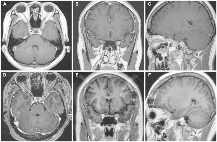
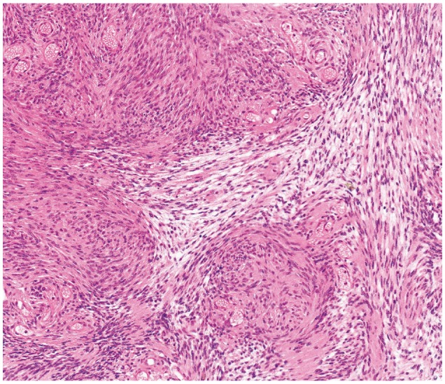
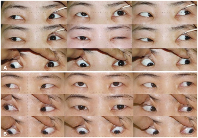
The patient underwent a major surgery via the left frontotemporal approach using a neuro-navigation system. We performed an intradural anterior clinoidectomy and unroofing of the superior orbital wall. The tumor was observed to originate from the ventral portion of the oculomotor nerve (Fig. 2), and was removed subtotally. The anatomical continuity of the oculomotor nerve was preserved. Histopathological examination of the tumor revealed a schwannoma (Fig. 3). There was no evidence of recurrence on the one-year follow-up MRI (Fig. 1D-F). After surgery, the left side ptosis and medial gaze limitation was evident (Fig. 4A), but the symptoms improved gradually (Fig. 4B).
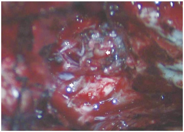 | Fig. 2Intaoperative photogram showing the tumor originated from ventral part of oculomotor nerve. V1: ophthalmic branch of trigeminal nerve, III: oculomotor nerve, II: optic nerve, arrows: the tumor.
|
Go to :
DISCUSSION
It is reported that intraorbital schwannomas account for 1-6% of all intraorbital tumors [2]. The accurate diagnosis, using imaging modalities, of oculomotor schwannoma may be difficult because of the complex orbital anatomy and its low incidence. Two clues leading to diagnosis are tumor location along the course of the oculomotor nerve, and oculomotor nerve palsy on neurologic examination [2,4]. The most common symptom of oculomotor schwannomas is oculomotor nerve palsy, but oculomotor nerve palsy is not always the initial symptom [4]. In the present case, optic nerve dysfunction was observed on neurologic examination without oculomotor nerve palsy. Thus, the optic nerve masses such as gliomas, schwannomas, or meningiomas appear to be differential preoperative diagnostic possibilities. However, we noted that the tumor origin was the oculomotor nerve during surgery. In patients with oculomotor schwannomas located in the intraorbital area without oculomotor nerve palsy, the operative finding may be important to confirm the tumor origin.
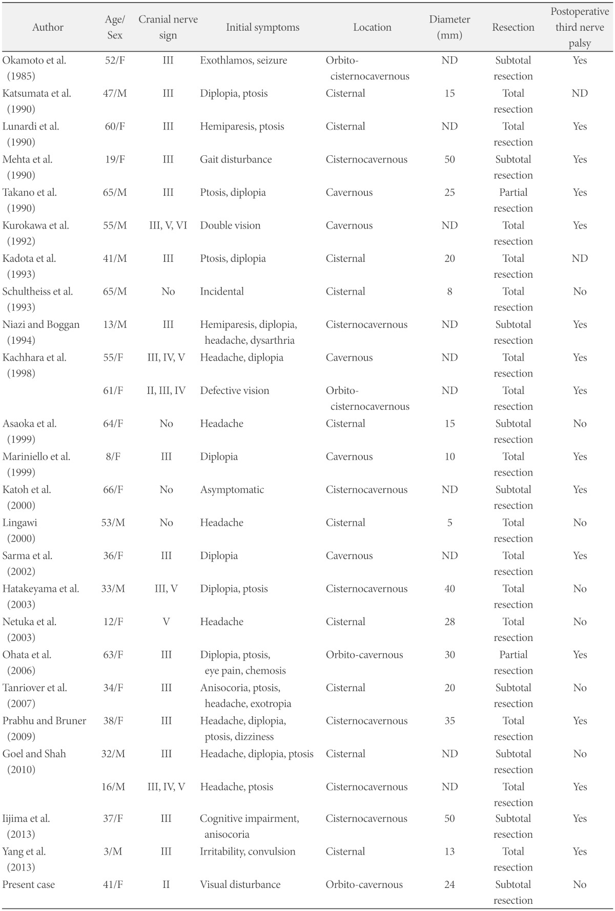
We reviewed 25 patients who received surgery for oculomotor nerve schwannomas (Table 1). Preoperative oculomotor nerve palsy manifested in 19 cases out of 25, and optic nerve dysfunction was shown in only 2 patients, including the present case. The tumor extended to the orbit region in 4 cases, and 2 of 4 patients manifested optic nerve dysfunction. Regardless of the radicality of the resection, postoperative oculomotor nerve palsy occurred in patients with oculomotor schwannomas in the orbital region, excluding our case.
Because of the tumor's benign property, total resection of the tumor is the treatment modality and adjunctive therapy is not needed. The oculomotor nerve is known to be fragile and is easily injured [4]. Previous studies reported that total resection of the tumor results in complete oculomotor nerve palsy [5,6,7]. Worsening of oculomotor nerve function may occur after subtotal or partial resection of the tumor [2,4]. The oculomotor nerve contains somatic motor fibers to many of the orbital muscles, but also carries parasympathetic fibers to the papillary muscle. Thus, oculomotor nerve palsy may worsen the patient's quality of life. It is difficult to decide on the best treatment strategy for oculomotor schwannomas. According to each case, several treatment strategies have been reported by researchers. Katoh et al. [5] recommend 'wait-and-see' policy for asymptomatic patients with oculomotor schwannoma. According to Kim et al. [8], Gamma Knife radio-surgery may be an effective and minimally invasive treatment modality without risk of cranial nerve palsy in treatment of patients with schwannomas originating from the oculomotor, trochlear, and abducence nerves. Radical resection inevitably results in worsened oculomotor function, almost invariably in complete palsy. Thus, Asaoka et al. [4] recommend subtotal resection except large tumors that cause intractable symptoms. On the other hand, there were some reports of total resection of oculomotor schwannoma without permanent nerve palsy [3,9]. It's location influences the radicality of tumor resection. The chance of oculomotor nerve injury after surgical resection may increase as the resection proceeds more anteriorly toward the superior orbital fissure [10]. In our review, past postoperative oculomotor nerve palsies occurred in all patients with tumors that extended to the orbital region, except our present case (Table 1), where the tumor was located in the superior orbital fissure and close to other neurovascular structures. Also, the preoperative oculomotor nerve function was preserved. Thus, we decided to perform a subtotal resection to avoid complete oculomotor nerve palsy, and planned an adjuvant frameless radiosurgery for the remnant tumor. During the operation, most of the mass was removed leaving the tumor capsule. In the postoperative MRI, the target lesion could not be detected so we decided not to perform the adjuvant radiosurgery. There is no evidence of tumor recurrence on the one-year follow-up MRI, and we planned for follow-up MRI annually.
In conclusion, we report a case of oculomotor schwannoma in which the preoperative diagnosis may have been very difficult due to the complex orbital anatomy and the low incidence of disease. The treatment strategy should be established considering the preoperative nerve functions and the tumor location. We removed the tumor subtotally using the fronto-temporal approach with resulting temporary oculomotor nerve palsy.
Go to :
 XML Download