

 PDF
PDF ePub
ePub Citation
 Print
Print


INTRODUCTION
Intracranial ependymal cysts (ECs) are congenital, benign, extracerebral lesions. These are rare, and mostly occur in the supratentorial area [1]. The cerebellopontine angle (CPA) is an extremely rare location for ECs. To the best of our knowledge, this is the 7th case of CPA EC worldwide. Six cases reported to date were symptomatic except ECs first reported case. Presenting symptoms were unknown (1, autopsy case), hearing disturbance (1), rhinolalia (1), hemifacial spasm (2), dizziness & vomiting (1, with hemorrhage) [2-7]. In this report, we report a case of ependymal cyst in left CPA presenting with syncope.
CASE REPORT
A 44-year-old man came to the emergency department in our hospital with suboccipital headache and paresthesia of the left arm. Examining his history, he was generally healthy and had no distinguishable medical history besides several near syncopes. However, he had not visited a hospital when he started to feel a near-syncope.
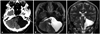
Two days before admission, the patient started to experience dizziness and syncope while he stood up in the pub. After recovery of consciousness, he felt a suboccipital headache and paresthesia of the left arm, so he visited the emergency department of another institute. At that time, since his symptoms had vanished, the hospital discharged him without complete medical checkup such as a computed tomography (CT) or magnetic resonance imaging (MRI). However, when he returned home, the headache and paresthesia reappeared, causing the patient to visit our hospital for accurate medical evaluation. The medical evaluation results are as follows: physical and neurological exam were normal except for paresthesia of the left arm. The CT scan revealed a 6.6×5.4 cm sized large cystic lesion of the left CPA with no abnormal enhancement by contrast medium (Fig. 1A). The MRI demonstrated a cystic lesion, similar to an arachnoid cyst, with shift of the cerebellar hemisphere, pons and medulla to the right side (Fig. 1B, C).
After admission, general evaluation followed to focus on the cardiogenic syncope. During the routine check-up within the emergency room, the electrocardiogram showed a sinoatrial node block. Consultation with a cardiologist followed to evaluate the sinoatrial node block and syncope. Furthermore, an echocardiogram, 24 hours Holter monitoring and flecainide provocation test was performed to rule out the possibility of Brugada syndrome. Despite these efforts, the result of cardiac evaluation did not show any abnormality which could cause the syncope.
We assumed that EC resulted in the syncope and therefore decided to intervene surgically. A left suboccipital retrosigmoid craniotomy was conducted to approach to the cyst, and which was followed by fenestration into the subarachnoid space. Partial cystic wall removal was also conducted for pathological diagnosis. The pathological examination with immunohistochesmistry study showed features of a typical ependymal cyst. The lining epithelium of the cyst consisted of simple cuboidal epithelial cells with no prominent cilia (Fig. 2A). The epithelial cells showed diffuse positivity for glial fibrillary acidic protein (Fig. 2B) and dot-like positivity for epithelial membrance antigen (Fig. 2C). Postoperatively, the hospital course was favorable, with the exception of nausea and vomiting which persisted for several days and which resolved later. He was discharged at 14 days after surgery. At the last follow-up at 4 months after surgery, he was neurologically normal and free from near syncope.
DISCUSSION
ECs are rare, benign, ependymal-lined cysts which are commonly located in the lateral ventricle or juxtaventricular area of the temporoparietal region and frontal lobe [1]. These cysts have a heterogenous histogenetic nature and therefore have been referred to as ECs, glioependymal cysts, neuroepithelial cysts, choroidal epithelial cysts, or simply as epithelial cysts [2].
The majority of clinical symptoms in supratentorial ECs are seizures, attacks of unconsciousness, or manifestations including a progression of localizing neurological deficits [1]. However, CPA ECs reported to date have showed symptoms related to involvement of a specific cranial nerve except for two cases (Table 1). Ho and Chason [2] reported an autopsy case, and Ho and Wu [7] reported a case presenting with hemorrhage leading to nausea and vomiting.
Interestingly, our patient's unique neurologic symptom, specifically syncope, has not been reported in any journals that dealt with CPA EC. Cardiologic evaluation showed no significant abnormality in our case. We speculated that his recurrent near syncope and syncope were due to the CPA EC. Prilipko et al. [8] reported a Chiari type I malformation case with orthostatic intolerance and syncope. Cardiologic evaluation of that patient showed results corresponding to postural orthostatic tachycardia syndrome (POTS). This study suggests that direct hindbrain compression may affect the baroreflex system and therefore causing POTS leading to syncope. Glossopharyngeal neuralgia may also cause cardiac syncope [9,10]. It is speculated that artificial synapses between the glossopharyngeal and vagus nerve, developed from irritative sensory phenomenon, result in near syncope and syncope [9,10].
The cyst in this patient case compressed the brain stem and cerebral hemisphere from just below tentorium to the pontomedullary junction level. We suggest that the mass effect in CPA may cause syncope according to a similar pathophysiology as above.
Several surgical treatment methods have been reported: total excision of cyst, shunting to the peritoneal space, cystic fluid aspiration, and fenestration into the subarachnoid space. We performed fenestration of the cyst into the subarachnoid space with cystic wall biopsy. For cysts with hemorrhage, surgical resection of cyst may be the best choice of surgery [7]. In other cases, the method of connection between the cystic and subarachnoid space would be physiologic, safe and effective.
It is noteworthy that no reports were found in literature describing the ependymal cyst in the CPA with syncope. Although various surgical methods were recommended for EC in the CPA, we suggest relative minimal invasive method such as fenestration of the cyst into the subarachnoid space is safe and enough to relieve patient's neurologic symptoms.

 XML Download