Journal List > J Lipid Atheroscler > v.6(2) > 1059596

Chung, Kim, Lim, Seo, Zo, Kim, and Kim: Double Right Coronary Arteries Originated from Separate Ostia with Total Occlusive Lesion in One of the Two Right Coronary Arteries

Abstract

Double right coronary arteries (RCA) are very rare congenital anomalies of coronary artery. We report a case of double RCA with total occlusion, incidentally found by collateral flows. A 71-year-old patient underwent percutaneous coronary intervention of left coronary arteries for angina, and the presence of double RCA was missed at initial coronary angiography (CAG). About 20 months later, second CAG was performed due to recurrent angina, and the CAG showed newly developed collateral flow suggesting the presence of the other missed RCA. There was a total occlusion at missed RCA and the lesion was successfully revascularized with drug eluting stent. If there was no collateral flow, the other RCA could not be found and its critical lesion could not be managed properly. Our case suggests that collateral flow can be a useful clue in detecting coronary anomaly. Besides, it is important to fully understand coronary anatomy, not to miss uncommon coronary lesion.

REFERENCES

1. Garg N, Tewari S, Kapoor A, Gupta DK, Sinha N. Primary congenital anomalies of the coronary arteries: a coronary: arteriographic study. Int J Cardiol. 2000; 74:39–46.
crossref
2. Topaz O, DeMarchena EJ, Perin E, Sommer LS, Mallon SM, Chahine RA. Anomalous coronary arteries: angiographic findings in 80 patients. Int J Cardiol. 1992; 34:129–138.
crossref
3. Yamanaka O, Hobbs RE. Coronary artery anomalies in 126,595 patients undergoing coronary arteriography. Cathet Cardiovasc Diagn. 1990; 21:28–40.
crossref
4. Harikrishnan S, Jacob SP, Tharakan J, Titus T, Kumar VK, Bhat A, et al. Congenital coronary anomalies of origin and distribution in adults: a coronary arteriographic study. Indian Heart J. 2002; 54:271–275.
5. Chen YF, Chien TM, Chen CW, Lin CC, Lee CS. Double right coronary artery or split right coronary artery? Int J Cardiol. 2012; 154:243–245.
crossref
6. Chien TM, Chen CW, Chen HM, Lee CS, Lin CC, Chen YF. Double right coronary artery and its clinical implications. Cardiol Young. 2014; 24:5–12.
crossref
7. Gupta SK, Abraham AK, Reddy NK, Moorthy SJ. Supernumerary right coronary artery. Clin Cardiol. 1987; 10:425–427.
crossref
8. Soydinc S, Sari I, Davutoglu V. The dilemma in diagnosing double right coronary artery: contribution of multidetector computed tomography. Int J Cardiol. 2008; 126:132–133.
crossref
9. Basso C, Maron BJ, Corrado D, Thiene G. Clinical profile of congenital coronary artery anomalies with origin from the wrong aortic sinus leading to sudden death in young competitive athletes. J Am Coll Cardiol. 2000; 35:1493–1501.
crossref
10. Davis JA, Cecchin F, Jones TK, Portman MA. Major coronary artery anomalies in a pediatric population: incidence and clinical importance. J Am Coll Cardiol. 2001; 37:593–597.
crossref
11. Ozeren A, Aydin M, Bilge M, Dursun A, Onuk T. Atherosclerotic double right coronary artery and ectasia of left coronary arteries in a patient with presented acute coronary syndrome and ventricular tachycardia. Int J Cardiol. 2005; 102:341–343.
crossref

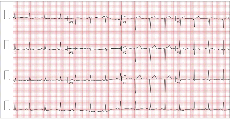
Fig. 1.
Patient’s electrocardiogram at first presentation.
jla-2017-6-2-97f1.tif
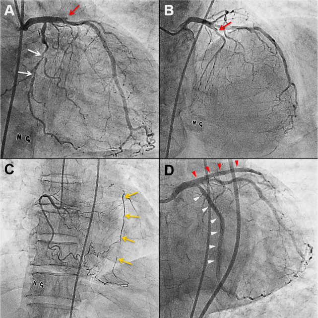
Fig. 2.
Coronary angiography at initial presentation. (A and B) Total occlusion of proximal left anterior descending artery (LAD) (red arrow) and significant stenosis in left circumflex artery (LCX) (white arrows), (C) Right coronary artery with collateral flow (yellow arrows) to LAD, and (D) revascularized LAD (red arrow heads) and LCX (white arrow heads) after percutaneous coronary intervention with drug eluting stents.
jla-2017-6-2-97f2.tif
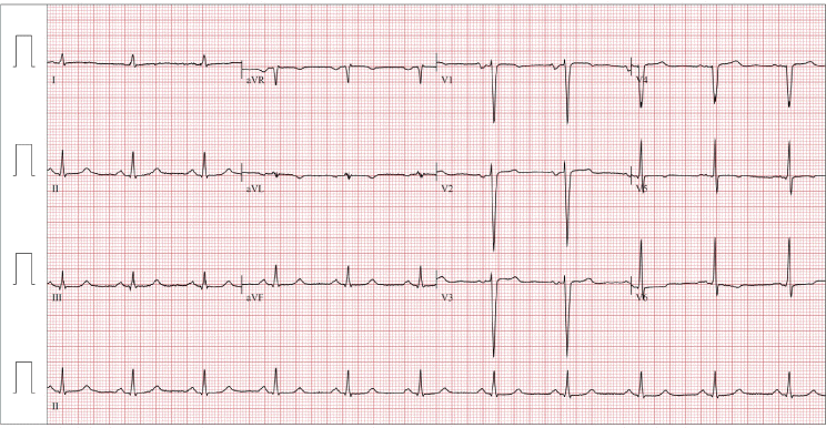
Fig. 3.
Patient’s electrocardiogram at second presentation.
jla-2017-6-2-97f3.tif
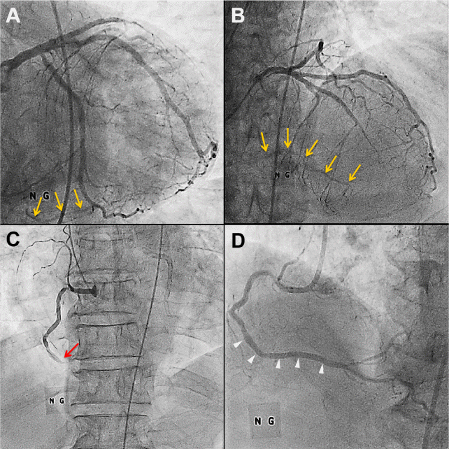
Fig. 4.
Coronary angiography at the second presentation. (A and B) Left coronary angiogram showing collateral arteries to right coronary artery (RCA) territory (yellow arrows), (C) detected total occlusive lesion in distal segment of the missed RCA (red arrow), and (D) revascularized missed RCA after percutaneous coronary intervention with drug eluting stents (white arrow heads).
jla-2017-6-2-97f4.tif
TOOLS
Similar articles