

 PDF
PDF ePub
ePub Citation
 Print
Print


INTRODUCTION
Comprehensive physiologic assessment can provide additional information about microvascular function in coronary artery disease. Three physiologic indices, fractional flow reserve (FFR), index of microcirculatory resistance (IMR) and coronary flow reserve (CFR) can be easily measured in culprit vessel of ST elevation myocardial infarction (STEMI) patient after successful primary revascularization. Here, we reports 2 cases of STEMI patients who underwent compressive physiologic assessment in order to determine microvascular function after successful primary revascularization.
CASE 1
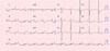
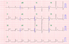
A 56 year old man presented with a 4 hours of ongoing chest pain and admitted via emergency department for further evaluation and management. He was non-smoker and he had been treated with hypertension and dyslipidemia in local clinic. He had been taking aspirin 100mg, rosuvastatin 10 mg, and olmesartan 40 mg once a day. He had no family history of cardiovascular disease. His initial electrocardiogram (EKG) showed ST segment elevation in lead II, III, aVF, accompanied with reciprocal changes in V4-6 with ST depression (Fig. 1).
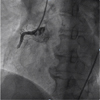
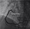
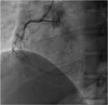
In coronary angiography, total thrombotic occlusion with TIMI 0 flow in mid right coronary artery (RCA) without any measurable collateral flow from non-culprit vessels was notified and the culprit lesion was successfully treated with thrombus suction and stent implantation (Synergy 4.0×28 mm) (Fig. 2). The door to balloon time was 55 minutes. The final angiogram showed TIMI 3 flow without residual stenosis (Fig. 3).
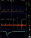
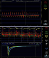
Post-percutaneous coronary intervention (PCI) physiologic study in culprit vessel was performed. FFR under maximal hyperemia using intracoronary nicorandil 2 mg bolus injection was 0.97, however, CFR was depressed to 1.1 and IMR was elevated up to 75 U which suggested the presence of overt microvascular damage in culprit vessel territory (Fig. 4).
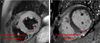
Cardiac magnetic resonance imaging (MRI), which was performed 5 days after primary PCI showed intramyocardial hemorrhage in T2 weighted image and microvascular obstruction was also notified in infarcted area after late gadolinium enhancement (Fig. 5).
CASE 2
A 41 year old man presented with a 1 hour history of ongoing resting chest pain and admitted via emergency department for further management. He was current smoker and he had no remarkable medical history. His initial EKG showed ST segment elevation in II,III,aVF with reciprocal changes with ST depression in V2-4 (Fig. 6).
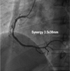
Initial coronary angiography showed thrombotic total occlusion with TIMI 0 flow in mid-RCA without any measurable collateral flow from non-culprit vessels. The culprit vessel was successfully revascularized after thrombus suction and stent implantation (Synergy 3.5x38 mm). The final angiogram showed TIMI 3 flow without residual stenosis (Fig. 7, 8). We also performed post-PCI comprehensive physiologic assessment in culprit vessel. FFR with use of nicorandil was 0.98, CFR was 2.2 and IMR was 24 U. The results from physiologic assessment implied well preserved microvascular function and non-significant flow limitation through epicardial coronary artery as well as microvascular beds (Fig. 9).
Cardiac MRI was taken 2 days after primary PCI and showed difference pattern with case 1. There were no intra-myocardial hemorrhage and edema in T2 weighted image and there was only subtle enhancement in infarcted area and we couldn't find microvascular obstruction in late gadolinium enhancement image (Fig. 10).
DISCUSSION
Previously, Fearon et al1 showed that elevated IMR value, more than 40 U, is the independent predictive value associated with worse clinical outcome in STEMI patients. And also elevated IMR at the time of STEMI represents larger degree of myocardial and microvascular damage and less recovery of left ventricular function that can be assessed by cardiac enzyme, cardiac MRI, echocardiography, or positron emission tomography.23 Another report from our group presented that not only elevated IMR, but also low CFR was significantly associated with worse clinical outcome in patients with stable coronary artery disease and functionally insignificant epicardial coronary stenosis.4 These findings emphasize the importance of comprehensive physiologic assessment in patients with coronary artery disease. Similarly another reports from Korea also presented the significant association between microvascular damage, which was defined with depressed CFR as well as elevated IMR, and long-term outcomes in STEMI patients after successful revascularization.56 In this case report, we presented the results from the comprehensive physiologic assessment in culprit vessel after primary PCI in STEMI patients and validated the results using cardiac MR findings. There is acute and overt inflammatory change in myocardial cell and vasculature, especially in acute phase of myocardial infarction. Nevertheless, the reproducibility of IMR measurement and prognostic implication in patients with STEMI, even in the acute phase has been presented by previous studies.789
Previous studies showed several mechanisms cause overt microvascular dysfunction. Individual susceptibility (e.g. underlying comorbidity, lack of preconditioning, collateral recruitability), degree and extent of ischemic injury and reperfusion injury cause difference in physiologic findings in these two cases. We think that degree of ischemic injury, especially duration of ischemic event (i.e. symptom to door time), is the one of the major causes of difference in these two cases.
In real world practice, most physicians do not perform the comprehensive physiologic assessment after revascularization of epicardial coronary stenosis. We wanted to emphasize the importance of comprehensive physiologic assessment for microvascular function by presenting these 2 cases with substantially different physiologic findings, even after the successful revascularization for the epicardial coronary stenosis.
In conclusion, comprehensive physiologic evaluation at the time of primary PCI at STEMI is feasible, and low CFR and high IMR highly suggests overt microvascular damage even after the successful revascularization of culprit vessel.

 XML Download