

 PDF
PDF ePub
ePub Citation
 Print
Print


INTRODUCTION
Coronary artery disease (CAD) is the leading cause of death worldwide.1 Coronary revascularization with percutaneous coronary intervention (PCI) has been validated as a highly effective therapy improving cardiovascular outcomes in patients with unstable CAD.2 Although PCI is safe, serious complications can sometimes occur during the procedure.3 One of the rare but devastating complications of PCI is inappropriate migration and loss of coronary stent.456789 Stents embolize only one site and do not move to another location in most cases. Herein, we report a case of a wandering coronary stent from the coronary artery to the femoral artery via the axillary artery. The migrated stent was successfully removed by percutaneous approach using a snare.
CASE REPORT
A 78-year old woman visited our emergency department due to dyspnea and stuporous mentality. Since one month prior to the presentation, she had complained of general weakness and sore throat, but had no cough, sputum, fever, dyspnea, or chest pain. One day before the hospital visit, she felt sudden dyspnea on exertion that progressively worsened to dyspnea at rest, but she did not complain of chest pain. Cough and sputum also occurred. She had been taking medications for her multiple coronary risk factors including hypertension, diabetes mellitus, and dyslipidemia.
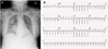
On admission, she was hemodynamically unstable: blood pressure was too low to measure, heart rate was variable with tachycardia over 100 per min, and respiratory rate was over 30 per min with 70% oxygen saturation by pulse oxymetry. She had no fever. On physical examination, she suffered from increased effort of breathing. Breathing sounds on both lower lung fields were decreased. There was no cardiac murmur or peripheral edema. Chest X-ray showed diffuse infiltration of both lungs with consolidation on the right upper lobe (Fig. 1A). Electrocardiogram showed sinus tachycardia with poor R progression and ST depression in multiple precordial and limb leads (Fig. 1B). Within two hours of hospital visit, CK-MB and troponin I were elevated up to 111.8 ng/mL (normal range, <6.6 ng/mL) and 25.9 ng/mL (normal range, <0.028 ng/mL), respectively. Brain natriuretic peptide was over 25,000 pg/mL (normal range, <100 pg/mL). C-reactive protein was elevated up to 7.0 mg/dL (normal range, <0.50 mg/dL). Arterial blood gas analysis revealed hypoxemia combined with severe respiratory acidosis (arterial blood pH 6.9). Transthoracic echocardiography showed a depressed left ventricular ejection fraction (LVEF) of 35% and extensive left anterior descending (LAD) coronary artery territory akinesia. She was diagnosed with acute myocardial infarction complicated by pulmonary edema and cardiogenic shock. Mechanical ventilation and vasopressor administration were started, and emergent coronary angiography (CAG) was performed.
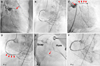
CAG showed total occlusion of the proximal LAD, moderate degree diffuse stenosis of the left circumflex artery (LCX) (Fig. 2A), and multiple severe stenosis at the right coronary artery (RCA) (Fig. 2B). After intra-aortic balloon pump (IABP) insertion, LAD revascularization was successfully performed using Orsiro Hybrid sirolimuseluting stents (2.5×26 mm and 3.0×30 mm sized) (Orsiro SES, Biotronik AG, Bulach, Switzerland) (Fig. 2C). Subsequently, Orsiro SES (3.0×30 mm sized) was placed in the distal RCA (Fig. 2D). While an additional Orsiro SES (3.0×30 mm sized) was being delivered to the proximal portion of the RCA, the strut of the stent was suddenly dropped out of the guiding catheter at the RCA ostium. The embolized coronary stent was found in the left axillary artery with oscillating movement (Fig. 2E and Video 1). The remaining stenosis of the RCA was successfully revascularized using 3.0×32 mm and 3.5×22 mm sized Promus elements drug-eluting stents (Boston, Scientific, Natick, MA, USA) (Fig. 2F). She became stabilized after PCI, and current pneumonia was treated with proper antibiotics. The IABP was removed on the third day of PCI.
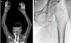
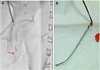
Two weeks later, computed tomography (CT) angiography of the upper extremities was performed to find the exact location of the lost coronary stent for surgical removal. To our surprise, there was no stent in the left axillary artery or any vessels of the upper extremities (Fig. 3A). The lost stent was found in the proximal portion of the left superficial femoral artery by fluoroscopic exploration (Fig. 3B). Although she did not have any symptoms of limb ischemia, we decided to remove the stent for the prevention of thrombosis. Then, the stent was successfully removed by the percutaneous approach using snare (5 mm Amplatz Goose Neck snares, eV3 Endovascular, Inc., Plymouth, MN, USA) (Fig. 4A and 4B). The patient is currently monitored regularly without complaints.
DISCUSSION
The present case showed wandering of the lost stent which started from the RCA ostium, passed through the upper extremity immediately, and finally arrived at the lower extremity. The stent was successfully retrieved by the percutaneous approach using a snare without complications. Although several case reports of stent loss have found in the literature, they have usually presented as single migration.456789 Wandering of a lost stent is extremely rare.48
A recent meta-analysis revealed that stent loss occurred in 1.3% of the PCI cases, and that its incidence decreased to 0.3% after 2005, probably due to improvement in manufacturing technology of coronary stents.4 Lodgment sites of lost stents are reported as the aorta, renal artery, brachial artery, and pedal artery.10111213 The axillary and femoral arteries are relatively rare sites of lost coronary stents. Although stent loss dose not increase mortality rate, the rates of emergency coronary bypass surgery and bleeding requiring transfusion are elevated.5 Unfavorable morphologies and lesion characteristics of the coronary artery, such as proximal tortuosity, calcification, and severe angulation are known to increase the incidence of stent loss.5 In our case, however, considering that stent delivery to the distal RCA was not difficult, the stenotic lesions of the RCA were not so tricky. However, resistance for stent advancement at RCA ostium was increased, because there was ostial stenosis of RCA and guiding catheter was not coaxial with proximal part of RCA. Together with this factor, technical and structural problem of stent itself might be also related to the stent loss in our case.
Although surgical retrieval is required in some cases,68 most of migrated stents can be retrieved by interventional methods.45 Percutaneous retrieval can be performed using the following methods: (1) an inflated balloon with smaller profile than the stent, (2) biliary forceps, (3) Cook retained fragment retriever, (4) two wires twisted around the stent, and (5) basket retrieval device.5 In our case, the patient was hemodynamically unstable to retrieve the stent at the time of recognition of the stent loss. Moreover, oscillating movement of the stent caused technical difficulty catching the stent with snares.
Mechanisms underlying movement of lost stents against antegrade arterial flow is still unknown. Although it was possible that body position and upper trunk movement might have an impact on stent migration, IABP might be the most important triggering factor for stent migration especially when the stent was moved from axillary to subclavian artery or aorta. A possible mechanism is oscillating movement of a stent at left axillary artery in accordance with the inflation-deflation cycle of IABP as shown in Video 1. Deflation of the balloon in the systolic phase may produce transient retrograde flow.14 In this case, the lost stent escaping from the left axillary artery might have a chance of embolization to major cerebral arteries. Therefore, any arterial foreign body in the upper extremities should be immediately retrieved if possible, especially in patients with IABP, to prevent further dangerous embolization.
In conclusion, we reported the unique case of re-mobilization of the lost coronary stent from the upper extremities to the lower extremities. The IABP may facilitate retrograde movement and provoke the migration of the lost stent. Immediate retrieval of lost stent is recommended for the prevention of secondary embolic events.
 XML Download