

 PDF
PDF ePub
ePub Citation
 Print
Print


INTRODUCTION
Endovascular aneurysm repair (EVAR) for an abdominal aortic aneurysm is a valuable treatment option in selected patients; it has acceptable morbidity and mortality rates.1 A stent graft with suprarenal fixation offers the potential advantages of a decreased incidence of distal migration and endoleak.2
However, when this device obstructs the renal ostia, renovascular hypertension can occur. When renal blood flow is reduced, juxtaglomerular cells in the kidneys activate prorenin and secrete renin directly into the circulation, causing high blood pressure. Decreased blood flow to the kidneys can decrease the glomerular filtration rate (GFR). If the stenosis is longstanding and severe, the GFR in the affected kidneys does not increase and results in renal failure.3
The incidence of renal artery occlusion following endovascular abdominal aortic aneurysm repair is low.3 Here, we describe a patient with elevated renin and aldosterone levels, as well as hypertension due to obstructed renal arteries after EVAR. The patient's hypertension and renal artery occlusion were resolved after renal artery stenting, and the levels of renin and aldosterone decreased.
CASE REPORTS
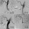
A 67-year-old male underwent EVAR due to a rapidly growing abdominal aortic aneurysm. He had a medical history of stable angina pectoris without hypertension, and his blood pressure was 120/70 mmHg. He received 300 mg of aspirin and 300 mg of clopidogrel on the day of the procedure. A common femoral artery access was utilized to perform the endovascular treatment. Heparin anticoagulation was maintained throughout the procedure with an activated coagulation time of 200-300 seconds. First, we deployed the plug for the right internal iliac artery due to the right common iliac artery aneurysm and we deployed the 23/13/120 mm main body via a right femoral approach. Then, we deployed a 16/20/95 mm contra-lateral limb via a left femoral approach and a 16/10/95 mm ipsilateral extension for the right external iliac artery via a right femoral approach. The final angiogram showed good distal flow without an endoleak (Fig. 1A). The patient was discharged and returned home after 9 days without symptoms or complications.
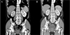
After discharge, the patient's blood pressure increased over time; one month after discharge, his blood pressure was 131/76 mmHg, and three months later his blood pressure was 161/82 mmHg. We monitored the patient's blood pressure for a total of 24 hours, 5 months after his discharge from the hospital; the daytime average was 180/106 mmHg, and the nighttime average was 172/100 mmHg. Further workup revealed an elevated serum renin level of 98.26 ng/ml/h (aldosterone 23.8 ng/dL). Abdominal CT angiography revealed that the right kidney had decreased in size with a delayed nephrogram (Fig. 2A), and a captopril renal scan showed that the right kidney exhibited decreased renal function with delayed excretion. These results indicated right renal artery occlusion, so angioplasty was performed.
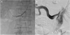
We performed a peripheral angiogram (PAG) using a pigtail catheter via the left brachial artery. The PAG showed no significant stenosis on an antero-posterior cranial view (Fig. 1B). However, it showed significant stenosis with a characteristic bird's-beak appearance in the os of the right renal artery caused by a previous stent graft on the left anterior oblique cranial view (Fig. 1C and 1D). We selected the right renal artery using a 5 Fr Judkins right number 4 guiding catheter. After the failure of a 0.035 inch Terumo guidewire due to tight stenosis, we inserted a 0.014 inch guidewire into the right renal artery and performed plain old balloon angioplasty (POBA) using a 4×20 mm balloon (Fig. 3A). However, the stent graft was re-expanded using the characteristics of a nitinol stent, so we were unable to insert a larger balloon and stent. After repeated POBA using a 4×20 mm balloon, we inserted a 4 Fr multipurpose catheter into the right renal artery and inserted a 0.035 inch Terumo guidewire. However, we were unable to insert a 5×40 mm balloon. Thus, we changed the guidewire to a coiled stiff wire and performed POBA using a 5×40 mm balloon. Despite POBA using a 5×40 mm balloon and a back-up stiff wire, we were unable to insert the stent and guiding catheter into the right renal artery. However, after changing the guiding catheter, we successfully inserted a 5 Fr shuttle catheter into the right renal artery. After positioning a 6×18 mm SCUBA® (Medtronic Minneapolis, MN, USA) stent for the right renal artery os on the inside of the guiding catheter, we extracted the guiding catheter and deployed the stent. The final PAG showed an optimal result for the right renal artery os with good distal flow and a normalized nephrogram (Fig. 3B).
This patient was discharged after 5 days without symptoms or complications. The patient's blood pressure was 120/70 mmHg without hypertensive medication, and his renin activity level was 5.19 ng/ml/h (aldosterone 7.7 ng/dL) at the time of discharge. The patient continued to exhibit a stable blood pressure at his 1-month follow-up appointment. Follow-up CT and renal scans showed no significant occlusion of the renal artery and a normalized nephrogram (Fig. 2B).
DISCUSSION
Endovascular abdominal aortic aneurysm repair is a safe, durable, and effective procedure. Previous studies have shown that EVAR is better than open repair in patients with an abdominal aortic aneurysm.14 However, if the renal ostia is obstructed, the patient's blood pressure will become elevated and the serum creatinine level will be elevated due to renal infarction. Renal artery occlusion secondary to a stent graft impingement remains an uncommon complication that is typically recognized early, and which is immediately treated during the procedure.3 Karmacharya et al.5 reported that 9 of 550 patients exhibited small renal infarcts associated with EVAR on follow-up CT scans.
Our case was a hypertensive patient that had elevated renin-aldosterone levels after the deployment of a stent graft to treat an abdominal aortic aneurysm. Abdominal CT angiography and captopril renal scans suggested a right renal artery obstruction. We performed angiography and found a partial obstruction in the renal artery os. Therefore, we deployed a stent into the renal artery, which subsequently reduced the patient's blood pressure and reduced the serum renin-aldosterone level.
Our case has two main points. First, the patient's blood pressure rose slowly over a long period of time after the initial EVAR procedure and was found by laboratory tests performed in the outpatient department. When renal blood flow is reduced, juxtaglomerular cells in the kidneys activate prorenin and secrete renin directly into the circulation, resulting in high blood pressure. In our patient, the renin level was increased 26 times over the normal range. Therefore, if patients with a history of EVAR visit the hospital with an elevated blood pressure, clinicians should consider renal artery occlusion and check the patient's renin level.
Second, we did not observe the partial obstruction of the renal artery when looking at the antero-posterior angiographic view. However, we did observe the obstruction from a different angle when the angiogram was redone. We were unable to confirm the cause of the right renal artery os obstruction following EVAR. The stent-graft may have migrated after the EVAR procedure, or it was positioned incorrectly during the procedure. The latter is a more likely cause of the obstruction. Therefore, this case study highlights the importance of viewing these scans from various angles, despite the low incidence of renal artery occlusion. Additionally, intravascular ultrasound imaging provides valuable additional information on endograft position and the patency of the renal artery orifice. Several reports have determined that adjunctive renal artery stenting during an endovascular abdominal aortic aneurysm repair is effective and safe.6 Thus, if the occlusion is confirmed during EVAR, adjunctive stenting will prevent the need to undergo a second medical procedure.
As EVAR gains popularity, the incidence of EVAR-associated renal occlusion may increase. Thus, the presence of a renal artery occlusion should be assessed before and after EVAR.
 XML Download