

 PDF
PDF ePub
ePub Citation
 Print
Print


Abstract
Objectives
The purpose of the study was to compare plaque characteristics by coronary computed tomography angiography (CCTA) with those by virtual histology-intravascular ultrasound (VH-IVUS).
Methods
We enrolled 50 asymptomatic patients with diabetes mellitus or more than two risk factors for coronary artery disease such as hypertension, smoking, and hyperlipidemia. If the patient had a coronary lesion (plaque with more than 50% stenosis or calcium score more than 100), we recommended coronary angiography and VH-IVUS and compared CCTA findings with VH-IVUS findings.
Results
35 patients (70%) had coronary lesions, and we performed both CCTA and VH-IVUS in 23 patients. All 23 patients had multiple risk factors, and the majority of target lesions were located at left anterior descending artery (73.9%), and calcium score of lesion site was 106±162 with plaque volume of 232±153 mm3 by CCTA. Calcium score of lesion site was significantly greater in diabetic patients (n=14) than non-diabetic patients (n=9) (118±159 vs. 88±175, p=0.038). By VH-IVUS, plaque volume was 174±127 mm3, absolute necrotic core (NC) volume was 22±21 mm3, and relative NC volume was 20.8±8.7%. Absolute dense calcium (DC) volume and absolute NC volumes were significantly greater in diabetic patients than non-diabetic patients (11.5±13.8 mm3 vs. 9.1±11.0 mm3, p=0.028, and 23.9±24.7 mm3 vs. 18.1±14.3 mm3, p=0.035, respectively). Plaque volume by CCTA correlated with that of VH-IVUS (r=0.742, p<0.001), and plaque volume by CCTA correlated with absolute NC volume by VH-IVUS (r=0.621, p<0.001), and calcium score of lesion site by CCTA correlated with absolute dense calcium volume by VH-IVUS (r=0.478, p=0.028).
Figures and Tables
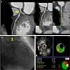 | Fig. 1Coronary computed tomography angiography (A), coronary angiography (B), and virtual histology-intravascular ultrasound findings (C) in patients with diabetes mellitus, hypertension, smoking, and hyperlipidemia.
|
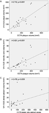 | Fig. 2Correlation between plaque volume by coronary computed tomography angiography (CCTA) with that by virtual histology-intravascular ultrasound (VH-IVUS) (A), and correlation between plaque volume by CCTA with absolute necrotic core volume by VH-IVUS (B), and correlation between lesion site calcium score by CCTA with absolute dense calcium volume by VH-IVUS (C).
|
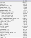
Table 1
Baseline characteristics in patients who underwent both computed tomography angiography and virtual histology-intravascular ultrasound
![]()
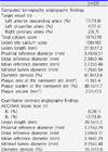
Table 2
Computed tomography angiography and quantitative coronary angiography findings in patients who underwent both computed tomography angiography and virtual histology-intravascular ultrasound
![]()
References
1. Newman WP 3rd, Freedman DS, Voors AW, Gard PD, Srinivasan SR, Cresanta JL, et al. Relation of serum lipoprotein levels and systolic blood pressure to early atherosclerosis. The Bogalusa Heart Study. N Engl J Med. 1986; 314:138–144.

2. Relationship of atherosclerosis in young men to serum lipoprotein cholesterol concentrations and smoking. A preliminary report from the Pathobiological Determinants of Atherosclerosis in Youth (PDAY) Research Group. JAMA. 1990; 264:3018–3024.
3. Newman AB, Arnold AM, Naydeck BL, Fried LP, Burke GL, Enright P, et al. "Successful aging": effect of subclinical cardiovascular disease. Arch Intern Med. 2003; 163:2315–2322.
4. Fuster V. Lewis A. Conner Memorial Lecture. Mechanisms leading to myocardial infarction: insights from studies of vascular biology. Circulation. 1994; 90:2126–2146.
5. Ross R. Atherosclerosis--an inflammatory disease. N Engl J Med. 1999; 340:115–126.
6. Zaman AG, Helft G, Worthley SG, Badimon JJ. The role of plaque rupture and thrombosis in coronary artery disease. Atherosclerosis. 2000; 149:251–266.
7. Hoffmann MH, Shi H, Schmitz BL, Schmid FT, Lieberknecht M, Schulze R, et al. Noninvasive coronary angiography with multislice computed tomography. JAMA. 2005; 293:2471–2478.
8. Herzog C, Zwerner PL, Doll JR, Nielsen CD, Nguyen SA, Savino G, et al. Significant coronary artery stenosis: comparison on per-patient and per-vessel or persegment basis at 64-section CT angiography. Radiology. 2007; 244:112–120.
9. Meijboom WB, Weustink AC, Pugliese F, van Mieghem CA, Mollet NR, van Pelt N, et al. Comparison of diagnostic accuracy of 64-slice computed tomography coronary angiography in women versus men with angina pectoris. Am J Cardiol. 2007; 100:1532–1537.
10. Wexler L, Brundage B, Crouse J, Detrano R, Fuster V, Maddahi J, et al. Coronary artery calcification: pathophysiology, epidemiology, imaging methods, and clinical implications. A statement for health professionals from the American Heart Association. Writing Group. Circulation. 1996; 94:1175–1192.
11. Rumberger JA, Simons DB, Fitzpatrick LA, Sheedy PF, Schwartz RS. Coronary artery calcium area by electronbeam computed tomography and coronary atherosclerotic plaque area. A histopathologic correlative study. Circulation. 1995; 92:2157–2162.
12. Blankenhorn DH. Coronary arterial calcification: a review. Am J Med Sci. 1961; 242:41–49.
13. Eggen DA, Strong JP, McGill HC Jr. Coronary calcification. Relationship to clinically significant coronary lesions and race, sex, and topographic distribution. Circulation. 1965; 32:948–955.
14. Rumberger JA, Sheedy PF, Breen JF, Schwartz RS. Electron beam computed tomographic coronary calcium score cutpoints and severity of associated angiographic lumen stenosis. J Am Coll Cardiol. 1997; 29:1542–1548.
15. Guerci AD, Spadaro LA, Popma JJ, Goodman KJ, Brundage BH, Budoff M, et al. Relation of coronary calcium score by electron beam computed tomography to arteriographic findings in asymptomatic and symptomatic adults. Am J Cardiol. 1997; 79:128–133.
16. Fallavollita JA, Brody AS, Bunnell IL, Kumar K, Canty JM Jr. Fast computed tomography detection of coronary calcification in the diagnosis of coronary artery disease. Comparison with angiography in patients < 50 years old. Circulation. 1994; 89:285–290.
17. Baumgart D, Schmermund A, Goerge G, Haude M, Ge J, Adamzik M, et al. Comparison of electron beam computed tomography with intracoronary ultrasound and coronary angiography for detection of coronary atherosclerosis. J Am Coll Cardiol. 1997; 30:57–64.
18. Tanenbaum SR, Kondos GT, Veselik KE, Prendergast MR, Brundage BH, Chomka EV. Detection of calcific deposits in coronary arteries by ultrafast computed tomography and correlation with angiography. Am J Cardiol. 1989; 63:870–872.
19. Mintz GS, Nissen SE, Anderson WD, Bailey SR, Erbel R, Fitzgerald PJ, et al. American college of cardiology clinical expert consensus document on standards for acquisition, measurement and reporting of Intravascular Ultrasound Studies (IVUS). A report of the American college of cardiology task force on clinical expert consensus documents. J Am Coll Cardiol. 2001; 37:1478–1492.
20. Rodriguez-Granillo GA, García-García HM, Mc Fadden EP, Valgimigli M, Aoki J, de Feyter P, et al. In vivo intravascular ultrasound-derived thin-cap fibroatheroma detection using ultrasound radiofrequency data analysis. J Am Coll Cardiol. 2005; 46:2038–2042.
21. Nair A, Kuban BD, Tuzcu EM, Schoenhagen P, Nissen SE, Vince DG. Coronary plaque classification with intravascular ultrasound radiofrequency data analysis. Circulation. 2002; 106:2200–2206.
22. Oh HJ, Kwon K, Park SH, Park SH, Lee YH, Yu MA, et al. CT coronary angiography using multidetector computed tomography in coronary artery disease : a comparative study to quantitative coronary angiography. Korean Circ J. 2004; 34:1167–1173.
23. Leber AW, Becker A, Knez A, von Ziegler F, Sirol M, Nikolaou K, et al. Accuracy of 64-slice computed tomography to classify and quantify plaque volumes in the proximal coronary system: a comparative study using intravascular ultrasound. J Am Coll Cardiol. 2006; 47:672–677.
24. Arad Y, Spadaro LA, Goodman K, Newstein D, Guerci AD. Prediction of coronary events with electron beam computed tomography. J Am Coll Cardiol. 2000; 36:1253–1260.
25. Reaven PD, Sacks J. Investigators for the VADT. Coronary artery and abdominal aortic calcification are associated with cardiovascular disease in type 2 diabetes. Diabetologia. 2005; 48:379–385.
26. Lee S, Choi EK, Chang HJ, Kim CH, Seo WW, Park JJ, et al. Subclinical coronary artery disease as detected by coronary computed tomography angiography in an asymptomatic population. Korean Circ J. 2010; 40:434–441.
 XML Download