

 PDF
PDF ePub
ePub Citation
 Print
Print


INTRODUCTION
The left internal thoracic artery (LITA) is generally accepted as a standard grafting method. Because of the long patency rates and resistance to atherosclerosis, LITA is the preferred graft for surgical revascularization of the left anterior descending artery (LAD).1,2 However, the presence of significant proximal left subclavian artery stenosis may result in reversal of LITA coronary graft flow and produce myocardial ischemia. Prevalence of significant stenosis of the subclavian artery in patients referred for coronary artery bypass graft (CABG) surgery was reported to be 0.2% to 6.8%.3,4,5 Subclavian artery stenoses are mainly of atherosclerotic origin. Here, we report a case of a 49-year-old female patient who complained of resting chest pain and left arm pain after CABG surgery using the LITA due to subclavian artery stenosis.
CASE REPORT
A 49-year-old female with end-stage renal disease (ESRD) from diabetes mellitus on peritoneal dialysis was admitted to the emergency room with a progressive dyspnea lasting 2 days and newly developed resting chest pain. She also complained of left shoulder pain radiating to her left forearm. Five years ago, she was diagnosed with multi-vessel coronary artery disease and underwent CABG surgery. LITA was anastomosed to the LAD and Y-composited graft using a harvested left radial artery was anastomosed to the obtuse marginal branch (OM). Because her native LAD and left circumflex coronary artery were totally occluded from their proximal portion, blood supply to the anterior and lateral wall was solely dependent on the grafts.
Upon admission, the initial electrocardiography showed a new ST segment depression on lateral leads (V4-6) and elevated serum troponin I (19.75 ng/mL), consistent with a non-ST segment elevation myocardial infarction (NSTEMI) involving the lateral wall. Echocardiographic examination showed global hypokinesia of the left ventricle and akinesia of the inferior wall with an ejection fraction < 20%. Brachial systolic blood pressure measured in the left arm (98/62 mmHg) was about 30 mmHg lower than that measured in the right arm (130/88 mmHg). Despite medical treatment, diabetes mellitus was poorly controlled with a hemoglobin A1c level of 8.3%. Her blood lipid profile was as follows: total cholesterol=143 mg/dL, triglycerides=134 mg/dL, high-density lipoprotein cholesterol=54 mg/dL and low-density lipoprotein cholesterol=63 mg/dL.
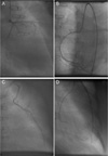
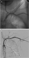
Urgent coronary angiography and bypass graft angiography did not reveal any critical stenosis in the CABGs connected with the LAD or OM (Fig. 1). However, during cannulation of the left subclavian artery from the aortic arch for selective LITA angiography, we had difficulty advancing the catheter through the subclavian artery. A 6-F guiding catheter (Cordis, genesis, Cordis, Miami Lakes, FL, USA) wash able to advance into the left subclavian artery ostium. Aortic arch angiography showed significant stenosis at the ostium of the left subclavian artery (Fig. 2A) with sluggish flow into the LITA. Therefore, we concluded that subclavian artery stenosis, not CABGs, might be the cause of impaired myocardial perfusion. Subclavian artery stenosis was successfully treated using an 8×25-mm stent (Cordis Genesis, Cordis, Miami Lakes, FL, USA) (Fig. 2B). After subclavian artery intervention, her chest pain and left arm pain subsided and ST depression improved. Blood pressure measured in both arms was approximately equal (right arm=130/70 mmHg, left arm=128/70 mmHg). Short-term follow-up echocardiography 2 weeks later showed significant improvement of left ventricular wall motion and systolic function with an ejection fraction of 40%.
DISCUSSION
CABG surgery is the treatment of choice for diabetic patients with left main artery disease, multi-vessel disease with impaired left ventricular function, or complex lesions (i.e., total occlusion, calcified lesions, or bifurcation lesions).6 A lower incidence of major adverse cardiovascular events and repeat revascularizations has been reported in diabetic patients with multi-vessel disease who underwent coronary CABG compared with percutaneous coronary intervention.7 Myocardial revascularization using the LITA has become the standard for CABG surgery due to its long-term graft patency and lower repeat revascularization rate compared to a saphenous vein graft. However, an occluded or stenosed CABG is a frequent cause of recurrent angina, particularly in patients with a heavy atherosclerotic burden, such as ESRD or poorly controlled diabetic mellitus.8
The prevalence of significant subclavian artery stenosis was reported to be 0.2-6.8% in patients treated with CABG surgery using LITA.3,4,5 Atherosclerosis is the most common cause of stenosis (95-97%), although arteriovenous fistula, Takayasu's arteritis, congenital aortic abnormalities, and thoracic outlet syndrome have also been described as possible causes.3 Progression of left subclavian artery stenosis can lead to ischemia of upper extremity and severe stenosis of the left subclavian artery before the origin of the LITA ostium can lead to decreased LITA flow. Chronic arterial insufficiency of the upper extremity can cause arm pain, particularly with upper extremity work. More than 20 mmHg difference in blood pressure is highly indicative of subclavian artery stenosis. Myocardial ischemia can also be aggravated by retrograde blood flow from the partially patent native coronary circulation through the LITA into the distal subclavian artery.4
Although operative reconstruction was previously considered to be the procedure of choice of subclavian artery stenosis9, recent studies10,11 have suggested endovascular intervention as the first-line therapy owing to equal effectiveness and fewer complications. Furthermore, patients who have already had CABG and developed subsequently with coronary-subclavian steal syndrome have been considered as good candidates for endovascular intervention.10
We have presented a case of 49-year-old female patient who complained of resting chest pain and left arm pain after CABG surgery using the LITA. Because significant left subclavian artery stenosis was not detected during preoperative evaluation, NSTEMI was caused by de novo left subtotal subclavian artery stenosis proximal to the LITA. ESRD and poor glycemic control can aggravate rapid progression of native left subclavian artery stenosis. If we had not discovered that subclavian artery stenosis developed after CABG surgery, we might have performed a less effective, and possibly harmful, intervention to the native coronary artery.
Coronary angiographies are increasingly performed using a radial approach; thus, left subclavian artery evaluation proximal to the LITA graft can easily be missed. However, since most patients who undergo CABGs have a large atheromatous burden in coronary arteries, as well as in overall vascular beds, atherosclerosis of the native carotid artery could progress proximal to the internal thoracic artery.
In conclusion, a careful physical evaluation, including blood pressure measurement in both arms and meticulous evaluation of the overall pathway from the aorta to the CABGs, must be conducted in patients who have undergone CABG surgery to provide valuable information regarding uncommon and unexpected culprit lesions beyond the CABGs.
 XML Download