

 PDF
PDF ePub
ePub Citation
 Print
Print


Foot-and-mouth disease (FMD) O, A, and Asia 1 type viruses have been circulating in Asian countries [1]. In Korea, FMD occurred once in 2000, once in 2002, and three times in 2010, making a total of five outbreaks since 2000 [23456]. All animals susceptible to FMD were vaccinated in early 2011 to help prevent recurrence of the disease. Since that time, due to concerns about a recurrence, vaccines against three serotypes, O, A, and Asia 1, have continued to be vaccinated (O1 Manisa, A Malaysia 97, and Asia 1 Shamir) [7]. In July-August 2014, FMD occurred on three pig farms in Uiseong and Goryeong in Gyeongbuk province and in Hapcheon in Gyeongnam province. This paper describes the outbreak, the diagnosis, the quarantine measures for FMD under the condition of vaccination and pathogenesis, genetic analysis of the isolated virus.
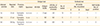
The outbreak of FMD among pigs was reported in Uiseong, Gyeongbuk province on July 23, 2014; the symptoms were detaching of the hooves and vesicles. Using a lateral flow device for detecting antigens [8] by the veterinarian of regional veterinary service in Gyeongbuk, the pigs were identified as being FMD-positive in the farm site. The susceptible animals were thoroughly examined using reverse transcriptase polymerase chain reaction and enzyme-linked immunosorbent assays (ELISAs), and FMD was confirmed (Table 1).
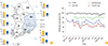
Four days later, on July 27, cases of suspected FMD were reported to have occurred on a pig infected premise (IP) in Goyrong, Gyeongbuk. The detachment of hooves and the formation of ulceration in the mouth were additionally reported 64 km away from the farm where FMD first occurred. The third report was made 10 days later on August 6 in Hapcheon, Gyeongnam, 25.7 km away from the second IP where FMD occurred; the symptoms included vesicles in the mouth, ulcers on the bridge of the nose, the inability to stand up, and lack of appetite. FMD was confirmed after a laboratory diagnosis (Fig. 1A).
At the first premise where FMD occurred, FMD case occurred in the herd where vaccinations had not been implemented prior to the FMD outbreak. At the second premise where FMD case occurred, the disease occurred in some pigs that had not been vaccinated. FMD virus (FMDV)-infected animals of a lower level were identified on this farm compared to the other farms. The owner in third IP recognized the FMD outbreak around the area and simultaneously vaccinated to the own pigs, but it is estimated that its animals were infected with FMD before protectable immunity induced. In the process of an epidemiological survey, a retrospective analysis of serum collected on 16 June was positive for the non-structural protein (NSP) antibody around the second IP, and finally NSP-positive antibody in an additional six farms were detected. Therefore, the first outbreak is estimated to have been before June. Although the etiology of FMD was uncertain, there was no outbreak of FMD from April 2011 for 3 years and 3 months.
In Korea, the average antibody prevalence rate by structural protein (SP)-ELISA was 98.9% for cattle and 50.5% for pigs. The antibody prevalence rate of cattle was much higher than the 80% immunity level, a transmission inhibition level in general [9], but in finisher pigs it was relatively low due to one shot vaccination (Fig. 1B). The antibody prevalence rate in cattle tested in Korea was high level, and therefore we estimated that it was difficult for field viruses to circulate. In pigs, the rate accounted for approximately 80% in breeding pigs and 40%-45% in finisher pigs. The possibility of virus infection is higher among finisher pigs than breeding pigs (Fig. 1B). Pigs with clinical symptoms were culled and pigs without clinical symptoms on farms where FMD occurred were vaccinated emergently. The movement of livestock on the farms that tested positive was restricted for 3 weeks and additional quarantine measures, such as disinfection, were taken. Additional tests and careful examinations of the environment or other risk factors were made until the risk disappeared.
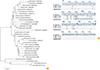
According to the results by full-length or VP1 sequence analysis of the FMDV, the outbreaks were of O type, SEA topotype, and Mya-98 lineage and were classified as the viruses most similar to those that occurred in East Asia in 2014 (Fig. 2A). According to the similarity analysis of the VP1 nucleotide, they were different by 3.13% from O/Primorskiy/RUS/2014 identified to be the viruses closest to a nucleotide sequence reported thus far (World Reference Laboratory for FMD), by 3.44% from O/GZ-MT/CHA/2013 in China, and by 4.23% from O/HKN/13/2010 in Hong Kong (Table 2Fig. 2A).
Surprisingly, we found a deletion of 69 nucleotides (23 amino acids [aa]) in 3A/3B1 region of the virus (5 aa of 3A region and 18 aa of 3B1 region) by genome analysis (Fig. 2B). This virus is a natural variant from the field. Experimentally, the reduced single-VPg viruses produced a mild disease in swine, indicating that the VPg copy number is an important determinant of host range and virulence. However, the deletion of individual 3B proteins in FMDV cause clinical disease in cattle [1011].
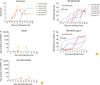
For the pathogenesis test, the footpads of pigs were inoculated with the FMDV (103.0 TCID50/0.1 mL) that occurred in first case in 2014, and a contact group was established at the same time point. A vaccination group was inoculated with commercial trivalent vaccine (O1 Manisa, A Malaysia 97, and Asia 1 Shamir) 5 days prior to the contact-challenge. Next, FMD specific clinical symptoms were observed to identify the transmission of FMD. According to the experimental results, the pigs that were inoculated started to show lameness, erosions at the coronary band, and vesicles in their hooves, and the one of contact group began to have the same symptoms 6 days after the start of the experiment. However, the one contacted and two vaccinated pigs did not appear any symptoms until 21 days of cohabiting, the ending time point of the experiment. In the animals with clinical signs during the experiment, viremia and virus excretion were identified, but in the vaccination group, small amount of virus after contact was only detected (Fig. 3A). The SP antibody was produced in 4-5 days post-inoculation in inoculated animals and in 8 days post-contact in the contact pigs (Fig. 3B). Therefore, it was proven that even though the immunized period was short after emergency vaccination, this vaccination was shown the protective capacity with no clinical symptom, virus excretion. In the field, decreases in transmission and the protection capability were identified when high potency vaccination (>6 PD50) carried out although result of vaccine matching of the isolated virus with vaccine strain, O1 Manisa was identified comparatively low as r1 value of 0.14. The O1 Manisa vaccine was able to protect against O/Andong/SKR/2010 [4] and O/SKR/01/2014, which was verified in the field.
The outbreak of FMD in Korea that has been determined to be closely related to the occurrence of FMD in neighboring countries is considered to have been affected from viruses of adjacent nations by genetic analysis in this case, too. However, the direct cause of this inflow has not been verified. This, O/SKR/01/2014, was new virus with the deletion of 23 amino acids in 3A/3B1 region and low pathogenic property.
 XML Download