

 PDF
PDF ePub
ePub Citation
 Print
Print


Introduction
Japanese encephalitis (JE) is a zoonotic disease that is characterized by acute inflammation in the central nervous system. The Japanese encephalitis virus (JEV) is an arthropod-borne virus that is kept in a transmission cycle between pigs and mosquitoes. However, it can cause encephalitis in horses and humans by incidental infection [1]. Global warming affects the activity of mosquitoes and as such, JE was considered a re-emerging disease associated with climate change. JE cases have been identified in most Asian countries, particularly in South Asia, Southeast Asia, and East Asia [2]. Recent studies estimated the occurrence of JE at 68,000 cases annually, resulting in 10,000-15,000 deaths [3456789].
JEV belongs to the Flaviviridae family of viruses and genus Flavivirus [10]. It is an enveloped virus with a single stranded positive sense RNA genome that is approximately 11 kb in length. Sequence analysis of the viral capsid/premembrane/envelop (E) gene indicated that JEV could be classified into five genotypes (G1 to G5) [11]. Since the replacement of JEV G3 with G1 was first identified in 1994 in Japan, G1 has become the dominant circulating JEV in many Asian countries, including China [12], Thailand [13], Vietnam [14], and Korea [15]. In Japan, before the 1950s, over several hundred JE cases were reported annually; however, vaccination and improvements in hygiene have dramatically reduced its incidence [6]. Since 1986, there has been no report of equine JE in Japan; however, one unvaccinated horse died from JE in 2003, and it was revealed that this isolate belonged to G1 [16]. In Korea, since the first identification of JEV in 1946, a number of JE cases were reported in domestic animals as well as in humans throughout the 1950s and 1960s [17]. A live attenuated JEV vaccine containing G3 was developed for the prevention of JEV infection in animals, and has been widely used for pigs and horses in Korea since the late 1980s [8]. Until recently, there were no reports on horses clinically infected with JEV in Korea. However, there are reports of other animals infected with G1 in Korea [1819]. The potential impact of JEV genotype changes on vaccine efficacy has been estimated using a mouse model and the various JEV genotypes [20].
In the present study, we investigated the antigenic relationship between JEV G1 and G3 using the hemagglutination inhibition antibody (HI) test and virus neutralization (VN) test. This study provides additional data to support the notion that genotype shift can adversely affect vaccine efficacy. In our study, we also conducted a serological survey to determine the prevalence of antibodies against JEV G1 and G3 in racehorses and conventional pigs in Korea.
Materials and Methods
Viruses and cells
The Anyang 300 (G3) and KV1899 (G1) viruses were used as antigens for the neutralizing antibody tests. These strains were officially obtained from the Animal and Plant Quarantine Agency of Korea. JEV was propagated in Vero cells, which were maintained in α-minimum essential medium (α-MEM) supplemented with 5% fetal bovine serum (FBS), penicillin (100 µg/mL), streptomycin (100 µg/mL), and amphotericin B (0.25 µg/mL). The virus was inoculated in this media for experiments. After absorption for 1 hour, α-MEM was added and incubated until a cytopathic effect (CPE) was observed. The KV1899 strain, with a titer of 107.2 50% tissue culture infectious doses (TCID50/100 µL), was used as a representative of the original JEV G3 strain. The Anyang 300 strain (107.3 TCID50/100 µL) was used as representative of JEV G1. Titration of the viral strains was performed in microtiter plates. Ten-fold serial dilutions of the viruses were prepared in quadruplicates in α-MEM and were added to Vero cells at a density of 1×10⁵ cells/well. After incubation at 37℃ for 4 days in a CO2 atmosphere, the titer was defined as the end point dilution showing CPE in 50% of the wells using the Karber method.
Collection of sera from pigs and horses
A total of 42 blood samples were taken from conventional sows in several provinces of Korea between April and November 2012, and 216 horse blood samples were collected from thoroughbred racehorses in the same year. Most of the sows and racehorses are vaccinated once a year with live JEV vaccine (Anyang 300 strain).
Guinea pig sera
Antisera against JEV G1 (KV1899) and G3 (Anyang 300) were produced in normal guinea pigs weighing 350 g. The antigen for guinea pig immunization was prepared in Vero cells cultured in α-MEM supplemented with 5% FBS. After 4 days of incubation at 37℃, the supernatant of the infected culture was collected, centrifuged at 5,000 ×g for 20 minutes, and then titrated in 96-well plates as described above. Each viral suspension was emulsified with the adjuvant Montanide IMS 3012 (Seppic, France) at a 1:2 ratio (vol/vol).
Each emulsion was used to immunize five guinea pigs of an average weight of 350 g (JEV G1 in group A guinea pigs, JEV G3 in group B guinea pigs). A total of 2 mL of emulsion per animal was administered in two separate intramuscular injections 2 weeks apart. Blood samples were taken from guinea pigs to determine the antibody titers at the time of the first inoculation (T0) and then 2 weeks after the second inoculation (T1). At T0 all guinea pigs tested seronegative for the two JEV genotypes (G1 and G3), as determined by the VN test. At the end of the study, all the guinea pigs were euthanized.
HI test
The HI test was carried out using the JEV HI test kit (Median Diagnostics Inc., Chuncheon, Korea) according to manufacturer's instructions. Prior to the HI test, sera were inactivated at 56℃ for 30 minutes. The KV1899 (G1) strain of JEV was used as the antigen for the HI test. Using the sucrose acetone extraction method, viral antigens were prepared from the brains of suckling mice infected with the Korean isolate of strain KV1899 [7]. An HI test was performed in 96-well microplates, using slightly modified standard methods to estimate the concentration of JEV antibodies in pig sera. Briefly, to remove non-specific inhibitors, 10 µL of serum and 50 µL of 4% bovine albumin were mixed with 40 µL of 25% kaolin (Sigma, St. Louis, MO, USA). The mixture was then incubated for 30 minutes. The kaolin was then removed by centrifugation at 3,000 ×g for 15 minutes. The resultant clear supernatant was mixed with 5 µL of packed goose erythrocytes to remove any natural agglutinins. After incubation at 37℃ for 1 hour, the treated sera were separated from the goose erythrocytes by centrifugation. The treated sera (25 µL) were then diluted two-fold from 1:10 to 1:10,240 in round-bottom 96-well microplates and reacted with 8 hemagglutination units of JEV. After incubation at 37℃ for 1 hour, 50 µL of 0.33% goose erythrocytes was then added to the microplates and incubated for 30 minutes at 37℃. To confirm test reliability, JEV positive and negative pig control sera were used in all HI tests. HI titer was expressed as the reciprocal of the highest dilution of serum showing complete HI, and an antibody titer higher than 1:10 was considered positive.
VN test
The VN test for JEV was carried out in 96-well microplates using Vero cells. A 50 µL aliquot of a two-fold serial dilution of heat-inactivated serum was mixed with an equal volume of 200 TCID50 of the virus and incubated at 37℃ for 1 hour. A total of 100 µL of Vero cells at a concentration of 2×10⁵ cells per ml in α-MEM containing 5% FBS were added to each well. The microplates were incubated for 5 days at 37℃ under 5% CO2, and then virus-induced CPE was visually evaluated. The VN titer was expressed as the reciprocal of the highest serum dilution that completely inhibited CPE in the wells. The serum dilution ranged from 1:5 to 1:640, and an antibody titer higher than 1:10 was considered positive. All experimental procedures were approved by an independent Animal Care and Use committee and followed the guidelines of Green Cross Veterinary Products.
Results
Seroprevalence of JEV in sows and horses
Around 80% of all sows tested were positive for JEV and the positive ratio in the first half of the year was higher than in the second half of the year (2012). The positive ratio of JEV, as determined by the HI test, was 81%. The positive ratio of JEV, as determined by the VN test against G3 (Anyang 300) was 83.3%, and 11.9% against G1 (KV1899). The difference between the positive ratio of JEV between the first half and the second half of the year (2012) was not significant (Table 1).
The positive ratio of JEV in racehorses was around 90%, as determined using both the HI and VN tests against the Anyang 300 strain. However, that of JEV, tested by the VN test against KV1899, was 4.2% (Table 2).
Cross-reactivity of guinea pig sera between two genotypes of JEV
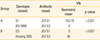
The geometric mean of the VN titers against the two JEV genotypes at T1 (2 weeks after the second immunization) is reported in Table 3. In guinea pigs immunized with JEV G1 (group A), the homologous VN titer (geometric mean) was 183.79 and the heterologous titer was 0. In guinea pigs immunized with JEV G3 (group B), the homologous VN titer (geometric mean) was 40.00 and the heterologous titer was 0. All differences were statistically significant (p<0.001).
Discussion
Most of the sows and racehorses raised in Korea have been vaccinated with an attenuated live JEV vaccine from March to May of each year since the vaccine was first developed in 1980. The sero-positive rate of JEV in racehorses identified in this study was higher than the rates reported in 2007 [9]. The HI titers in this study ranged from 1:20 to 1:160 in sows and racehorses (data not shown). Yang et al. [9] reported that most of the antibody titers induced by the Korean JEV vaccine are less than 1:160 since this vaccine is an attenuated live vaccine. However, the JEV killed vaccine produced in Japan is known to induce an HI titer of less than 1:640. Yang et al. [18] also reported that artificially inoculated pigs had a HI titer of 1:320 to 1:2,560 against JEV. The serum neutralizing (SN) antibody titer in this study ranged from 1:40 to 1:640 (data not shown). Yamanaka et al. [16] reported that animals infected with JEV induced SN antibody titers from 1:160 to 1:640. However, most of the Korean sows and racehorses had previously been vaccinated with JEV live vaccine, therefore, antibodies induced by JEV infection could not be differentiated from those induced by JEV vaccination.
The seropositive rate of JEV against JEV G1 was different from that against JEV G3, as determined by the VN test. Although the HA antigen used for the HI test was JEV G1, the positive rate of JEV assessed by the VN test against JEV G3 was similar to that obtained using the HI test.
For evaluating the antigenic relationship between JEV G1 and G3, we inoculated guinea pigs with the JEV G1 (KV1899) and G3 (Anyang 300) strains. Guinea pigs, unlike pigs and horses, are not a natural host of JEV infection and therefore may not have experienced previous exposure to JEV. Cross-antigenic evaluation of the JEV genotypes revealed that there was no cross neutralizing activity between the two genotypes. It is improbable that this may affect protection against the JEV G1 wild virus in animals that is prevailing in farms. This finding deserves particular attention, as HI is the gold standard test used in diagnostic laboratories for evaluation of humoral immunity to JEV.
The viral envelope (E) protein is known to play an important role in tissue tropism, cell fusion and infection, virus maturation, and protection [21]. Phylogenetic analysis of the E gene demonstrated that KV1899 was clustered into G1 and Anyang 300 belonged to G3 [22]. The homology of the E gene nucleotide sequence between these two strains is 87.1%. The Anyang 300 strain was isolated in 1969 and has been the live vaccine strain for animals in Korea. The KV1899 strain was isolated from a fatten pig in 1999 in Korea. In recent years, JEV G1 has displaced G3 as the dominant virus genotype throughout Asia [5]. Moreover, the genotype of JEV in Korea changed from G3 into G1 in 1991 [22]. A previous study demonstrated that sera from a patient immunized with the Taiwanese JE vaccine were unable to efficiently neutralize certain local JEV isolates [23].
The reasons for the emergence of JEV G1 in Korea are uncertain, and some investigators reported that circulation of vector-borne zoonotic viruses is largely determined by an overlap in the geographical distribution of virus-competent vectors and reservoir hosts. It is also reported that the viral molecular evolution is associated with climate change [4]. Based on the fact that the original JEV G3 no longer exists in the field and the proposition that the antigenic differences might decrease the efficacy of the vaccine, a new vaccine using the G1 strain should be developed and licensed.
In conclusion, the findings of this study indicate a discrepancy between the HI and SN titers, suggesting that HI is not adequate to accurately evaluate the protective immunity of the JE vaccine in pigs and horses, in particular against different genotypes of the JE virus. As is routinely done for influenza virus and rotavirus vaccines, the development of vaccines that match the circulating virus in the field could improve the effectiveness of prophylaxis for JEV infection. It is therefore necessary that future studies investigate the protective efficacy of the live attenuated JEV vaccine (Anyang 300) against experimental infection with a virulent JEV G1 strain (KV1899) in pigs.


 XML Download