

 PDF
PDF ePub
ePub Citation
 Print
Print


Introduction
Recorded outbreaks of Japanese encephalitis (JE) in Asia date back to the late 19th century. JE is a major cause of viral encephalitis and is endemic in several regions of Asia and the Pacific, causing an estimated 50,000 infections and 10,000 deaths annually [12]. JE virus (JEV) is maintained in a natural cycle between Culex mosquitoes and water birds, and is amplified in pigs. Accidental infection occurs in dead-end hosts, including humans and horses. The most important mosquito vector in Korea is Culex tritaeniorhynchus [34]. In regions where JE is endemic, vaccination against JEV is the most effective measure to control infection in humans and domestic pigs [5].
JEV can infect humans and a several other animals. However, most animals show no overt signs of infection, except for humans and horses. In horses, most infections are inapparent, and the mortality rate of viral encephalitis is less than 5%. Pigs are affected only during pregnancy; JEV infection manifests as abortion, mummified fetuses, and stillborn or weak piglets, depending on the stage of pregnancy [6]. JEV infection is an important economic disease in the domestic swine industry.
Japanese Encephalitis Virus
JEV, a member of the genus Flavivirus in the family Flaviviridae, clusters in the JE serocomplex group. JEV has an 11-kb encapsidated RNA genome composed of a single large open reading frame that encodes three structural and seven non-structural viral proteins (Fig. 1) [78]. The outer membrane of the virion consists of 90 envelope (E) protein homo-dimers, which play roles in the attachment to cellular receptors and membrane fusion [8]. Although the E protein was reported to be the major target for protection, animal experiments have shown that protection from disease is also mediated by antibodies against NS1, which is secreted from infected cells [910].
The Current Status of JE in Korea
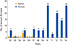
From 1984 to 2009, few cases (a mean of 2.1) of JE in humans were reported annually due to the national vaccination policy for Korean infants [11]. In 2010 and 2012, however, there were human outbreaks involving 26 cases with 7 deaths and 20 cases, respectively [12]. There has been no official notification of an outbreak in the Korean pig population since 2007 (Fig. 2). Swine JE is the second notifiable disease in the Act on the Prevention of Contagious Animal Disease [13]. A JE vaccination program for the entire Korean sow population has been performed annually to prevent reproductive disorders. In addition, regular serosurveillance has been conducted in the pig population to evaluate herd immunity in sows and monitor JEV in fattening pigs.
Epidemiology of JE: Genotype Shift Phenomenon
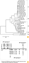
JEV can be classified into five genotypes based on similarities in the E gene (Fig. 3A) [14]. The replacement of JEV genotype 3 (G3) by genotype 1 (G1) was first reported in 1994 in Japan [15]. Similar to many other Southeast Asian countries, G1 viruses were introduced into Korea in 1993 and subsequently completely replaced G3 viruses as the dominant circulating JEV genotype (Fig. 3B) [316]. In addition, a JE G5 virus was first reported from a Culex mosquito in 2011; it is closely related to the Muar strain found in Malaysia in 1952 [17]. The genotypes differ by 10%-20% and 2%-6% at the nucleotide and amino acid levels, respectively. It has been suggested that JEV comprises a single serotype due to its relatively limited diversity at the amino acid level [18]. A mouse experiment demonstrated that vaccine strains induced significant, but lower, cross-neutralization against heterologous viruses [819]. However, Fan et al. [5] reported recently that live attenuated G3 swine vaccine (at222 strain) was partially protective against G1 viruses with very low cross-protective antibody titers. It is widely accepted that the current JEV vaccines derived from G3 strains are also able to protect against the now predominant G1 viruses. However, it is controversial whether the vaccine strain (G3) can induce a protective immune response against the now dominant JEV G1 strain.
The Current JE Vaccine for Animals
History of sequential attenuation of the JE vaccine strain
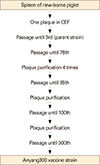
The JEV Anyang strain was isolated from the spleen of a new-born piglet in 1969 [20], and was sequentially attenuated in chicken embryo fibroblast primary cells (CEF) over 300 passages (Fig. 4). During the attenuation process, chorioallantoic and amniotic fluid from chicken embryos was added instead of fetal bovine serum. After seven plaque purification steps in which the plaques had a small, blurred border, the 300 virus formed a small homogenous plaque. The vaccine strain was designated Anyang300 [21]. For commercialization, the vaccine seed virus was propagated in duckling embryo fibroblast primary cells at 30℃, which resulted in greater viral growth [22].
Pathogenicity of the JE vaccine strain in various experimental animals
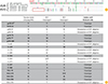
Mice are highly susceptible to wild-type JEV when inoculated via an intracerebral or peripheral route. The neurovirulence of the parent and an intermediately attenuated strain (Anyang285) was tested using a mouse model with virus titers of 107 pfu/mL. Weaning mice inoculated with the attenuated virus via a subcutaneous route did not die, although the LD50 of the parent virus was 0.01 pfu. In 3-week-old mice, the attenuated virus also did not cause death via an intraperitoneal route, although the LD50 of the parent virus was 80 pfu. Via an intracranial route, the attenuated virus was 9×10⁵ times less virulent than the parent virus. The attenuated virus still had weak neurovirulence, but it had completely lost the capacity for neuroinvasion (Table 1) [23].
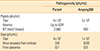
The pathogenicity in pigs was evaluated at two life stages. In piglets, the test focused on the duration of viremia and production of neutralizing antibodies. Compared to the parent virus, the vaccine strain (Anyang300) did not show any viremia and resulted in low titers of neutralizing antibodies. In pregnant sows, the vaccine strain was not recovered from embryos or placentas, while the parent virus was recovered from two embryos. These results indicate that the vaccine strain induced protective immunity, but completely lost its pathogenic features and the capacity for mosquito transmission via pigs (Table 2) [23].
Immunity studies of the vaccine strain in animals
In a preliminary test of piglets, the vaccine strain induced significant neutralizing antibody titers (>1:100; data not shown). In comparison, in a field test of sows, the first inoculation of the vaccine strain induced only low neutralizing antibody titers, close to the minimum protective titer of 1:10. The second inoculation increased the neutralizing antibody titer significantly. Based on a field test, the standard program for vaccinating sows against JE was established as two injections at a 3-week interval (Fig. 5A) [24]. In horses, the vaccine strain induced significant neutralizing antibodies with a single injection (Fig. 5B) [25].
Molecular Characterization of the Vaccine Strain
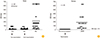
When the genome sequence of the vaccine strain was compared with those of other JEV strains, it was grouped with G3 viruses. At the nucleotide level, it showed high homology with other G3 viruses but differed clearly from G1 viruses [26]. At the start of the 3'-untranslated region, the genome of G3 viruses is characterized by specific nucleotide insertions right after the stop codon [16]. At the deduced amino acid level, there are several differences between G3 and G1 viruses scattered throughout the genome, including nonstructural proteins. There are also several differences from all other JEV strains, which were thought to be related to the viral attenuation or cell adaptation that occurred during the many passages in CEF cells. In the prM and E protein genes, which are important for protective immunity, there are four amino acid differences compared to the G1 virus group, especially two amino acid differences related to the induction of neutralizing antibodies in the E domain III region. These results indicate that the vaccine strain clearly has the molecular character of JEV G3, with several amino acid differences from the JEV G1 strains that now prevail in Korea (Fig. 6).
Development of New JE Vaccines for Animals
Inactivated JE vaccine
A live attenuated JE vaccine (Anyang300 strain, G3) has been used in veterinary medicine in Korea for more than 30 years. However, the low immunogenicity of the current vaccine in pigs and the recent genotype shift phenomenon (G3 to G1) in Asia have raised questions regarding the protective immunity generated with the current vaccine strain. New JEV G1 vaccines with enhanced immunogenicity are required for pigs. A new inactivated G1 JEV, which originated from a virus obtained from the blood of an infected piglet in 1999 (KV1899 strain), is now being developed for pigs, and its protective ability and safety are being evaluated in various animal species. The new vaccine induced significantly high neutralizing antibody levels in pigs and will be commercialized for pigs soon.
Live attenuated JE vaccine
To induce significantly high protective immunity in animals and considering the economic efficiency of the vaccine, a new live attenuated JE vaccine should be developed. To replace the current G3 vaccine, a recent G1 isolate, KV1899, was sequentially attenuated in African monkey kidney cells (Vero cells) until the virus completely lost its pathogenicity in various animals. The development and evaluation of this new live attenuated G1 virus are now in progress.
Other new JE vaccine candidates
For human vaccines, many new JE vaccine candidates have been reported, including recombinant virus-like particles [27], DNA vaccines containing the E and NS1 genes [28], and chimeric flavivirus vaccines [2930]. Unfortunately, few researchers are attempting to develop JE veterinary vaccines using advanced technologies. We anticipate that novel, effective veterinary vaccines will be developed using reverse genetics systems in the near future.
Conclusion
In the JE endemic region, vaccination against JEV is the most effective measure for controlling JEV infection in humans and domestic pigs. This article reviewed the history of JEV vaccine development and the molecular characterization of the vaccine strain in animals. The low immunogenicity of the current vaccine in pigs and the recent genotype shift phenomenon (G3 to G1) in Korea have increased the demand for new G1 vaccines. New inactivated and live attenuated G1 vaccines have been developed to induce more effective immunity in animals. Furthermore, novel, effective vaccines should be developed for animals using advanced technologies so that we can deal promptly with newly emerging JE genotype viruses.


 XML Download