

 PDF
PDF ePub
ePub Citation
 Print
Print


Introduction
Tuberculosis (TB) is a chronic infectious disease caused by Mycobacterium tuberculosis. It is estimated that about one-third of the world's population is latently infected with M. tuberculosis and that TB causes the death of 1.5 million people per year [1,2]. Unfortunately, the efficacy of the current TB vaccine is limited, thus development of an effective vaccine against TB is urgently required.
The M. bovis bacillus Calmette-Guérin (BCG) strain is used in the current TB vaccine, which has been used since 1921 [3,4]. Whereas BCG is effective for the prevention of severe forms of TB in children, such as miliary TB and TB meningitis [5,6], reports of its efficacy for the prevention of adult pulmonary TB are inconsistent [7,8]. In fact, the efficacy varies from 0% to 80% between studies, and it is the lowest in developing countries where protection against TB is the most needed [7,8].
For effective control of M. tuberculosis infection, T cell-mediated immune responses are essential [9,10] and thus, T cell activation following vaccination is considered to be a key requirement for generating protective immunity against TB. In particular, CD4+ T helper 1 (Th1) cells play a major role in immune responses against TB by secreting cytokines such as interferon-γ (IFN-γ) and tumor necrosis factor-α (TNF-α) [11, 12, 13, 14], which activate macrophages. In addition, CD8+ T cells also make important contributions to the protective immune responses against TB, as they not only secrete IFN-γ and TNF-α, but also lyse infected macrophages and epithelial cells [15, 16, 17].
Recently, the importance of activating polyfunctional T cells, which simultaneously produce multiple cytokines, has been emphasized in vaccine development. Initially, the significance of polyfunctional T cells was proven by a study that showed that the degree of vaccine protection against Leishmania major was predicted by the frequency of polyfunctional Th1 cells simultaneously secreting IFN-γ, TNF-α, and interleukin-2 (IL-2) [18]. In TB, the presence of polyfunctional CD4+ Th1 cells correlates with protection against M. tuberculosis challenge in mice [19,20]. In addition, the presence of polyfunctional CD8+ T cells simultaneously secreting IFN-γ and IL-2 was found to be associated with natural protection against TB and protection following anti-mycobacterial therapy in TB patients [21,22]. In view of these facts, it is expected that effective TB vaccines will also elicit polyfunctional T cell responses.
In the development of new TB vaccines, resuscitation-promoting factor (Rpf) is considered a promising target antigen [23,24]. Rpfs, first identified in Micrococcus luteus, are bacterial proteins that promote the recovery of bacteria from latency to a replicating phase [25]. M. tuberculosis expresses five different Rpf proteins, RpfA, B, C, D, and E [26], and they have resuscitation activity [27,28]. Indeed, deletion of rpf genes results in delayed reactivation of M. tuberculosis in a murine dormancy model [29]. Given that Rpf proteins are expressed during growth of M. tuberculosis, Rpfs-specific T cell immunity was expected to be effective in the control of M. tuberculosis growth [23,24]. Moreover, Rpfs were shown to be immunogenic in protein [23] or DNA [24] vaccination of mice.
In the present study, we examined if polyfunctional T cell responses are elicited by DNA immunization of M. tuberculosis RpfB. Specifically, we immunized mice with RpfB-encoding plasmid DNA and analyzed the polyfunctionality of T cells.
Materials and Methods
RpfB DNA plasmid
The rpfB gene of M. tuberculosis H37Rv (NCBI Gene ID: 886048) was codon-optimized for mammalian expression and synthesized by GenScript (Piscataway, NJ, USA). The synthesized rpfB gene was cloned into the pcDNA3.1(+) mammalian expression vector (Invitrogen, Carlsbad, CA, USA), where the cloned gene is expressed under the control of the cytomegalovirus IE-1 promoter. The sequence of the cloned rpfB gene was confirmed by sequencing. Large-scale production of plasmid DNA was performed using the Plasmid Giga Kit (Qiagen, Valencia, CA, USA).
Recombinant RpfB protein
The rpfB gene was amplified by polymerase chain reaction from genomic DNA of M. tuberculosis H37Rv and cloned into the pET-28a bacterial expression vector (Novagen, Darmstadt, Germany), which carries an N-terminal His-tag for protein purification. The sequence of the cloned rpfB gene was confirmed by sequencing. The RpfB protein was overexpressed in BL21 (DE3) Escherichia coli and purified using nickel-nitrilotriacetic acid agarose resin. Protein size and purity were confirmed by sodium dodecyl sulfate polyacrylamide gel electrophoresis. The purity was >90%.
Mouse immunization
Female C57BL/6 mice were housed in a specific pathogen-free facility in accordance with our institutional guidelines and used for immunization at the age of 5-6 weeks. For RpfB DNA vaccination, mice were immunized three separate times, at 3-week intervals, intramuscularly in both quadriceps muscles with 100 µg of RpfB plasmid DNA. For RpfB protein vaccination, mice were immunized three separate times, at 3-week intervals, subcutaneously with 50 µg of recombinant RpfB (rRpfB) protein. In protein immunization, incomplete Freund's adjuvant (Sigma, St. Louis, MO, USA) was used. For comparison, control mice were vaccinated subcutaneously with 2×10⁵ colony-forming unit of BCG (Pasteur 1173).
Synthetic peptides
Seventy one pentadecamer peptides (Mimotopes, Clayton, Australia), overlapping by 10 amino acids each and spanning the complete M. tuberculosis (H37Rv) RpfB protein sequence (NCBI Gene ID: 886048) were used. The overlapping peptides (OLPs) were resuspended at 20 mg/mL in dimethyl sulfoxide and further diluted to 1 mg/mL with phosphate-buffered saline (PBS). RpfB OLPs were mixed into two separate aliquots. The first aliquot included peptides from OLP-1 to OLP-35 and was designated as OLP mix 1. The other aliquot included peptides from OLP-36 to OLP-71 and was designated as OLP mix 2. In T-cell assays, either each single OLP or OLP mixes were used for T-cell stimulation.
Indirect enzyme-linked immunosorbent assay
Serum was collected 4 weeks after the final immunization, and anti-RpfB antibody was assessed by indirect enzyme-linked immunosorbent assay (ELISA). A Maxisorp microtiter plate (Nunc, Roskilde, Denmark) was coated with 10 µg/mL of rRpfB protein and blocked with 5% fetal bovine serum (FBS)-0.05% Tween-phosphate buffered saline (PBST). Sera of the immunized mice were serially diluted and dispensed into each well. After 2 hours of incubation, horseradish peroxidase-conjugated goat anti-mouse IgG (Millipore, Bedford, MA, USA) was added. One hour later, color reaction was performed with 3,3', 5,5'-tetramethylbenzidine (Sigma) for 15 minutes. The reaction was stopped by 2 M H2SO4, and optical density was read at 450 nm.
IFN-γ enzyme-linked immunosorbent spot assay
A 96 well MultiScreen enzyme-linked immunosorbent spot (ELISpot) plate (Millipore) was coated with anti-mouse IFN-γ antibody (clone AN-18, eBioscience, San Diego, CA, USA). The antibody-coated plate was blocked with 1% bovine serum albumin (Bovogen Biologicals, Essendon, Australia). Freshly-isolated splenocytes were dispensed into each well (500,000 cells/well), and 10 µg/mL of each single OLP was added. After 20 hours of incubation, the plate was washed with PBS and 0.05% PBST. Biotin-conjugated anti-mouse IFN-γ antibody (clone RA-6A2, eBioscience) and alkaline phosphatase-streptavidin (BD Pharmingen, San Diego, CA, USA) were sequentially added. A color reaction was then performed using AP color reagent (Bio-Rad, Hercules, CA, USA). The number of IFN-γ spot forming units was counted using an ELISpot reader (Cellular Technology Ltd., Cleveland, OH, USA). To evaluate CD8+ and CD4+ T cell responses separately, T cells were isolated from splenocytes using CD8 or CD4 microbeads (Miltenyi Biotec, Auburn, CA, USA), and IFN-γ ELISpot assays were performed with isolated T cells and T cell-depleted splenocytes [30].
Intracellular cytokine staining and polyfunctional T cell assay
Freshly-isolated splenocytes were resuspended in RPMI 1640 containing 10% FBS and 2 mM L-glutamine and stimulated with a single immunodominant OLP (10 µg/mL), OLP mix (1 µg/mL for each OLP) or rRpfB protein (5 µg/mL). To evaluate cytotoxic degranulation activity of T cells, anti-CD107a-PE-Cy7 (BD Biosciences, San Jose, CA, USA) was added into the culture medium. Brefeldin A (GolgiPlug, BD Biosciences) and monensin (GolgiStop, BD Biosciences) were added 2 hours later. After another 10 hours of incubation, splenocytes were first stained with ethidium monoazide (Sigma) and then stained with anti-CD3-V500, anti-CD4-V450 and anti-CD8-APC-H7 (all from BD Biosciences). The stained cells were permeabilized using Cytofix/Cytoperm kit (BD Biosciences) and then stained with anti-IFN-γ-APC, anti-TNF-α-PE and anti-IL-2-FITC (all from BD Biosciences). Fluorescence-activated cell sorting (FACS) analysis was performed using an LSRII flow cytometer (BD Biosciences) and the data were analyzed using FlowJo software (Treestar, San Carlos, CA, USA). T cells positive for the various combinations of cytokines and degranulation were analyzed and quantified using a Boolean gating function in FlowJo software [31].
Results
Humoral immune responses induced by RpfB immunization
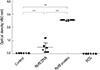
First, we assessed the RpfB-specific humoral immune responses after RpfB immunization. RpfB-specific immunoglobulin (Ig) was quantified in serially-diluted sera by indirect ELISA, and both DNA and protein immunization of RpfB were found to significantly increase the anti-RpfB Ig titer, though protein immunization was more potent (Fig. 1). In contrast, anti-RpfB Ig was not induced by BCG immunization (Fig. 1). These data show that both RpfB DNA and protein are immunogenic and capable of eliciting humoral immune responses.
Identification of the immunodominant T-cell epitopes in RpfB
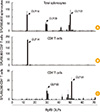
Next, we identified the immunodominant T-cell epitopes in RpfB. Splenocytes were isolated from RpfB DNA-immunized mice and stimulated with each of 71 OLPs spanning RpfB followed by assessment using an IFN-γ ELISpot assay. IFN-γ production was observed by direct ex vivo stimulation following stimulation with OLPs including OLP-14, OLP-30, and OLP-51 (Fig. 2A). To identify which T cell subset is responsible for IFN-γ production following stimulation with each epitope, CD8+ or CD4+ T cells were isolated from splenocytes, and isolated T cells and T cell-depleted splenocytes were subjected to IFN-γ ELISpot assays. OLP-14-stimulated IFN-γ production was observed in isolated CD8+ T cells (Fig. 2B), while OLP-30- and OLP-51-stimulated IFN-γ production was observed in CD4+ T cells (Fig. 2C). Taken together, OLP-14 (AGVQVHDADTIVLRR) is a dominant epitope for CD8+ T cells, and OLP-30 (GGLVRTVHLPAPNVA) and OLP-51 (LPVANVVVTPAHEAV) are dominant epitopes for CD4+ T cells; however, it should be noted that the minimal epitope within each peptide was not determined. Thus, we used these OLPs for T cell stimulation in further analyses.
T cell immune responses elicited by RpfB DNA immunization
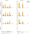
We evaluated T cell responses after RpfB immunization by intracellular cytokine staining (ICS). In ICS, splenocytes were stimulated with OLP mixes or dominant-epitope OLPs, and CD4+ and CD8+ T cell responses were separately analyzed by gating each subset during FACS analysis. CD4+ T cell responses to OLP mix 1 and OLP mix 2 were potently induced by RpfB DNA immunization as shown by IFN-γ ICS whereas they were scarcely induced by BCG or RpfB protein immunization (Fig. 3A). Similar results were observed when cells were stimulated with the CD4+ T-cell epitopes, OLP-30 and OLP-51 (Fig. 3A). RpfB DNA immunization also strongly induced RpfB-specific TNF-α production by CD4+ T cells (Fig. 3B). Moreover, cytotoxic degranulation activity (represented by CD107a) of CD4+ T cells was increased following RpfB DNA immunization (Fig. 3C). However, an IL-2 response was not elicited (Fig. 3D).
In CD8+ T cells, IFN-γ was potently induced in response to OLP mix 1 and the CD8+ T-cell epitope OLP-14 following RpfB DNA immunization, but not after BCG or RpfB protein immunization (Fig. 3E). RpfB DNA immunization also strongly induced TNF-α production (Fig. 3F) and cytotoxic degranulation activity (Fig. 3G) of CD8+ T cells whereas an IL-2 response was not elicited (Fig. 3H). Cytokine production in response to OLP mix 2 was not detected in CD8+ T cells and thus, it is not presented.
Taken together, RpfB DNA immunization induced vigorous responses in both CD4+ and CD8+ T cells, in particular IFN-γ and TNF-α production and cytotoxic degranulation activity.
Polyfunctionality of RpfB-specific T cells
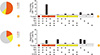
Finally, we assessed the polyfunctionality of RpfB-specific T cells by Boolean gating analysis of the ICS data presented in Fig. 3. To assess the polyfunctionality of T cells, we analyzed four different functions: production of IFN-γ, TNF-α, IL-2 and expression of a marker of cytotoxic degranulation activity, CD107a. First, the OLP mix 1-stimulated CD8+ T cell response was analyzed after RpfB DNA vaccination, and these CD8+ T cells were determined to be polyfunctional as more than half of the cells exerted three functions simultaneously (representative data from a single mouse are presented in Fig. 4A and B). The most predominant functional phenotype was that of IFN-γ+/TNF-α+/IL-2-/CD107a+ (Fig. 4B), and this polyfunctional T cell response was observed only following RpfB DNA immunization, and not following BCG or RpfB protein immunization (data not shown). A similar pattern of polyfunctionality was observed against the CD8+ T cell epitope, OLP-14 (data not shown).
In CD4+ T cells, the response to the OLP mix 2 was assessed for polyfunctionality. Compared to CD8+ T cells, CD4+ T cells were less polyfunctional (Fig. 4C). In fact, the most predominant functional phenotype was that of IFN-γ+/TNF-α+/IL-2-/CD107a- (Fig. 4D), and more than half of OLP mix 2-specific CD4+ T cells were monofunctional (Fig. 4C). However, RpfB DNA immunization induced more triple-positive and double-positive CD4+ T cells than did BCG or RpfB protein immunization (data not shown). A similar pattern of polyfunctionality was observed against OLP mix 1 and the CD4+ T cell epitopes, OLP-30 and OLP-51 (data not shown).
Thus, all mice immunized with Rpf DNA showed similar patterns of T cell polyfunctionality: a highly polyfunctional CD8+ T cell response and a less polyfunctional CD4+ T cell response.
Discussion
In the present study, we investigated the immunological response to M. tuberculosis RpfB DNA immunization. RpfB DNA immunization induced not only an RpfB-specific antibody response, but also CD8+ and CD4+ T cell responses in mice. Further, the induced CD8+ T cell response was determined to be polyfunctional as the major population of RpfB-specific CD8+ T cells were triple-positive (IFN-γ+/TNF-α+/CD107a+). Previously, T cell immunogenicity of Rpf DNA immunization was reported [24]; however, T cell polyfunctionality has never been studied after Rpf DNA immunization.
Polyfunctional T cells simultaneously exert multiple functions such as cytokine secretion and cytotoxic activity. Recently, T cell polyfunctionality has been investigated in several infectious diseases of human and mice. It was reported that polyfunctional human immunodeficiency virus (HIV)-specific CD8+ T cells were maintained in HIV long-term nonprogressors [32]. Furthermore, the degree of Th1 cell polyfunctionality correlated with vaccine efficacy in a Leishmania vaccination study [18]. In addition, the efficacy of the smallpox vaccine has been attributed to polyfunctionality of virus-specific CD8+ T cells [33]. Further, in a recent primate study, polyfunctional hepatitis C virus (HCV)-specific T cells were associated with vaccine-induced control of HCV [34]. Taken together, the generation of polyfunctional T cells has been emphasized in vaccine development.
Polyfunctional T cells have been also studied in TB. The presence of polyfunctional CD8+ T cells was associated with natural protection against TB and protection following anti-mycobacterial therapy in TB patients [21,22]. Furthermore, vaccine-induced polyfunctional CD4+ Th1 cells correlated with protection against TB in mice challenged by M. tuberculosis [19,20]. Induction of polyfunctional T cells was also proven by experimental TB vaccines in human studies [35,36,37]. Altogether, polyfunctional T cells are considered to provide vaccine-induced immunity and mediate protection against TB.
In the present study, RpfB DNA immunization elicited a polyfunctional CD8+ T cell response; however, the CD4+ T cell response was somewhat less polyfunctional with more than half of RpfB-specific CD4+ T cells being monofunctional (Fig. 4D) in RpfB DNA-immunized mice. Although RpfB DNA immunization induced more triple-positive or double-positive CD4+ T cells than did BCG or RpfB protein immunization (Fig. 4F), this level of polyfunctionality of CD4+ T cells might not be sufficient for protection against TB considering the importance of the CD4+ Th1 response during TB protection [11,12,13,14]. Thus in future studies, increasing the polyfunctionality of CD4+ T cells will need to be addressed in order to improve RpfB DNA vaccination. This might be achieved by in vivo electroporation during RpfB DNA vaccination as it is known that electroporation enhances DNA vaccine-induced immune responses [38,39,40].
In summary, RpfB DNA immunization elicits polyfunctional T cell response, especially in the CD8+ T cell subset, and this polyfunctional CD8+ T cell response was dominated by the IFN-γ+/TNF-α+/IL-2-/CD107a+ phenotype. These results suggest that RpfB DNA immunization might induce protective immunity against TB, and M. tuberculosis-challenging studies are warranted in the future.
 XML Download