

 PDF
PDF ePub
ePub Citation
 Print
Print


Introduction
Global warming can increase the activity of mosquito carrying out infectious virus to animals and human. Japanese encephalitis (JE) considered as re-emerging disease associated with climate change is an acute central nervous inflammatory disease bitten by mosquitoes containing Japanese encephalitis virus (JEV). JEV has been recognized recently as reemerging pathogen and the geographical distribution of JEV is expending to southwest India and Torres Strait of Northern Australia [1]. Approximately 50,000 human cases of JE per year have been reported in southern Asia and resulted in about 10,000 death and neuropsychiatric defects among survivors [2]. JEV has caused a swine disease showing abortion and weak piglets and caused occasional fatal outcome in horses [3].
JEV belongs to a member of family Flaviviridae and genus Flavivirus containing an encapsulated 11 kb of positive single strand RNA. The envelope (E) possess three domains designated I, II, and III which involved important biological functions such as virulence and induction of neutralizing antibody [4]. Based on the nucleotide sequence analysis of the E gene in JEV, JEV can be clustered into five genotypes (G1-5) [5]. Since the replacement of JEV G3 with G1 was first identified in 1994 in Japan, G1 has become the dominant circulating JEV in many Asian countries including China, Thailand, Vietnam, and Korea [6,7]. The potential impact of JEV genotype change on vaccine potency has been estimated using a mouse model and different JEV genotypes [8]. It was indicated that the vaccine consisting of JEV G3 showed similar protections against both G1 and G3, but low level of strain specific cross neutralization was observed in mice and pigs. For the prevention of JEV infection in sow, live attenuated JEV vaccine containing G3 was developed and has applied to pig farms since the late 1980's in Korea. However, the live JEV strain, Anyang 300, should be propagated in chicken or duck embryonic cell that cultivated in media adjusted to pH 8.0. The previous study revealed that the vaccine induced low level of antibody titer in pigs [9].
Several genetic engineered vaccines have currently been reported, including a yellow fever virus-based novel JE vaccine, recombinant adenoviruses expressing immune-dominant epitopes against JEV, and the plasmid based DNA vaccine [10,11,12]. In order to increase the immunogenicity of the vaccine, an alternative approach is to co-deliver adjuvants with antigens to up-regulate the immune response of vaccine, and to include interleukin-2, flagellin and granulocyte monocyte-colony stimulating factor (GM-CSF) [6,13,14]. GM-CSF is a pleiotropic cytokine and has been used as adjuvant to enhance immune response of many vaccine antigens [13]. GM-CSF is one of the discrete families of cytokines that provides a link between innate and acquired immunity and plays a role as one of the first lines of the body's defensive barriers [15].
In this study, to develop more effective JEV G1 vaccine for pigs, the humoral immune responses and efficacy of inactivated JEV G1 (KV1899 strain) vaccine containing recombinant porcine GM-CSF (reporGM-CSF) protein was evaluated in the mice, guinea pigs, and fattening pigs.
Materials and Methods
Viruses and cells
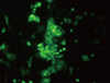
The KV1899 strain of JEV G1, which had undergone 10 serial passages in Vero cell culture, was used for the preparation of vaccine. The JEV was propagated in Vero cells and checked by indirect fluorescent assay test using monoclonal antibody (MEDIAN diagnostic, Chuncheon, Korea) against JEV (Fig. 1) [9]. Vero cells were regularly maintained in α-minimum essential medium (MEM) supplemented with 5% fetal bovine serum (FBS), penicilline (100 µg/mL), streptomycine (100 unit/mL) and amphotericin B (0.25 µg/mL). To propagate the JEV, Vero cells grown in α-MEM were washed three times with phosphate buffered saline (PBS; pH 7.2) and the virus was inoculated. After adsorption, α-MEM was added and incubated until cytopathic effect (CPE) showed 80-90%. In order to harvest the virus, the bulks were thawed and frozen three times and centrifuged at 5,000 ×g for 30 minutes to get rid of cell debris.
Inactivation of JEV
JEV was inactivated with binary ethyleneimine (BEI) by method of Barteling and Cassim [16]. In brief, BEI was prepared from 2% 2-bromo-ethylamine hydrobromide in solution of 0.2 N NaOH and treated the solution in incubator at 37℃ 1 hour, and then prepared 0.1 M BEI. The final concentration of BEI was adjusted to 0.001 M of bulk and pH of bulks also was adjusted to 8.0 with 1 N NaOH. Inactivation was done at 37℃ for 10 hours and was stopped with 2 mM sodium thiosulfate. For verifying virus inactivation, supernatant from the final bulk was dialyzed in PBS for 24 hours and inoculated into Vero cells, and CPE of the cells inoculated with the supernatant were observed for 7 days. After confirming the inactivation of viruses, bulks were used for preparation of vaccine.
Construction and expression of reporGM-CSF baculovirus
Porcine GM-CSF gene was synthesized based on the GenBank accession number U67175 and cloned into pGEM-T easy vector. For the construction of reporGM-CSF baculovirus, Bac-N-Blue DNA (Invitrogen, Carlsbad, CA, USA) and 10 µg/µL of purified pBlueporGM-CSF plasmid DNA were mixed with Cellfectin, commercial lipid-based transfection reagent (Invitrogen), in Grace's insect medium without supplement. After incubation for 15 minutes at room temperature, transfection mixture was added into the 60 mm dish in which Spodoptera frugiperda (Sf-9) cells had been cultivated at 27℃. After 3 days, supernatant was harvested and the cells were incubated continuously by adding fresh medium containing FBS. Plaque assay to purify recombinant baculovirus was performed in 1% agarose medium containing 150 µg/mL of X-gal. Polymerase chain reaction (PCR) assay against reporGM-CSF baculovirus was carried out to confirm the isolation of a pure plaque using specific baculovirus primers (Table 1). Passage of reporGM-CSF baculovirus was conducted three times using Sf-9 cell infected with 0.1 multiplicity of infection. The third passage number was used as viral stock for expression. As for the vaccine adjuvant, the bulk from infected Sf-9 cells were frozen and thawed three times and centrifuged at 5,000×g for 30 minutes to get rid of cell debris. One dose of the reporGM-CSF baculovirus was composed of virus titer of 107.0 TCID50/mL and added to the test vaccine formula as 10% volume (v/v).
Formula of experimental vaccines
One dose of the vaccine was composed of virus titer of 107.0 TCID50/mL. Two kinds of inactivated JEV vaccines were prepared with or without reporGM-CSF protein and IMS1313 adjuvant (Seppic, Paris, France) was used as adjuvant for the preparation of the inactivated vaccine against JEV. The first experimental vaccine consisting of JEV antigen and IMS1313 adjuvant were blended with 7:3 ratios under agitation. The second vaccine comprising JEV antigen, IMS1313 and reporGM-CSF protein was 7:2:1 ratio.
Hemagglutination inhibition test
Before the hemagglutination inhibition (HI) test, the sera were inactivated at 56℃ for 30 minutes. The KV1899 (G1) strain of JEV was used as the antigen for the HI test, which was isolated from Korean pig blood in 1999. An HI test was performed in 96-well microplates, using slightly modified standard methods to estimate the JEV antibody in the pig sera. Using a sucrose-acetone extraction method, viral antigens were prepared from the brains of suckling mice infected with the Korean isolate of JEV strain KV1899. Briefly, to remove non-specific inhibitors, 10 µL of serum and 50 µL of 4% bovine albumin were mixed with 40 µL of 25% kaolin (Sigma, St. Louis, MO, USA) and it was incubated for 30 minutes. After pipetting, the kaolin was removed by centrifugation at 3,000 ×g for 15 minutes. The resultant clear supernatant was mixed with 5 µL of packed goose erythrocytes to remove any natural agglutinins. After incubation at 37℃ for 1 hour, the treated sera were separated from the goose erythrocytes by centrifugation. The treated sera (25 µL) were diluted two-fold from 1:10 to 1:10,240 in round-bottom 96-well microplates and reacted with 8 HA units of JEV. After incubation at 37℃ for 1 hour, 50 µL of 0.33% goose erythrocytes was added to the microplates and they were incubated for 30 minutes at 37℃. To confirm test reliability, positive and negative JEV infection pig control sera were used in all HI tests. HI titer was expressed as the reciprocal of the highest dilution of serum showing complete HI.
Plaque reduction neutralization test by 90 percent
Plaque reduction neutralization test (PRNT) was performed by using monolayers of Vero cells in 24-well plates seeded with 3×10⁴ Vero cells per well in α-MEM (Gibco BRL, Grand Island, NY, USA) with 5% heat-inactivated FBS, and 100 units of penicillin and streptomycin (Gibco BRL). Cells were incubated for 2 days at 37℃. Test sera were heat inactivated at 56℃ for 30 minutes. The same volume (200 µL) of the test sera with 2-fold dilutions (from 1:10 to 1:320) and KV1899 virus diluent (200 pfu) were mixed and then it was incubated for 90 minutes at 37℃. Virus-sera mixture was inoculated (0.1 mL per well) and absorbed for 1 hour at 37℃ at which point the inoculums were removed. Media for the first overlay consisted of 0.5 mL of 1.0% low-melting point agarose and MEM containing 2.5% heat-inactivated FBS. The second overlay contained 1.0% low-melting point agarose in α-MEM with neutral red. Plaques were counted at 2 days of the second overlay. The antibody titer in serum was determined as that reduced the number of plaques by 90% of the control without serum.
Safety and immunogenicity
The inactivated JEV vaccines were inoculated into mice, guinea pig and 90-day-old pigs to check safety including the change of behavior and feeding activity. Ten mice were inoculated with 0.5 mL of vaccine intraperitoneally (IP), four guinea pigs with 2 mL and 1 mL of vaccine, IP and intramuscularly (IM), respectively. Two doses (6 mL) of vaccine were inoculated into fattening pigs at lateral ear site. Mice, guinea pigs and pigs inoculated with vaccines were observed for 14 and 21 post inoculation days. In order to evaluate immunogenicity in pigs, one dose of two kinds of vaccine was inoculated into eight pigs twice with 2 weeks interval and then bloods were collected at 2 weeks after second immunization. Control pigs remained with any treatment except for taking blood.
Efficacy of vaccine in mice
The potency of vaccines was performed according to World Organization for Animal Health (OIE) manual [17]. In short, groups (n=10) of 3-week-old mice were immunized IM with 0.1 dose of vaccine. Booster doses were given subsequently three days later with the same dose of vaccine. A control group of mice remained without any treatment. All mice were challenged eight days post-immunization with a highly lethal dose (100 LD50) of virulent JEV (KV1899 strain), which was administered intracerebrally, since adult mice aren't vulnerable to peripheral JEV infection and observed for 15 days.
Results
Expression of reporGM-CSF protein in insect Sf-9 cells
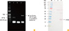
Porcine GM-CSF gene was cloned into pGEM-T and pBlu-Bac4.5/V5- His vector, which contains six-histidine tag in the C-terminal region. After transfection into Sf-9 cells, the plaque purified reporGM-CSF baculovirus was identified by CPE and reverse transcription-PCR (Table 1, Figs. 2, 3A). The reporGM-CSF protein was identified with Western blotting using specific monoclonal antibodies against six-histidine and the molecular weight was found to be approximately 24 kDa (Fig. 3B). The reporGM-CSF baculovirus propagated in Sf-9 cell was added to inactivated JEVgenotype 1 vaccine as adjuvants.
Safety and immune response in animals
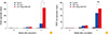
Mice and guinea pigs inoculated with the vaccines did not any clinical symptoms for 14 days after inoculation. The fattening pigs immunized with two doses of inactivated JEV vaccines via intramuscular route did not show any symptoms related to encephalitis for period of observation. Virus-neutralizing antibody is important to protection against JE and over a titer of 1:10 is indicative of protective immunity [8,9]. The anti-JEV antibody titers in pigs inoculated with the inactivated JEV vaccine were compared with those immunized with the inactivated JEV vaccine containing reporGM-CSF protein. Fig. 4 showed that HI antibody titers were significantly higher in pigs immunized with the vaccine containing reporGM-CSF protein than in pigs inoculated with the vaccine alone and ranged from 1:80 to 1:640. The fattening pigs inoculated with the vaccine containing reporGM-CSF protein had JEV neutralizing antibody titers of between 1:16 and 1:512 and mean PRNT titer was also higher in pigs inoculated with the vaccine containing reporGM-CSF protein than in pigs inoculated with the vaccine alone. Moreover, after the second vaccination at day 14, the HI and PRNT antibody titers were significantly increased in the vaccinated pigs (HI, p<0.05). These findings indicated that immunization with inactivated JEV vaccine containing reporGM-CSF protein could induce the higher level of HI and PRNT antibody titers than those of immunization with inactivated JEV vaccine only.
Potency of inactivated JEV vaccines in mice
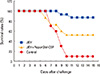
Groups of mice vaccinated with 0.1 dose of inactivated JEV vaccine in the presence or absence of reporGM-CSF protein, were challenged via intracranial route with the low passaged KV1899 strain of JEV at eight days after completion of the vaccination schedule (Fig. 5). Whilst 90% of naïve mice succumbed to the virus challenge, 87% of mice immunized with the inactivated JEV vaccine survived without showing the characteristic signs of JE such as ruffled fur, hunched posture, tremors and hind-leg paralysis. Mice immunized with the inactivated JEV vaccine containing reporGM-CSF protein showed 53% survival rate.
Discussion
JE continues to be one of the economically significant reproductive disorders in the swine industry and has been one of important zoonoses in Asia. JEV G3 was the most major genotype in many South and Southeast Asian countries, but genotype change G3 into G1 has occurred in Japan, China, and Korea since the 1990s [8,18,19]. In Korea, JEV G1 strain (K9307) was first identified in 1993 and became the dominant circulating genotype based on molecular epidemiological analysis of isolated JEVs [7,9,19]. It was reported that mice received an inactivated JEV G3 vaccine had reduced strain-specific neutralizing antibody titer against G1 [20]. In accordance with replacement of JEV G3 by G1, development of JEV vaccine containing G1 for swine has been required in Korea.
It has been known that GM-CSF is a glycoprotein involved in the recruitment of antigen presenting cells, differentiation and maturation of dendritic cells [6]. GM-CSF is produced by T cells, epithelial cells, and macrophages in response to a number of inflammatory stimuli such as lipopolysaccharide and tumor necrosis factor-α. Dodecamer assembly of GM-CSF signaling complex on hematopoietic cells predominantly activates the JAK2/STAT signal pathway [15]. The GM-CSF is one of the most studied cytokines used both therapeutically and diagnostically, as well as in vaccine development and adjuvant technology [14,21]. Mice inoculated with simian immunodeficiency virus VLPs containing GM-CSF increased CD4+ and CD8 T cell response and other study reported that the highest level of foot and mouth disease virus specific humoral immune response was induced in mice immunized with recombinant adenovirus expressing both VP1 and GM-CSF [22,23,24]. Thus, preparation of recombinant GM-CSF protein obtained from each species may be helpful to elicit high immune response. Therefore, we anticipated that reporGM-CSF protein plays a similar role in inducing adequate humoral immunity in swine. In this study, we cloned porcine GM-CSF gene in baculovirus transfer vector and expressed reporGM-CSF protein in insect cells. Furthermore, we prepared the inactivated JEV G1 vaccine for swine and evaluated safety and immunogenicity in experimental animals. The results indicated that experimental animals inoculated with two doses of inactivated vaccines did not show any adverse effects such as loss of body weight, death and local reactions, even after second IM injection in fattening pigs.
Humoral immune response is responsible for protecting animals from the JEV challenge [25]. HI and PRNT antibody titers were measured in pigs inoculated with the inactivated JEV vaccine in presence or absence of reporGM-CSF protein. Given that the antibody responses induced by vaccination appear to play the important protective role [26], pigs inoculated with JEV vaccine with or without reporGM-CSF protein generated HI and PRNT antibody titers, suggesting that pigs may be protected from wild JEV infection. A single inoculation of inactivated JEV vaccine induced low level of HI and PRNT antibodies raging from 1:10 to 1:20, but boosting significantly increased circulating HI antibody levels between 1:40 and 1:640. According to our results, it was speculated that reporGM-CSF stimulate more on hemagglutination antibody response which showed serologically high cross reaction within flaviviruses than neutralizing antibody response. Pigs inoculated with JEV vaccine containing reporGM-CSF protein showed higher HI and PRNT antibody titers, indicating that the reporGM-CSF protein may activate T cells, macrophages and endothelial cells in pigs.
Potency of the inactivated JEV G1 vaccine was observed in immunized mice with or without reporGM-CSF protein in accordance with minimum requirements for the inactivated JEV vaccine. Given that the survival rate should be more than 40% in the immunized group [17], the both groups of mice inoculated with the inactivated JEV vaccine with or without reporGM-CSF protein fulfilled the OIE's requirements.
On the contrary to the above results in pigs, the inactivated JEV vaccine without reporGM-CSF protein was proved to be a better immunogen in protection against virulence JEV challenge than the vaccine containing reporGM-CSF protein. Considering the efficacy test result in mice, GM-CSF still remained the controversy as an effective adjuvant. As to our knowledge, the biological activity of the GM-CSF generally may depend on species specific, due to the low genetic homology among animal species. It was reported that human GM-CSF gene showed approximately 50% homology to murine GM-CSF gene at the deduced amino acid level and recombinant human GM-CSF had no activity in the murine cells in vitro [27]. It was also reported that administration JEV DNA vaccine expressing a GM-CSF gene revealed suppressive effects on the immune response and protective immunity in mice [6]. In this study, JEV vaccine containing porcine GM-CSF protein expressed in insect cell was evaluated the vaccine efficacy in different animal species, mice. Therefore, to evaluate the efficacy of the vaccine containing reporGM-CSF protein, it should be developed other methods such as evaluating the cellular immune response in pigs.
In conclusion, our results demonstrated that two kinds of the inactivated JEV G1 vaccines with or without reporGM-CSF protein was safe in mice, guinea pig, and pigs and induced protective immune responses that were capable of protecting pigs against JEV. This result may provide helpful preventive measures against JEV. Our data indicated that inactivated JEV vaccine containing reporGM-CSF protein enhanced humoral immune responses in pigs. In further study, the effects of the reporGM-CSF protein on the cellular immune response should be investigated in pigs.

 XML Download