

 PDF
PDF ePub
ePub Citation
 Print
Print


Introduction
Orthomyxoviridae is a family of RNA viruses comprising 3 genera of influenza viruses: A, B, and C. Influenza is an economically significant disease that affects pigs, horses, and fowls [1]. Influenza A viruses are important pathogens in both mammalian including human and avian hosts. The canine influenza virus (CIV) H3N2 has been reported in dogs in South Korea and China, especially in veterinary clinics located near the capital city of Seoul. Recent virological and serological surveillance of the avian-derived H3N2 CIV in dogs in Korea has shown that the epidemiology resembles that of the horse-derived H3N8 CIV currently circulating in dog populations of the United States [2-4].
Glucocorticoids are commonly used in dogs as first-line therapy for immune-mediated diseases for induction and maintenance of remission, including immune-mediated haemolytic anemia, immune-mediated thrombocytopenia, polyarthritis, inflammatory bowel disease, dermatologic disease, systemic lupus erythematosus and asthma or allergic bronchitis [5]. Prednisolone is the active form of the metabolic precursor prednisone. And the dose of prednisolone used determines the pharmacological response. An anti-inflammatory response occurs at 0.5-1.0 mg/kg/day, and an immunosuppressive response occurs at 2.0-4.0 mg/kg/day. In this study, we evaluated the effects of glucocorticoids on the viral shedding pattern of the CIV in dogs.
Materials and Methods
Seven-week-old conventional beagle puppies were divided into 2 experimental groups: the control group (C group; n=4) and the immunocompromised group (IC group; n=4). All puppies were inoculated intranasally with 2 mL of H3N2 CIV with a titre of 106 50% egg infectious dose (EID50)/0.1 mL. Dogs of the IC group were administered 3.0 mg/kg prednisolone (PO; 5-mg Solondo tabs, YuHan Corp., Seoul, Korea) for 7 consecutive days starting on study day 1, as previously described [6]. Strict bioisolation was maintained throughout the study to protect the personnel who were in contact with the dogs. The dogs were acclimated to the housing facilities for a minimum of 3 weeks before the beginning of the study. Before inoculation, the animals were sedated by intramuscular injection of 0.1 mg of acepromazine maleate (Bayer, Seoul, Korea), as previously reported [4]. Clinical signs, including fever, nasal discharge, and cough, were monitored for 2 weeks. Nasal discharges and rectal swabs were examined daily for virus shedding by real-time reverse transcriptase polymerase chain reaction (RT-PCR) for 2 weeks after inoculation [4]. All experimental procedures were approved by an independent Animal Care and Use Committee, and followed the guidelines of Korea Research Institute of Bioscience and Biotechnology.
Results
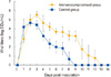
We detected influenza viral RNA in nasal swabs of C group dogs 1-8 days post inoculation (dpi) and in nasal swabs of IC group dogs at 1-13 dpi (p<0.01). Viral shedding peaked at 2 dpi (C group) and 3 dpi (IC group) (Fig. 1). One of the IC dogs was dead 7 days after inoculation. After necropsy and histopathological examination, exacerbated bronchopneumonia with severe multilobular or diffuse necrotizing bronchiolitis and alveolitis was observed (Fig. 2). The H3N2 virus was not detected in rectal swabs during the entire experimental period, as assessed by real-time RT-PCR. Clinical signs-sneezing, severe cough, and nasal discharge-were observed in both groups at 2-8 dpi.
Discussion
Glucocorticoids are widely used in veterinary medicine, especially in small animal practice for cats and dogs, because of their anti-inflammatory and immunosuppressive properties. However, several reports have suggested a correlation between prolonged viral shedding and glucocorticoid treatment in influenza as well as in other viral infections. Interestingly, a veterinary clinician has noted that the severity of the clinical signs, the prognosis, and mortality of influenza-infected dogs are worse than those of specific pathogen-free beagles inoculated with influenza virus only (personal communication).
Prolonged virus shedding is a predictor of major morbidity and is associated with complications in influenza infection [7]. Recently, Lee et al. [8] suggested prolonged virus shedding as a marker of disease severity in patients infected with seasonal influenza, for it was related with older age, chronic comorbidity, and therapy with systemic corticosteroids. Comorbidity or concomitant systemic corticosteroid administration may slow down viral clearance. However, assays for the influenza virus antigen and culture of stool samples are only occasionally performed in some studies. To date, few studies have considered the detection of viral RNA in faecal specimens as a method to diagnose gastrointestinal tract infection in immunocompromised patients [9]. Therefore, we attempted to detect the virus in rectal swabs of influenza-inoculated dogs. However, no positive signal was detected in stool samples of either C or IC groups dogs.
Immunosuppressive therapeutic agents can facilitate viral infection [10] or reactivate the latent virus [6] through modulation of the normal host defence mechanisms. Healthy dogs treated with glucocorticoids show mild or no clinical signs of infection after artificial inoculation of the West Nile virus, yet prolonged viremia occurs [10].
The clinical significance of prolonged viral shedding and viral load dynamics has not yet been assessed in hospitalized animals infected with H3N2 CIV. The present study showed that PO-induced immunosuppression in dogs is associated with prolonged shedding of the H3N2 CIV. Veterinary and human patients undergoing glucocorticoid therapy inevitably have one or more concurrent diseases. Chronic illness requires that the patients be treated with glucocorticoids for a prolonged period. Therefore, the effect of glucocorticoids on viral shedding might affect the susceptibility to influenza infection.

 XML Download