

 PDF
PDF ePub
ePub Citation
 Print
Print


Introduction
Presently, more than 100 different human papillomavirus (HPV) types have been discovered [1,2]. Among these, at least 30 different types of HPVs are known to cause cancers in the cervix, vagina, anus and penis as well as genital warts in the form of sexually transmitted diseases [1,2]. In particular, a high oncological risk group, type 16, and to a lesser degree type 18 are responsible for more than 70% of cervical cancers [3]. Other high risk groups of HPV include types 31, 33, 35, 39, 45, 51, 52, 56, 58, 59, 68, 73, and 82 [4]. On the other hand, low oncological risk groups, types 6 and 11, are known to cause 90% of all genital warts [5]. Moreover, HPV has been also reported to be linked to the incidence of cancers in the oropharynx, although smoking and drinking are other well-known risk factors. In oral epithelial dysplasia, all 20 of the patients in a previous study were positive for high oncological risk HPVs as determined by in situ hybridization [6]. Epidemiologic evidence has also shown that HPV 16 and 18 are associated with incidence of oral cancers as an independent risk factor [7]. In the study, HPV was transmitted by sexual behaviors (i.e., oral sex practices). It has been thought that HPV does not enter into the blood stream but attaches to the exposed epithelial cells of the mucus and skin. In this context, HPV is highly tropic for the epithelial cells of the anogenital and oropharygeal regions. It is likely that HPV exposure to the epithelial cells might lead to subsequent viral infection. However, overall incidence of head and neck cancers (cancers of the oral cavity, the oropharynx, the hypopharynx, the larynx, the nasopharynx and the sinonasal tract) is not clearly associated with HPV infection. It was reported that a far smaller portion of head and neck squamous cell carcinoma (HNSCC) was caused by HPV when compared with cervical cancer [8]. In the previous study, HPV-positive HNSCC had male predominance [8]. Recently, clinical evidence has shown that HPV-positive HNSCC has a better prognosis than HPV-negative HNSCC, and that different risk factors exist between these two cancer types [9, 10], suggesting a different biology. This is supported by a more recent study of 117 cases of head and neck cancers (96% SCC types) in Senegal, only 4 (3.4%) of which were found to possess HPV DNA [11]. These findings suggest that HPV-targeted vaccines might be also beneficial for protecting from HPV-positive HNSCC.
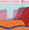
HPV has double-stranded circular DNA which is surrounded by a capsid protein structure [12]. HPV genome codes for six early regulatory proteins (E1, E2, E4, E5, E6, E7) and two structural proteins (L1 as a major structural protein and L2 as a minor structural protein) [12]. It is well known that E6 and E7 proteins are associated with oncogenesis through their inhibition of the cellular proteins (p53 and pRb) involved in the cell cycle. For instance, E6 and E7 proteins are highly synthesized by the disruption of E2, in which E6 proteins inactivate p53 while E7 proteins inactivate pRb proteins, resulting in an increment of the mitotic activity [13-15]. It is also known that the expression of viral early regulatory genes is limited in the basal cells, but infectious virus particles tend to be produced from the outer epithelial cells (Fig. 1). This strong dependence of viral replication on the status of cellular differentiation status makes it difficult to propagate HPV in vitro. When HPV-infected basal cells are not monitored by the host immune system, they tend to progress to the precancerous state, cervical intraepithelial neoplasia (CIN). The precancerous diseases CIN 2 and 3 spontaneously regress in about 20% of HPV infected patients [16]. Although there is presently no cure for HPV, the ultimate goal of designing therapeutic vaccines against HPV infection is to regress CIN and cure invasive cancers. Prophylactically, HPV infection is preventable by inducing neutralizing antibodies against viral coat proteins, while viral-infected cells can be recognized and lysed by cytotoxic T lymphocytes (CTLs) specific for viral early regulatory proteins. This is well illustrated in Fig. 1.
Prophylactic Vaccines
Presently, the two prophylactic vaccines, Gardasil® (Merck) and Cervarix® (GSK), have been licensed against HPV infection and cervical cancer. Gardasil® provides complete protection from infection with HPV types 6, 11, 16, and 18, which are responsible for approximately 70% of cervical cancers and 90% of genital warts. On the other hand, Cervarix® provides protective immunity against HPV types 16 and 18. These two vaccine types utilize the major structural protein present in different HPV types, L1 as a target antigen in a form of virus-like particle (VLP). In fact, VLP is unable to infect vaccinated persons as it doesn't possess an HPV genome but behaves as an actual virus particle, which allows for the induction of antibodies against L1 protein, a component of VLP. Furthermore, a possible application of these prophylactic vaccines might be also beneficial for prevention of orophryngeal cancer. This is based on the notion that the vaccines should work against these same HPVs in different locations, such as the oropharynx. In addition to these commercially available vaccines, different types of vaccines targeting the structural proteins are being tested in animals. A recent work from Roden's group showed that DNA vaccines coding for L1 proteins induced high titers of neutralizing antibodies against HPV [17]. Despite this result, it is too early to conclude that an L1 DNA vaccine type might be effective at preventing HPV infection. Based on these results, prophylactic vaccines are thought to reduce type-specific HPV infections, thus preventing the incidence of cervical cancer or its precancerous diseases.
DNA Vaccines and Other Vaccine Types as Therapeutic Vaccines against HPV
DNA vaccines can be manufactured simply and are stable at ambient temperature. Thus, they are likely less costly in production and distribution processes. They can also carry multiple antigens without vector-induced immune responses, which allow for repeated immunizations. Numerous studies using DNA vaccines demonstrated that they are indeed safe and tolerable in a human. One additional advantage of DNA vaccines is that the DNA vector backbone itself has an adjuvant effect via its inherent immunostimulatory elements. The unmethylated CpG motifs of DNA vectors can be recognized by Toll-like receptor (TLR)-9 [18], a microbial pattern recognition receptor, and trigger innate inflammatory responses [19,20]. DNA vectors in the cytoplasm can be also recognized and stimulate Absent In Melanoma 2 (AIM2) [21], and STimulator of IFN Genes (STING) pathways [22]. Consistent with this, we also observed in an HPV 16 E7 DNA vaccine model that the TLR9-MyD88 signaling pathway is essential for increased induction of Ag-specific immune responses and antitumor resistance [23]. In this context, it is likely that DNA vaccines have a number of advantages over other vaccine types. In the development of therapeutic vaccines against HPV infection, CIN and cervical cancer, E6 and E7 viral proteins have been considered potential targets because they play a critical role in tumor formation and maintenance in virus-infected epithelial cells. To date, E6- or E7-specific cellular responses including CTLs are known to be associated with clinical control of HPV-associated diseases [24-26]. Similarly, immunization with E7 DNA vaccines [27-32] as well as other vaccine types [33-36] resulted in controlling E7-expressing tumors through induction of Ag-specific CTL responses in animals. The possible clinical efficacy of HPV DNA vaccines can be also expected based on the magnitude of Ag-specific CTL responses induced by E6 or E7 DNA vaccines in both animal and human studies [32,37,38], and this is further supported by a recent clinical study showing that one-half of 20 patients with HPV 16-associated grade 3 vulvar intraepithelial neoplasia completely regressed after therapeutic vaccination with HPV 16 E6/E7 synthetic long peptides (50% complete regression rates by immunization vs. 1.5% spontaneous regression rates) [39]. However, the long peptide vaccines neither induced tumor regression nor prevented progressive disease when tested in patients with advanced or recurrent HPV 16-induced cervical cancers [40]. In detail, 11 of the 13 tested patients displayed vaccine-induced ELISPOT counts, suggesting that the vaccines may act to induce Ag-specific cellular responses without showing any clinical benefits. In this case, it is possible that cancer cells might have developed immune evasion schemes, such as a lack or loss of class I antigen expression, induction of regulatory T cells or myeloid derived suppressor cells and their production of immune inhibitory molecules (interleukin-10, transforming growth factor-β, cytotoxic T-lymphocyte antigen 4, etc.). These molecules as well as the immune cell types are well known to be involved in suppression of immune induction [41,42]. In cervical cancer patients, moreover, a lack or loss of class I antigen expression has been previously reported [43]. Table 1 shows HPV therapeutic vaccines tested in clinics and their clinical and immunologic efficacy. For immunological treatment of cancers, therefore, the alteration of tumor microenvironments by conventional therapy modalities, such as surgical excision, chemotherapy and radiotherapy, might be needed. Previously, our group reported that, when combined with chemotherapy, E7 subunit vaccines or adoptive T cell transfer were dramatically more effective at increasing tumor cure rates and long-term antitumor memory responses in a TC-1 tumor model [44,45]. A similar finding was also observed when E7 subunit vaccines were combined with radiotherapy [46]. In these studies, increased sensitivity of chemo- or radio-treated tumor cells to E7-specific CTL-mediated tumor killing was responsible for increased tumor cure rates. Along with these strategies to alter tumor microenvironment, it is also considered important to augment the magnitude of CD8+ CTL responses to HPV-associated antigens. In HPV DNA vaccines, numerous strategies have been utilized to improve their immunologic efficacy. Wu's group first reported that intracellular targeting of E7 antigens to the endosomal/lysosomal compartments of cells by conjugating E7 genes to lysosome-associated membrane proteins-1 (LAMP-1) genes resulted in enhancement of Ag-specific antitumor immunity in a TC-1 tumor model [47]. Moreover, conjugation of E7 genes to DNAs coding for calreticulin, bacterial toxin, heat shock protein 70 and viral protein 22 has been also shown to increase antitumor efficacy against TC-1 tumor cell challenges [27-30]. Our group also demonstrated that both codon optimization of E7 genes and their endosomal/lysosomal targeting approach were essential for enhancing antitumor protective immunity against TC-1 cells, and that this was mediated by more antigen production and subsequent augmentation of E7-specific CTLs [31]. Moreover, Wu's group also reported that co-injection of E7 DNA vaccines with plasmid DNAs encoding anti-apoptotic proteins or serine protease inhibitors increased antitumor activity by increasing dendritic cells survivals for longer antigen presentation [48,49]. In our study, however, co-injection of endolysosome-targeting E7 DNA vaccines with IL-12 DNAs resulted in dramatic suppression in Ag-specific CTL and antitumor protective responses [50], suggesting that an appropriate use of molecular adjuvants as well as the types of antigens and their intracellular locations should be evaluated prior to their combined use. More recently, it was reported that conjugation of an IgE leader sequence to codon-optimized E6 and E7 genes resulted in greater induction of Ag-specific humoral and cellular responses including CTL in animals and humans [37, 38]. In the study, moreover, oncogenic portions on the E6 and E7 DNA sequences were also modified to minimize the potential roles of E6 and E7 proteins in the tumor cell formation. More recently, our unpublished data have shown that intramuscular (IM) delivery of E7 DNA vaccines plus IL-2 cDNA in combination with adoptive transfer of anti-4-1BB antibodies dramatically increases tumor cure rates and long-term antitumor memory responses through the augmentation of Ag-specific CTL lytic activity. This study clearly shows that proper use of immune-potentiating antibodies is also beneficial for achieving better tumor control. Taken together, HPV DNA vaccines appear to be safe, tolerable, less costly and highly immunogenic in humans and small animals. Furthermore, DNA vaccines might be more beneficial for treating patients with HPV-associated diseases when they are modified or used together with other therapy protocols.
Electroporation as a DNA Vaccine Delivery Method
Electroporation (EP), the administration of electrical pulses to muscle or skin following DNA injection, for enhancement of the immunogenicity of DNA vaccines has been tested in a wide variety of small and large animal models. The clinical efficacy of EP delivery of DNA vaccines has been also suggested and demonstrated [38,51,52]. To date, DNA vaccine studies have mainly utilized muscle or skin as a vaccination target. In IM delivery, myocytes and antigen presenting cells (APCs) ingest DNA and then generate antigens by antigen processing-dependent, endogenous processing pathway, which can be recognized by naïve CD8+ T cells in the context with MHC class I molecules [53-55]. In this case, only bone marrow-derived APCs, but not myocytes play a dominant role in presenting antigens on MHC class I molecules to naïve CD8+ T cells [56,57]. Soluble and secreted vaccine antigens are phagocytosed by APCs and then gain entry into the MHC class II pathway for subsequent presentation to naïve CD4+ T cells [58]. This suggests that normally bone marrow-derived cells are responsible for antigen presentation to prime the adaptive immune response while muscle cells can serve as an antigen depot to expand activated effector cells. In contrast, skin is the first barrier of the body from infectious pathogens and other foreign substances. Keratinocytes in the skin are associated with induction of innate and adaptive immune responses through the release of cytokines [59]. Moreover, the epidermis and dermis of the skin also have immunological functions. They have resident APCs, such as Langerhans cells and dermal dendritic cells, which can take up antigens and present them to the draining lymph nodes for induction of adaptive immune responses [60]. When DNA was delivered to skin, the gene expression was detected in Langerhans cells and dermal dendritic cells [61], suggesting the important role of these cells for subsequent antigen presentation. Skin vaccination through intradermal delivery was once utilized during the smallpox eradication campaign and is still in use in BCG vaccination. Previously, it was reported that a single intradermal injection of DNA vaccines coding for influenza virus nucleoprotein could induce Ag-specific antibody and CTL responses and provide resistance to challenge with a heterologous strain of influenza virus, and that the antibody titers were higher than those induced by IM injection [62]. A similar finding was also observed when intradermal delivery of DNA vaccines by EP was more effective at inducing neutralizing antibodies against influenza virus and at reducing viral loads following viral challenge compared to that of IM-EP delivery [63]. In the study, however, IM-EP more highly induced Ag-specific cellular responses than intradermal delivery with EP. Based on these previous findings, it is highly likely that skin vaccination is one potential strategy to increase vaccine immunogenicity toward antibody responses. Contrary to adenoviral vector delivery of antigens, DNA vaccines are known to be less immunogenic [64]. This tendency is more prominent in humans than in small animals [65-67]. The reason why DNA vaccines fail to induce the expected level of immune responses in humans is still unclear. However, this might result from the fact that the amount of DNA vaccines relative to body mass and the level of TLR9 expression are simply lower in a human than in small animals [64]. However, the application of EP in the delivery of DNA vaccines likely overcomes the lack of vaccine immunogenicity in large animals including humans. IM delivery of DNA vaccines is known to attract APCs at the injection site [68-69]. However, EP augments the attraction of macrophages and dendritic cells to the DNA injection site [70]. This local effect of EP has been thought to contribute to the magnitude and longivity of the responses to DNA vaccines in larger animals, such as rabbits and humans [71]. EP also increases the permeability of muscle cells, thereby facilitating DNA uptake and subsequent antigen production [72-75]. Recently, we reported that IM-EP of E7 DNA vaccines elicited antitumor activity against established TC-1 tumors dramatically more than IM injection alone, and that this was mediated by more antigen expression at and more immune cell attraction to the sites of DNA injection [32]. A recent phase I clinical study also showed that HPV 16 and 18 E6 and E7 DNA vaccines delivered by EP induced a significant level of Ag-specific humoral and cellular responses including CTL responses [38]. This result is in contrast to the data obtained without use of EP [76]. Collectively, these studies support the notion that using EP as a DNA delivery method has the high potential to augment Ag-specific immune responses in both humans and animals. In this context, EP delivery of DNA vaccines targeting HPV antigens might be beneficial for treating HPV-associated diseases.
Conclusion
It is evident that therapeutic DNA vaccines can contribute to curtailing HPV-associated diseases commonly observed in oral, pharyngeal, anal and genital parts of the body. In this case, EP has a critical role in augmenting vaccine immunogenicity through increased attraction of APCs and other immune cells to and more antigen expression at the injection sites. Contrary to intradermal delivery with EP, IM-EP delivery of DNA vaccines could serve to better engender Ag-specific CTL responses, which are more importantly involved in killing of HPV-infected epithelial cells. Moreover, current immunological evidence obtained from IM-EP of E6/E7 DNA vaccines sheds some light on possible impact of DNA vaccines on the treatment of HPV-infected patients. However, this benefit is likely increased when such treatments are used in combination with chemotherapy, radiation, surgery or antibody therapy.

 XML Download