Journal List > Allergy Asthma Respir Dis > v.5(5) > 1059275

Lee, Park, Yang, Kim, Chun, Yoon, Kim, and Kim: Usefulness of flexible bronchoscopy in children with suspected pulmonary tuberculosis who have difficulty in sputum expectoration

Abstract

Purpose

To assess the usefulness of flexible bronchoscopy in patients with suspected pulmonary tuberculosis (PTB) who have difficulty in sputum expectoration.

Methods

The subjects of this study were patients who were suspected of PTB and visited the Division of Pediatric Pulmonology at a tertiary hospital from April 2006 to March 2016. PTB suspects were determined by clinical symptoms, radiologic findings, and immunologic studies. We aimed to examine the value and safety of bronchoscopy in diagnosis and differential diagnosis of PTB in PTB-suspected patients. The diagnostic criteria for PTB were defined when Mycobacterium tuberculosis was cultured in the sputum specimen or in the bronchial washing fluid.

Results

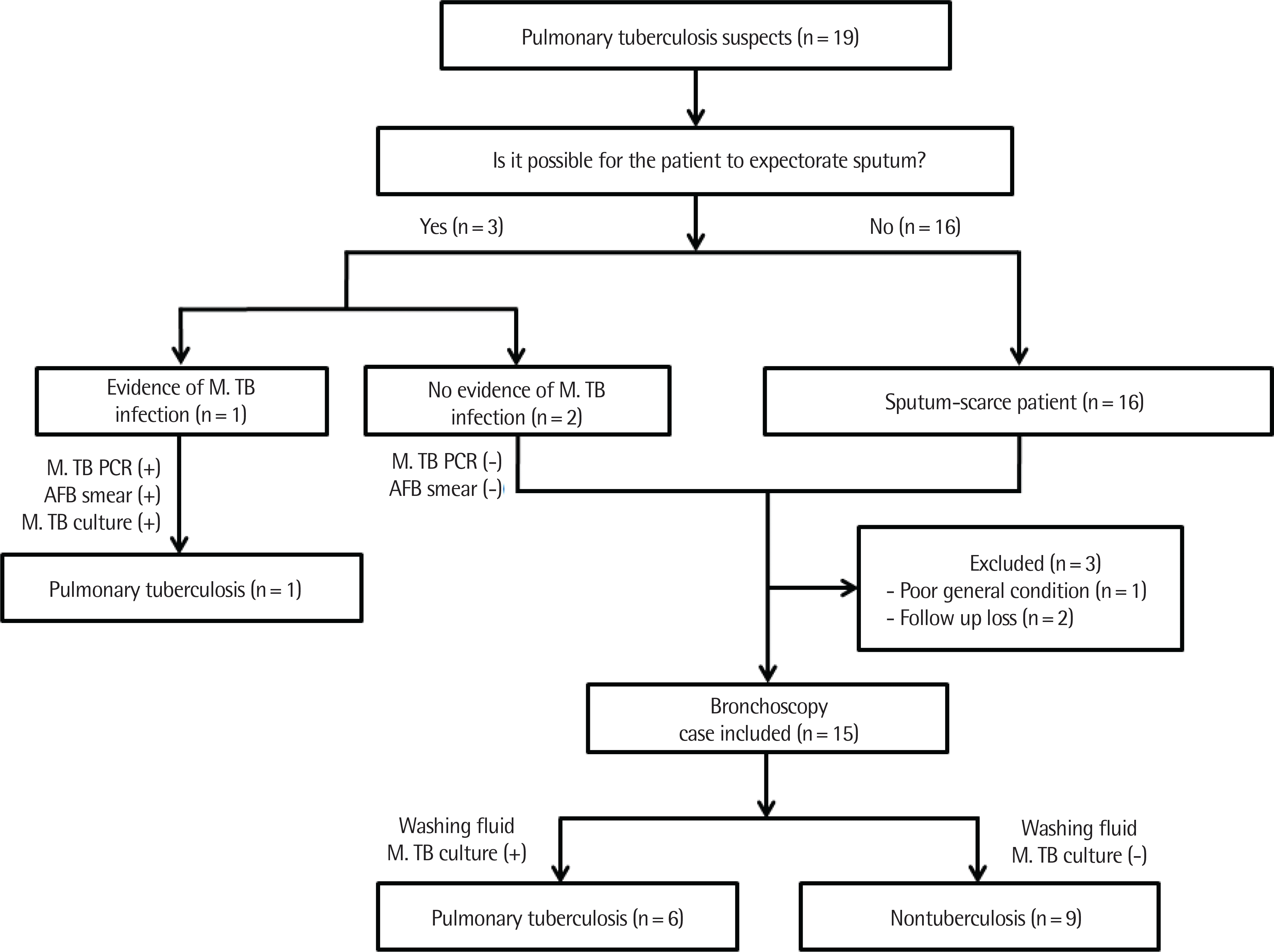
A total of 19 PTB suspects were included. One patient was diagnosed with PTB by using the sputum study. However, the re-maining 18 patients could not expectorate sputum or showed no evidence of Mycobacterium tuberculosis infection from the sputum study. Of the 18 patients, 15 underwent bronchoscopy. After bronchoscopy, 6 patients were diagnosed with PTB and 9 patients were diagnosed with Mycoplasma, viral, or fungal pneumonia, and tumors. For antituberculous drug resistance, there were 1 case of isoniazid (INH) resistance and 1 case of concurrent resistance to INH and prothionamide. There was no multidrug-resistant tuberculosis. None of the patients had significant complications due to bronchoscopy.

Conclusion

Flexible bronchoscopy appears to be a definitive and safe procedure for the differential diagnosis of patients suspecting PTB in children who have difficulty expectorating sputum.

REFERENCES

1. Global tuberculosis report 2015. 20th edition [Internet]. Geneva: World Health Organization;c2015. [cited 2016 Nov 23]. Available from. http://apps.who.int/iris/bitstream/10665/191102/1/9789241565059_eng.pdf.
2. Steingart KR, Sohn H, Schiller I, Kloda LA, Boehme CC, Pai M, et al. Xpert® MTB/RIF assay for pulmonary tuberculosis and rifampicin resistance in adults. Cochrane Database Syst Rev. 2013; (1):CD009593.
crossref
3. Dodd PJ, Gardiner E, Coghlan R, Seddon JA. Burden of childhood tuberculosis in 22 high-burden countries: a mathematical modelling study. Lancet Glob Health. 2014; 2:e453–9.
crossref
4. Jenkins HE, Tolman AW, Yuen CM, Parr JB, Keshavjee S, Pérez-Vélez CM, et al. Incidence of multidrug-resistant tuberculosis disease in children: systematic review and global estimates. Lancet. 2014; 383:1572–9.
crossref
5. Siddiqi K, Lambert ML, Walley J. Clinical diagnosis of smear-negative pulmonary tuberculosis in low-income countries: the current evidence. Lancet Infect Dis. 2003; 3:288–96.
crossref
6. Shin JA, Chang YS, Kim TH, Kim HJ, Ahn CM, Byun MK. Fiberoptic bronchoscopy for the rapid diagnosis of smear-negative pulmonary tuberculosis. BMC Infect Dis. 2012; 12:141.
crossref
7. Yoo R, Kim JI, Kim S, Lee J. Discordance between tuberculin skin test and interferon-gamma release assays for diagnosis of tuberculosis infection in Korean children. Pediatr Infect Vaccine. 2016; 23:18–24.
crossref
8. Jo YS, Park JH, Lee JK, Heo EY, Chung HS, Kim DK. Discordance between MTB/RIF and realtime tuberculosis-specific polymerase chain reaction assay in bronchial washing specimen and its clinical implications. PLoS One. 2016; 11:e0164923.
crossref
9. Tamura A, Shimada M, Matsui Y, Kawashima M, Suzuki J, Ariga H, et al. The value of fiberoptic bronchoscopy in culture-positive pulmonary tuberculosis patients whose pre-bronchoscopic sputum specimens were negative both for smear and PCR analyses. Intern Med. 2010; 49:95–102.
crossref
10. Lee SJ, Yoon DK, Kim SH, Park IW, Choi BW, Hue SH, et al. Diagnosis of suspected active pulmonary tuberculosis by flexible fiberoptic bronchoscopy. Tuberc Respir Dis. 1989; 36:22–7.
crossref
11. Le Palud P, Cattoir V, Malbruny B, Magnier R, Campbell K, Oulkhouir Y, et al. Retrospective observational study of diagnostic accuracy of the Xpert® MTB/RIF assay on fiberoptic bronchoscopy sampling for early diagnosis of smear-negative or sputum-scarce patients with suspected tuberculosis. BMC Pulm Med. 2014; 14:137.
crossref
12. Schoch OD, Rieder P, Tueller C, Altpeter E, Zellweger JP, Rieder HL, et al. Diagnostic yield of sputum, induced sputum, and bronchoscopy after radiologic tuberculosis screening. Am J Respir Crit Care Med. 2007; 175:80–6.
crossref
13. Dasgupta K, Menzies D. Cost-effectiveness of tuberculosis control strategies among immigrants and refugees. Eur Respir J. 2005; 25:1107–16.
crossref
14. Kang SJ, Kim YH, Jung CY, Lee HJ, Hyun MC. Clinical characteristics and radiologic patterns of adelescents with pulmonary tuberculosis: relevance to the reactive tuberculosis. Pediatr Allergy Respir Dis. 2012; 22:163–70.
crossref
15. Rapid diagnostic tests for tuberculosis: what is the appropriate use? American Thoracic Society Workshop. Am J Respir Crit Care Med. 1997; 155:1804–14.
16. Iyer VN, Joshi AY, Boyce TG, Brutinel MW, Scalcini MC, Wilson JW, et al. Bronchoscopy in suspected pulmonary TB with negative induced-sputum smear and MTD(®) Gen-probe testing. Respir Med. 2011; 105:1084–90.
17. Bai GH, Park YK, Choi YW, Bai JI, Kim HJ, Chang CL, et al. Trend of anti-tuberculosis drug resistance in Korea, 1994-2004. Int J Tuberc Lung Dis. 2007; 11:571–6.
18. Kahn MA, Whitcomb ME, Snider GL. Flexible fiberoptic bronchoscopy. Am J Med. 1976; 61:151–5.

Fig. 1.
Flow diagram of patients included in the study. M. TB, Mycobacterium tuberculosis; PCR, polymerase chain reaction; AFB, acid fast bacilli.
aard-5-287f1.tif
Table 1.
Demographic and clinical characteristics of all patients
Characteristic All suspects (n=16) TB (n=7) Non-TB (n=9)
Age (yr) 13.6±4.0 14.7±2.0 12.8±5.0
Male sex 8 (50.0) 1 (14.3) 7 (77.8)
Previous TB history y 4 (25.0) 0 (0) 4 (44.4)
Contact TB history 5 (31.3) 3 (42.9) 2 (22.2)
Immunologic test      
 IGRA positivity 6/11 (54.5) 5/6 (83.3) 1/5 (20)
 TST positivity 5/10 (50) 4/6 (66.7) 1/4 (25)
Symptom      
 Cough 10 (62.5) 4 (57.1) 5 (55.6)
 Sputum 5 (31.3) 2 (28.6) 3 (33.3)
 Fever 5 (31.3) 2 (28.6) 3 (33.3)
 Hemoptysis 2 (12.5) 1 (14.3) 1 (11.1)
 Night sweats 1 (6.3) 1 (14.3) 0 (0)
 Dyspnea 1 (6.3) 1 (14.3) 0 (0)
Radiologic findings      
 Consolidation 11 (68.8) 4 (57.1) 7 (77.8)
 Lymphadenopathy y 8 (50.0) 3 (42.9) 5 (55.6)
 Nodule 6 (37.5) 5 (71.4) 2 (22.2)
 Cavity 1 (6.3) 1 (14.3) 0 (0)
 Effusion 4 (25.0) 1 (14.3) 3 (33.3)
 GGO 6 (37.5) 2 (28.6) 4 (44.4)
 Tree in bud 2 (12.5) 2 (28.6) 0 (0)
 Atelectasis 1 (6.3) 0 (0) 1 (11.1)

Values are presented as mean±standard deviation or number of patients (%) TB, tuberculosis, IGRA, interferon-γ release assays; TST, tuberculin skin test; GGO, ground-glass opacity.

Table 2.
The results of laboratory tests for the 7 pulmonary TB patients
Case   Sex/age (yr) Previous TB Hx Contact TB Hx Symptom Radiologist reading suspecting TB Radiologic finding Bronchoscopic finding TST (mm) IGRA Washing fluid laboratory results M. TB sensitivity Treatment Outcome
M. TB PCR AFB smear M. TB Cx. Other result
A 1 F/16 (-) (-) Cough (+) Nodule, cavity Not done 0 (+) Sputum (+) Sputum (+) Sputum (+) (-) All S HERZ+HER Improved
B 1 F/15 (-) (+) Cough, sputum, hemoptysis (+) Consolidation, LAP, nodule, tree in bud RLL bronchus swelling 0 NA (+) (+) (+) (-) INH R+ HERZ F/U loss
  2 F/11 (-) (-) Cough, fever (+) Consolidation, nodule, GGO RLL bronchus swelling 10 (-) (+) (+) (+) (-) All S HERZ+HER Improved
  3 F/14 (-) (-) Cough, sputum, fever, night sweats (+) Consolidation, LAP, GGO, effusion N/S 25 (+) Sputum (-)(+) Sputum (-) (-) Sputum (+) (+) Rhino virus All S HERZ+HR Improved
  4 F/14 (-) (+) Dyspnea (+) Nodular opacity RUL bronchus inflammation NA (+) (+) (-) (+) (-) All S HERZ+HR Improved
  5 M/17 (-) (+) (-) (+) Nodule, tree in bud N/S 10 (+) (-) (-) (+) (-) INH R+, PTH R+ H ERZ(9) Improved
  6 F/16 (-) (-) Cough (+) Consolidation, LAP, nodule Right main bronchus narrowing 17 (+) (+) (+) (+) I Influenza virus All S HERZ+HR Improved

TB Hx, tuberculosis history; TST, tuberculin skin test; IGRA, interferon-γ release assays; M. TB, Mycobacterium tuberculosis; PCR, polymerase chain reaction; AFB, acid fast bacilli; Cx., culture; LAP, lymphadenopathy; RLL, right lower lobe; NA, not available INH, isoniazid; HERZ, isoniazid+ethambutol+rifampicin+pyrazinamid; F/U, follow-up; GGO, ground-glass opacity; N/S, nonspecific; HR, isoniazid+rifampicin; RUL, right upper lobe; PTH, prothionamide. ∗A, pulmonary tuberculosis (PTB) patient confirmed by only sputum test;

B, PTB patients confirmed by bronchoscopy.

Table 3.
The results of laboratory tests for the 9 nonpulmonary TB patients
Case Sex/ age (yr) Previous TB Hx Contact TB Hx Symptom Radiologist reading suspecting TB Radiologic finding Bronchoscopic finding TST (mm) IGRA Washing fluid laboratory results Diagnosis Treatment Outcome
M. TB PCR AFB smear M. TB Cx. Other result
1 F/13 (-) (+) Cough, sputum (+) Consolidation, nodule, GGO LLL bronchus swelling (-) NA (-) (-) (-) Mycoplasma PCR (+) MPP Roxithromycin Improved
2 M/17 (+) (-) Sputum, hemoptysis (-) LAP, atelectasis N/S NA NA (-) (-) (-) (-) (-) Observation Improved
3 M/12 (-) (-) Cough (+) Consolidation, effusion, GGO RML bronchus swelling NA NA (-) (-) (-) RSV Pneumonia AMX-CLA, prednisolone Improved
4 M/0 (+) (+) Cough, fever (-) Consolidation N/S (+) (-) (-) (-) (-) Adenovirus, E. facium Pneumonia Cefotaxime, clindamycin, prednisolone Improved
5 M/14 (-) (-) Neck mass (+) Consolidation, LAP LUL bronchus swelling NA (-) (-) (-) (-) Hodgkin's lymphoma Hodgkin's lymphoma Chemotherapy Improved
6 M/10 (+) (-) Cough, sputum, fever (-) Consolidation, LAP, effusion, GGO LUL bronchus mucus plug (-) (+) Sputum (-)(-) Sputum (-) (-) Sputum (-) (-) P.aeruginosa, Asp Ag (+) Fungal pneumonia Voriconazole Improved
7 M/15 (+) (-) Cough, fever (+) Consolidation, endobronchial TB RUL bronchus mass NA NA (-) (-) (-) Carcinoid tumor Carcinoid tumor VATS lobectomy Improved
8 F/17 (-) (-) (-) (-) Consolidation, LAP, effusion, GGO N/S NA (-) (-) (-) (-) Mycoplasma PCR (+) MPP Roxithromycin Improved
9 M/16 (-) (-) (-) (+) LAP, nodule RUL bronchus mucus (-) (-) (-) (-) (-) (-) (-) Observation Improved

TB Hx, tuberculosis history; TST, tuberculin skin test; IGRA, interferon-γ release assays; M. TB, Mycobacterium tuberculosis; PCR, polymerase chain reaction; AFB, acid fast bacilli; Cx., culture; LLL, left lower lobe; NA, not available; MPP, Mycoplasma pneumo-eumonia; LAP, lymphadenopathy; GGO, ground-glass opacity; LUL, left upper lobe; RUL, right upper lobe; AMX-CLA, amoxicillin-clavulanate; N/S, nonspecific; E. facium, Enterococcus faecium; LUL, left upper lobe; RUL, right upper lobe; VATS, video as-ed thoracoscopy.

TOOLS
Similar articles