

 PDF
PDF ePub
ePub Citation
 Print
Print


Abstract
Purpose
The aim of this study was to assess the relationship between serum 25-hydroxyvitamin D3 levels and the severity of atopic dermatitis (AD), markers of atopy (total IgE, total eosinophil count, and eosinophil cationic protein) in AD children according to allergen sensitization.
Methods
This cross-sectional study was carried out on 160 AD patients aged 1 to 18 years between March 2012 and August 2014. The AD patients (AD group) were subdivided into 2 categories according to the results of the allergic skin prick and Unicap tests: the allergic and nonallergic AD groups. We compared 25-hydroxyvitamin D3 levels between the AD and control groups. We also investigated relationships between serum 25-hydroxyvitamin D3 levels, the severity of AD, and markers of AD (total IgE, total eosinophil count, and eosinophil cationic protein) in the allergic and nonallergic AD groups.
Results
The average 25-hydroxyvitamin D3 levels were 30.6±11.7 and 23.7±10.0 ng/mL, respectively, in the control and AD groups (P<0.001). The average 25-hydroxyvitamin D3 levels were 19.7±8.6 and 27.5±9.8 ng/mL, respectively, in the allergic and nonallergic AD groups, with clinical implications (P<0.001). The 25-hydroxyvitamin D3 levels were not significantly associated with SCORing Atopic Dermatitis index in the allergic (P=0.004, r=-0.092) or nonallergic (P=0.610, r=-0.58) AD groups. The 25-hydroxyvitamin D3 levels were not significantly associated with the aforementioned markers of atopy in the AD group.
Figures and Tables
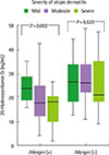 | Fig. 2The 25-hydroxyvitamin D3 levels of allergen and nonallergen patients according to the severity of atopic dermatitis.
|
Table 1
Comparison of characteristics in the study subjects

![]()
Table 2
Comparison of SCORAD index and atopic markers in atopic dermatitis patients
![]()
Table 3
The 25-hydroxyvitamin D3 levels in the study subjects

![]()
Table 4
The 25-hydroxyvitamin D3 levels according to age in the study subjects

![]()
Table 5
Relation between 25-hydroxyvitamin D3 levels and factors in atopic dermatitis patients with or without allergen

![]()
References
1. Holick MF. Vitamin D deficiency. N Engl J Med. 2007; 357:266–281.

2. Binkley N, Novotny R, Krueger D, Kawahara T, Daida YG, Lensmeyer G, et al. Low vitamin D status despite abundant sun exposure. J Clin Endocrinol Metab. 2007; 92:2130–2135.
3. Bischoff-Ferrari HA, Giovannucci E, Willett WC, Dietrich T, Dawson-Hughes B. Estimation of optimal serum concentrations of 25-hydroxyvitamin D for multiple health outcomes. Am J Clin Nutr. 2006; 84:18–28.
4. Suh SH. Prevalence of allergic diseases in Korean children, 2010. Public Health Wkly Rep. 2011; 4:425–431.
5. Bieber T. Atopic dermatitis. N Engl J Med. 2008; 358:1483–1494.
6. Benson AA, Toh JA, Vernon N, Jariwala SP. The role of vitamin D in the immunopathogenesis of allergic skin diseases. Allergy. 2012; 67:296–301.
7. Roider E, Ruzicka T, Schauber J. Vitamin D, the cutaneous barrier, antimicrobial peptides and allergies: is there a link? Allergy Asthma Immunol Res. 2013; 5:119–128.
8. Mutgi K, Koo J. Update on the role of systemic vitamin D in atopic dermatitis. Pediatr Dermatol. 2013; 30:303–307.
9. Bikle DD. Vitamin D metabolism and function in the skin. Mol Cell Endocrinol. 2011; 347:80–89.
10. Wang TT, Nestel FP, Bourdeau V, Nagai Y, Wang Q, Liao J, et al. Cutting edge: 1,25-dihydroxyvitamin D3 is a direct inducer of antimicrobial peptide gene expression. J Immunol. 2004; 173:2909–2912.
11. Peroni DG, Piacentini GL, Cametti E, Chinellato I, Boner AL. Correlation between serum 25-hydroxyvitamin D levels and severity of atopic dermatitis in children. Br J Dermatol. 2011; 164:1078–1082.
12. Chiu YE, Havens PL, Siegel DH, Ali O, Wang T, Holland KE, et al. Serum 25-hydroxyvitamin D concentration does not correlate with atopic dermatitis severity. J Am Acad Dermatol. 2013; 69:40–46.
13. Reitamo S, Rustin M, Harper J, Kalimo K, Rubins A, Cambazard F, et al. A 4-year follow-up study of atopic dermatitis therapy with 0.1% tacrolimus ointment in children and adult patients. Br J Dermatol. 2008; 159:942–951.
14. Park JH, Choi YL, Namkung JH, Kim WS, Lee JH, Park HJ, et al. Characteristics of extrinsic vs. intrinsic atopic dermatitis in infancy: correlations with laboratory variables. Br J Dermatol. 2006; 155:778–783.
15. Pugliarello S, Cozzi A, Gisondi P, Girolomoni G. Phenotypes of atopic dermatitis. J Dtsch Dermatol Ges. 2011; 9:12–20.
16. Schmid-Grendelmeier P, Simon D, Simon HU, Akdis CA, Wüthrich B. Epidemiology, clinical features, and immunology of the "intrinsic" (non-IgE-mediated) type of atopic dermatitis (constitutional dermatitis). Allergy. 2001; 56:841–849.
17. Hanifin JM, Raika G. Diagnostic features of atopic dermatitis. Acta Derm Venereol Suppl (Stockh). 1980; 92:44–47.
18. Oranje AP, Glazenburg EJ, Wolkerstorfer A, de Waard-van der Spek FB. Practical issues on interpretation of scoring atopic dermatitis: the SCORAD index, objective SCORAD and the three-item severity score. Br J Dermatol. 2007; 157:645–648.
19. Adinoff AD, Rosloniec DM, McCall LL, Nelson HS. Immediate skin test reactivity to Food and Drug Administration-approved standardized extracts. J Allergy Clin Immunol. 1990; 86:766–774.
20. Rosen CJ. Clinical practice. Vitamin D insufficiency. N Engl J Med. 2011; 364:248–254.
21. El Taieb MA, Fayed HM, Aly SS, Ibrahim AK. Assessment of serum 25-hydroxyvitamin d levels in children with atopic dermatitis: correlation with SCORAD index. Dermatitis. 2013; 24:296–301.
22. Hilger J, Friedel A, Herr R, Rausch T, Roos F, Wahl DA, et al. A systematic review of vitamin D status in populations worldwide. Br J Nutr. 2014; 111:23–45.
23. Choi EY. 25(OH)D status and demographic and lifestyle determinants of 25(OH)D among Korean adults. Asia Pac J Clin Nutr. 2012; 21:526–535.
24. Choi HS, Oh HJ, Choi H, Choi WH, Kim JG, Kim KM, et al. Vitamin D insufficiency in Korea: a greater threat to younger generation: the Korea National Health and Nutrition Examination Survey (KNHANES) 2008. J Clin Endocrinol Metab. 2011; 96:643–651.
25. Akan A, Azkur D, Ginis T, Toyran M, Kaya A, Vezir E, et al. Vitamin D level in children is correlated with severity of atopic dermatitis but only in patients with allergic sensitizations. Pediatr Dermatol. 2013; 30:359–363.
26. Lee SA, Hong S, Kim HJ, Lee SH, Yum HY. Correlation between serum vitamin d level and the severity of atopic dermatitis associated with food sensitization. Allergy Asthma Immunol Res. 2013; 5:207–210.
27. Noh GW, Lee KY. Blood eosinophils and serum IgE as predictors for prognosis of interferon-gamma therapy in atopic dermatitis. Allergy. 1998; 53:1202–1207.
28. Gutgesell C, Heise S, Seubert A, Stichtenoth DO, Frolich JC, Neumann C. Comparison of different activity parameters in atopic dermatitis: correlation with clinical scores. Br J Dermatol. 2002; 147:914–919.
29. Gebhardt M, Wenzel HC, Hipler UC, Herrmann D, Wollina U. Monitoring of serologic immune parameters in inflammatory skin diseases. Allergy. 1997; 52:1087–1094.
30. Kim MN, Shin BJ, Tak WJ, Ro BI, Park AJ. Eosinophil counts in peripheral blood, serum total IgE, eosinophil cationic protein, IL-4 and soluble e-selectin in atopic dermatitis. Korean J Dermatol. 2002; 40:1367–1373.
31. Simon D, Braathen LR, Simon HU. Eosinophils and atopic dermatitis. Allergy. 2004; 59:561–570.
32. Gleich GJ, Adolphson CR. The eosinophilic leukocyte: structure and function. Adv Immunol. 1986; 39:177–253.
33. Nahm DH, Kim ME, Shin YS, Ye YM, Park HS. Prevalence of vitamin D deficiency and clinical efficacy of vitamin D supplementation in patients with atopic dermatitis [abstract]. In : Program and Abstract, the Annual Spring Meeting of the Korean Academy of Asthma, Allergy and Clinical Immunology; 2012 May 25-26; Seoul, Korea. Seoul: the Korean Academy of Asthma, Allergy and Clinical Immunology;2012.
 XML Download