

 PDF
PDF ePub
ePub Citation
 Print
Print


Abstract
Purpose
Atopic dermatitis (AD) is a chronic inflammatory skin disorder with a association of genetic, environmental, and immunologic factors in the development of AD. And AD can be classified into IgE mediated and non-IgE mediated. We investigated a difference of clinical characteristics and immune response between the two groups.
Methods
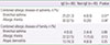
From January 2008 to December 2011, we enrolled 125 children who visited Dong-A University Medical Center and Busan Medical Center, and were diagnosed as AD with the Haniffin and Rajka's criteria. We checked the history of combined asthma and allergic rhinitis and allergic disease of family in patients. We measured serum total IgE, specific IgE by ImmunoCAP or skin prick test. We measured serum interleukin (IL) 4 (IL-4), interferon-γ (IFN-γ), IL-10, and IL-17, which are associated with chronic inflammatory disorder by flow cytometry method (Luminex).
Results
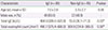
Eighty (64%) were included in the IgE mediated group, while forty-five (36%) were included in the non-IgE mediated group. The frequency of combined allergic disorder and serum total eosinophil count were relatively higher in IgE mediated group (P=0.023, P=0.032). The incidence of a family history in allergic disease and the mean of SCORing Atopic Dermatitis index had no difference between the two groups. Serum IL-4, IFN-γ, IL-10, IL-17 were higher in the IgE mediated group, but there were no statistically significant differences between two groups (P>0.05).
Figures and Tables


References
1. Bohme M, Svensson A, Kull I, Nordvall SL, Wahlgren CF. Clinical features of atopic dermatitis at two years of age: a prospective, population-based case-control study. Acta Derm Venereol. 2001; 81:193–197.

2. Park YM. Epidemiologic study and risk factors of atopic dermatitis. Pediatr Allergy Respir Dis. 2011; 21:74–77.
3. Oh JW, Pyun BY, Choung JT, Ahn KM, Kim CH, Song SW, et al. Epidemiological change of atopic dermatitis and food allergy in school-aged children in Korea between 1995 and 2000. J Korean Med Sci. 2004; 19:716–723.
4. Linna O, Kokkonen J, Lahtela P, Tammela O. Ten-year prognosis for generalized infantile eczema. Acta Paediatr. 1992; 81:1013–1016.
5. Wüthrich B. Atopic dermatitis flare provoked by inhalant allergens. Dermatologica. 1989; 178:51–53.
6. Hamid Q, Naseer T, Minshall EM, Song YL, Boguniewicz M, Leung DY. In vivo expression of IL-12 and IL-13 in atopic dermatitis. J Allergy Clin Immunol. 1996; 98:225–231.
7. Molet S, Hamid Q, Davoine F, Nutku E, Taha R, Page N, et al. IL-17 is increased in asthmatic airways and induces human bronchial fibroblasts to produce cytokines. J Allergy Clin Immunol. 2001; 108:430–438.
8. Schmidt-Weber CB, Akdis M, Akdis CA. TH17 cells in the big picture of immunology. J Allergy Clin Immunol. 2007; 120:247–254.
9. Lee JS, Kim TH, Cho GL, Jung JA, Kim JH. The classification between IgE and non-IgE mediated atopic dermatitis in Korean children. Pediatr Allergy Respir Dis. 2005; 15:352–358.
10. Chung HL. Clinical significance of serum IgE. Korean J Pediatr. 2007; 50:416–421.
11. Park JH, Choi YL, Namkung JH, Kim WS, Lee JH, Park HJ, et al. Characteristics of extrinsic vs. intrinsic atopic dermatitis in infancy: correlations with laboratory variables. Br J Dermatol. 2006; 155:778–783.
12. Novembre E, Cianferoni A, Lombardi E, Bernardini R, Pucci N, Vierucci A. Natural history of "intrinsic" atopic dermatitis. Allergy. 2001; 56:452–453.
13. Uehara M, Izukura R, Sawai T. Blood eosinophilia in atopic dermatitis. Clin Exp Dermatol. 1990; 15:264–266.
14. Furue M, Ohtsuki M, Ogata F, Ishibashi Y. Responsiveness to interleukin 4 and interleukin 2 of peripheral blood mononuclear cells in atopic dermatitis. J Invest Dermatol. 1991; 96:468–472.
15. Novak N, Kruse S, Kraft S, Geiger E, Kluken H, Fimmers R, et al. Dichotomic nature of atopic dermatitis reflected by combined analysis of monocyte immunophenotyping and single nucleotide polymorphisms of the interleukin-4/interleukin-13 receptor gene: the dichotomy of extrinsic and intrinsic atopic dermatitis. J Invest Dermatol. 2002; 119:870–875.
16. Akdis CA, Akdis M, Simon D, Dibbert B, Weber M, Gratzl S, et al. T cells and T cell-derived cytokines as pathogenic factors in the nonallergic form of atopic dermatitis. J Invest Dermatol. 1999; 113:628–634.
17. Reinhold U, Wehrmann W, Kukel S, Kreysel HW. Evidence that defective interferon-gamma production in atopic dermatitis patients is due to intrinsic abnormalities. Clin Exp Immunol. 1990; 79:374–379.
18. Grassegger A, Hopfl R. Significance of the cytokine interferon gamma in clinical dermatology. Clin Exp Dermatol. 2004; 29:584–588.
19. Hanifin JM, Schneider LC, Leung DY, Ellis CN, Jaffe HS, Izu AE, et al. Recombinant interferon gamma therapy for atopic dermatitis. J Am Acad Dermatol. 1993; 28(2 Pt 1):189–197.
20. Rebane A, Zimmermann M, Aab A, Baurecht H, Koreck A, Karelson M, et al. Mechanisms of IFN-γ-induced apoptosis of human skin keratinocytes in patients with atopic dermatitis. J Allergy Clin Immunol. 2012; 129:1297–1306.
21. Grewe M, Gyufko K, Schopf E, Krutmann J. Lesional expression of interferon-gamma in atopic eczema. Lancet. 1994; 343:25–26.
22. Akdis M, Trautmann A, Klunker S, Daigle I, Kucuksezer UC, Deglmann W, et al. T helper (Th) 2 predominance in atopic diseases is due to preferential apoptosis of circulating memory/effector Th1 cells. FASEB J. 2003; 17:1026–1035.
23. McKenzie BS, Kastelein RA, Cua DJ. Understanding the IL-23-IL-17 immune pathway. Trends Immunol. 2006; 27:17–23.
24. Koga C, Kabashima K, Shiraishi N, Kobayashi M, Tokura Y. Possible pathogenic role of Th17 cells for atopic dermatitis. J Invest Dermatol. 2008; 128:2625–2630.
25. Albanesi C, Scarponi C, Cavani A, Federici M, Nasorri F, Girolomoni G. Interleukin-17 is produced by both Th1 and Th2 lymphocytes, and modulates interferon-gamma- and interleukin-4-induced activation of human keratinocytes. J Invest Dermatol. 2000; 115:81–87.
26. Schwartz RH. Models of T cell anergy: is there a common molecular mechanism? J Exp Med. 1996; 184:1–8.
27. Borish L. IL-10: evolving concepts. J Allergy Clin Immunol. 1998; 101:293–297.
28. Laouini D, Alenius H, Bryce P, Oettgen H, Tsitsikov E, Geha RS. IL-10 is critical for Th2 responses in a murine model of allergic dermatitis. J Clin Invest. 2003; 112:1058–1066.
29. Prasse A, Germann M, Pechkovsky DV, Markert A, Verres T, Stahl M, et al. IL-10-producing monocytes differentiate to alternatively activated macrophages and are increased in atopic patients. J Allergy Clin Immunol. 2007; 119:464–471.
30. Barnes PJ. IL-10: a key regulator of allergic disease. Clin Exp Allergy. 2001; 31:667–669.
 XML Download