

 PDF
PDF ePub
ePub Citation
 Print
Print


Abstract
Objective
The conscious patients with a small amount of acute subdural hematoma had no neurological deterioration are managed conservatively. Most of them are resolved spontaneously in several weeks without surgery. In our experience, however, some progressed to chronic stage requiring surgical treatment in a few days, unlike chronic subdural hematoma derived from acute hematoma following several weeks or months after head trauma. We aimed to analyse this phenomenon and associated the risk factor comparing with the chronic subdural hematomas.
Methods
Retrospective analysis of 175 alert patients with unilateral acute subdural hematoma identified among 661 patients diagnosed the acute subdural hematoma from October 2009 to September 2012 was performed. Univariate and multivariate analyses were performed to describe the relationships between progression to chronic stage requiring surgery from small amount of acute subdural hematoma and clinical characteristics and radiologic features.
Results
Eighteen patients (10.3%) showed neurological deterioration due to progression to chronic stage of acute subdural hematoma and underwent a surgical treatment. The mean time interval between the head trauma and development of neurological symptoms was 12.7 days. Univariate and multivariate analyses found that depth of hematoma and degree of brain swelling were a risk factor for progression to chronic stage requiring surgery from the acute subdural hematoma.
Go to : 
References
1. Adams JH, Graham DI, Scott G, Parker LS, Doyle D. Brain damage in fatal non-missile head injury. J Clin Pathol. 33:1132–1145. 1980.

2. Andrews PJ, Citerio G. Intracranial pressure. Part one: historical overview and basic concepts. Intensive Care Med. 30:1730–1733. 2004.
3. Baechli H, Nordmann A, Bucher HC, Gratzl O. Demographics and prevalent risk factors of chronic subdural haematoma: results of a large single-center cohort study. Neurosurg Rev. 27:263–266. 2004.
4. Bullock R, Teasdale GM. Head injuries-surgical management: traumatic intracranial haematomas in Braakman R (eds): Vinken and Bruyn's Handbook of clinical neurology, head injury. Amsterdam: Elsevier Science Publishers;Vol. 24:pp2. p. 49–298. 1991.
5. Chen JC, Levy ML. Causes, epidemiology, and risk factors of chronic subdural hematoma. Neurosurg Clin N Am. 11:399–406. 2000.
6. Creasy JL. The general appearance of edema and hemorrhage on ct, mr and us (including a general introduction to ct, mr and us scanning): Dating neurological injury. Nashville, USA:. Springer, pp43–58;2011.
7. Croce MA, Dent DL, Menke PG, Robertson JT, Hinson MS, Young BH, et al. Acute subdural hematoma: nonsurgical management of selected patients. J Trauma. 36:820–826. ; discussion 826–827,. 1994.
8. Cuatico W, Yamamoto R, Howeiler B, Smith R. Spontaneous resolution of subdural hematomas. J Neurosurg Sci. 35:139–145. 1991.
9. Forster MT, Mathé AK, Senft C, Scharrer I, Seifert V, Gerlach R. The influence of preoperative anticoagulation on outcome and quality of life after surgical treatment of chronic subdural hematoma. J Clin Neurosci. 17:975–979. 2010.
10. Inamasu J, Nakamura Y, Saito R, Kuroshima Y, Mayanagi K, Ohba S, et al. Rapid resolution of traumatic acute subdural hematoma by redistribution. Am J Emerg Med. 20:376–377. 2002.
11. Ito H, Yamamoto S, Saito K, Ikeda K, Hisada K. Quantitative estimation of hemorrhage in chronic subdural hematoma using the 51Cr erythrocyte labeling method. J Neurosurg. 66:862–864. 1987.
12. Kang HJ, Lee YS, Suh SJ, Lee JH, Ryu KY, Kang DG. Clinical outcomes of patients with good neurological scores in spite of significant amounts of acute subdural hematoma. Korean J Neurotrauma. 9:12–16. 2013.
13. Labadie EL, Glover D. Local alterations of hemostatic-fibrinolytic mechanisms in reforming subdural hematomas. Neurology. 25:669–675. 1975.
14. Lee CH, Kang DH, Hwang SH, Park IS, Jung JM, Han JW. Spontaneous rapid reduction of a large acute subdural hematoma. J Korean Med Sci. 24:1224–1226. 2009.
15. Lee KS, Bae HG, Yun IG. Small-sized acute subdural hematoma: operate or not. J Korean Med Sci. 7:52–57. 1992.
16. Lee KS, Bae WK, Doh JW, Bae HG, Yun IG. Origin of chronic subdural haematoma and relation to traumatic subdural lesions. Brain Inj. 12:901–910. 1998.
17. Lee YB. Risk factors related to prognosis in patients with isolated traumatic subdural hematoma. J Korean Neurotraumatol Soc. 7:12–18. 2011.
18. Lindvall P, Koskinen LO. Anticoagulants and antiplatelet agents and the risk of development and recurrence of chronic subdural haematomas. J Clin Neurosci. 16:1287–1290. 2009.
19. Mathew P, Oluoch-Olunya DL, Condon BR, Bullock R. Acute subdural haematoma in the conscious patient: outcome with initial non-operative management. Acta Neurochir (Wien). 121:100–108. 1993.
20. Maurice-Williams RS. Chronic subdural haematoma: an everyday problem for the neurosurgeon. Br J Neurosurg. 13:547–549. 1999.
21. Mckissock W, Richardson A, Bloom WH. Subdural haematoma: A review of 389 cases. Lancet. 275:1365–1369. 1960.
22. Munro D, Merritt HH. Surgical pathology of subdural hematoma based on a study of one hundred and five cases. Arch Neurol Psychiatry. 35:64–79. 1936.
23. Park JK, Lee KS, Bae KG, Yun IG, Lee IS. Thin acute subdural hematoma: part 3: result of conservative treatment. J Korean Neurosurg Soc. 19:937–944. 1990.
24. Putnam T, Cushing H. Chronic subdural hematomaits pathology, its relation to pachymeningitis hemorrhagica and its surgical treatment. Arch Surg. 11:329–393. 1925.
25. Sato M, Nakano M, Sasanuma J, Asari J, Watanabe K. Rapid resolution of traumatic acute subdural haematoma in the elderly. Br J Neurosurg. 19:58–61. 2005.
26. Tokmak M, Iplikcioglu AC, Bek S, Gökduman CA, Erdal M. The role of exudation in chronic subdural hematomas. J Neurosurg. 107:290–295. 2007.
27. Winn HR, Youmans JR. Youmans neurological surgery, ed 6. Philadelphia, PA: Saunders, Vol 2, pp424;2011.
28. Wintzen AR. The clinical course of subdural haematoma. A retrospective study of aetiological, chronological and pathological features in 212 patients and a proposed classification. Brain. 103:855–867. 1980.
29. Yamamoto T, Katayama Y, Tsubokawa T, Sasaki J, Kumagara H, Sugitani M. Features of chronic subdural haematoma developed from definitely identified acute subdural haematoma. Brain Inj. 4:135–146. 1990.
30. Yamashima T, Yamamoto S. Clinicopathological study of acute subdural haematoma in the chronic healing stage. Clinical, histological and ultrastructural comparisons with chronic subdural haematoma. Neurochirurgia (Stuttg). 27:98–105. 1984.
31. Yamashima T, Yamamoto S. How do vessels proliferate in the capsule of a chronic subdural hematoma? Neurosurgery. 15:672–678. 1984.
Go to :
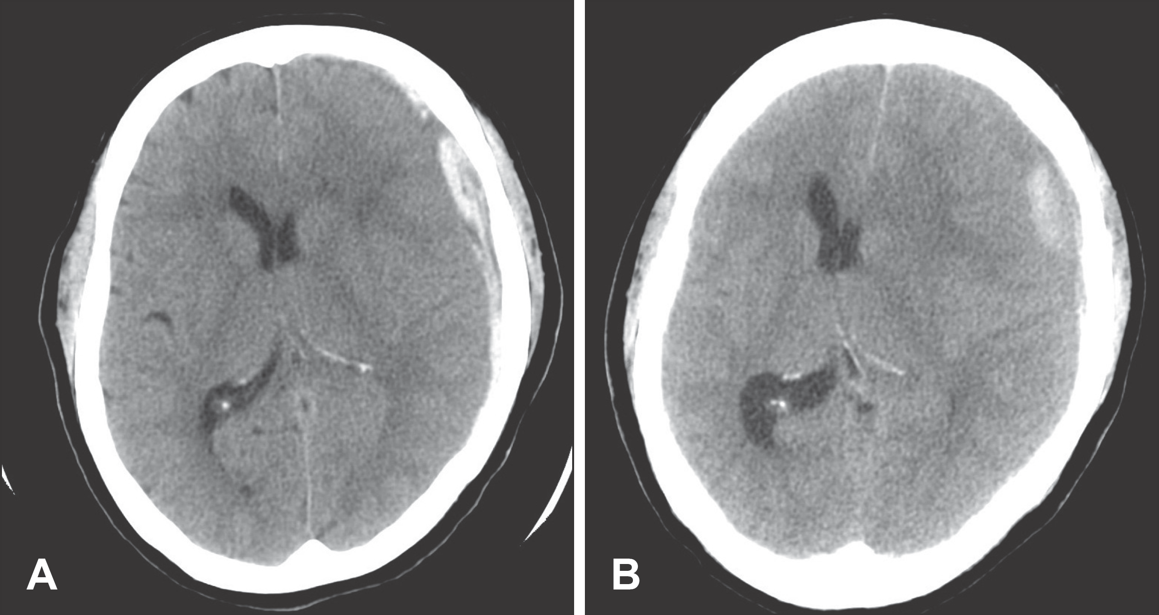 | FIGURE 1.Brain CT image of case 2. A: Initial CT scan on admission revealed a subdural hematoma with a maximal thickness of approxima-tely 11 mm in the left fronto-temporo-parietal con-vexity with a complete effacement of sulci and the decrease the ventricle in size. B: Eight days later, she was drowsy mental status with right hemiple-gia, CT scan disclosed a increase of the subdural hematoma of mixed density, accompanied by a severe midline shift. |
TABLE 1.
Grade of Cerebral swelling
The cerebral swelling is to cause the involved gyri to expand and the intervening sulci to decrease in size. As the brain continues to swell, not only do the sulci decrease, but all of the CSF spaces of the hemispheres decrease as well. The more brain swelling progress, it is to cause the ventricles to decreased in size. As the brain tissues swell, in order for the total intracranial volume to remain constant, the ventricles and extraaxial CSF spaces must decrease in total volume (6)
TABLE 2.
Factors related to progressed to chronic stage requiring surgical treatment: univariate analysis
| Factor | No. of patients (%) | p value | |
|---|---|---|---|
| Case | Control | ||
| Age (years) | 61.1±13.9∗ | 59.5±20.3∗ | 0.737 |
| Max depth of ASDH | 0.000 | ||
| ≥7 mm | 15 (83.3%) | 40 (25.5%) | |
| <7 mm | 3 (16.7%) | 117 (74.5%) | |
| Brain swelling | 0.001 | ||
| Grade 1 | 2 (11.1%) | 59 (37.6%) | |
| Grade 2 | 4 (22.2%) | 63 (40.1%) | |
| Grade 3 | 8 (44.4%) | 27 (17.2%) | |
| Grade 4 | 4 (22.2%) | 8 (5.1%) | |
| Hypertension | 6 (33.3%) | 33 (21.0%) | 0.240 |
| Diabetes mellitus | 4 (22.2%) | 30 (19.1%) | 0.478 |
| Cerebravascular disease | 3 (16.7%) | 9 (5.7%) | 0.111 |
| Cardiac disease | 4 (22.2%) | 7 (4.5%) | 0.017 |
| Liver disease | 1 (5.6%) | 8 (5.1%) | 0.633 |
| Kidney disease | 1 (5.6%) | 3 (1.9%) | 0.355 |
| Antiplatelet drug | 4 (22.2%) | 21 (13.4%) | 0.296 |
| Alcohol (bottle per month) | 7.89±14.8∗ | 6.22±14.6∗ | 0.426 |
| Smoking (PYS) | 6.11± 9.3∗ | 7.06±13.9∗ | 0.935 |
TABLE 3.
Multivairate logistic regression analysis of factors related to progressed to chronic stage requiring surgical treatment
 XML Download