Abstract
Temporal fossa hollowing can represent a serious cosmetic concern to patients after post-traumatic surgery, oncological resection, or surgical dissection for obtaining access to the temporal area. Various methods have been described to augment temporal fossa hollowing, such as use of autogenous bone and cartilage implants, high-density polyethylene implants, and dermal fat grafts. We report a case of 22-year-old man with temporal fossa hollowing after post-traumatic surgery, including temporal muscle resection, whose defect was augmented by using titanium mesh even though long after cranioplasty.
Go to : 
References
1. Atherton DD, Joshi N, Kirkpatrick N. Augmentation of temporal fossa hollowing with Mersilene mesh. J Plast Reconstr Aesthet Surg. 63:1629–1634. 2010.

2. Badie B. Cosmetic reconstruction of temporal defect following pterional [corrected] craniotomy. Surg Neurol. 45:383–384. 1996.
3. Cheung LK, Samman N, Tideman H. The use of mouldable acrylic for restoration of the temporalis flap donor site. J Craniomaxillofac Surg. 22:335–341. 1994.

4. Ducic Y, Pontius AT, Smith JE. Lipotransfer as an adjunct in head and neck reconstruction. Laryngoscope. 113:1600–1604. 2003.

5. Figi FA. Chronic stenosis of the larynx with special consideration of skin grafting. 1940. Ann Otol Rhinol Laryngol 103 (4 Pt 1): 249–264. 1994.
6. Frodel JL, Lee S. The use of high-density polyethylene implants in facial deformities. Arch Otolaryngol Head Neck Surg. 124:1219–1223. 1998.

7. Guo J, Tian W, Long J, Gong H, Duan S, Tang W. A retrospective study of traumatic temporal hollowing and treatment with titanium mesh. Ann Plast Surg. 68:279–285. 2012.

8. Kim S, Matic DB. The anatomy of temporal hollowing: the superficial temporal fat pad. J Craniofac Surg. 16:760–763. 2005.
9. Maas CS, Merwin GE, Wilson J, Frey MD, Maves MD. Comparison of biomaterials for facial bone augmentation. Arch Otolaryngol Head Neck Surg. 116:551–556. 1990.

10. McNichols CH, Hatef DA, Cole P, Hollier LH, Thornton JF. Contemporary techniques for the correction of temporal hollowing: augmentation temporoplasty with the classic dermal fat graft. J Craniofac Surg. 23:e234–e238. 2012.
11. Rapidis AD, Day TA. The use of temporal polyethylene implant after temporalis myofascial flap transposition: clinical and radiographic results from its use in 21 patients. J Oral Maxillofac Surg. 64:12–22. 2006.

12. Schick B, Draf W, Schauss F. [Augmentation of the temporal fossa with Ionos bone cement]. HNO. 44:467–470. 1996.
13. Weidenbecher M, Waller G, Lehmann W. [Mersilene mesh in reconstruction of the osseous walls of the pneumatised skull (author's transl)]. HNO. 24:351–353. 1976.
14. Welling DB, Maves MD, Schuller DE, Bardach J. Irradiated homologous cartilage grafts. Long-term results. Arch Otolaryngol Head Neck Surg. 114:291–295. 1988.

15. Worley CM, Strauss RA. Augmentation of the anterior temporal fossa after temporalis muscle transfer. Oral Surg Oral Med Oral Pathol. 78:146–150. 1994.

16. Wright S, Bekiroglu F, Whear NM, Grew NR. Use of Palacos R-40 with gentamicin to reconstruct temporal defects after maxillofacial reconstructions with temporalis flaps. Br J Oral Maxillofac Surg. 44:531–533. 2006.
Go to : 
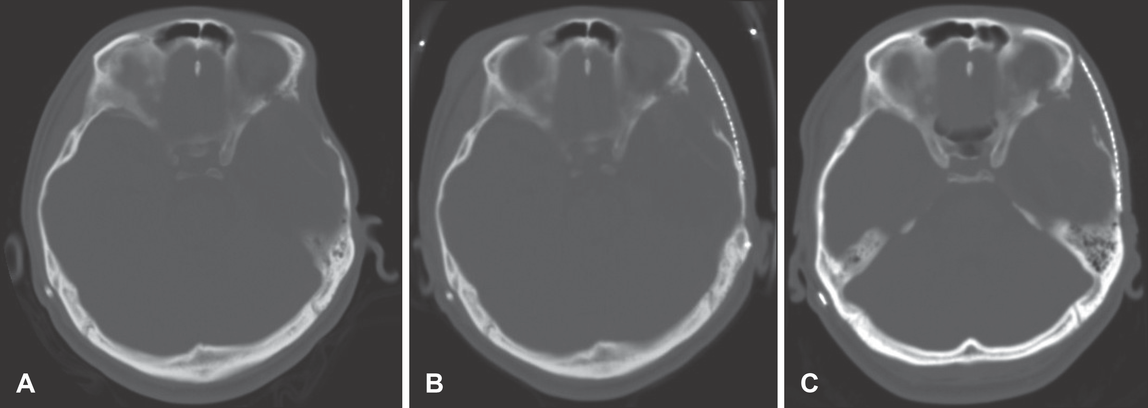
 | FIGURE 1.Sequential brain computerized tomography (CT) scan. A: Before the surgery for temporal hollowing, the left temporal area shows a defect compared to the right temporal area, because of previous temporal muscle resection. B: Two months after operation, the scan shows temporal fossa hollowing disappeared by augmentation using titanium mesh. C: Six months after operation, the cosmetic improvement persisted throughout the outpatient follow-up period. |


 PDF
PDF ePub
ePub Citation
 Print
Print


 XML Download