

 PDF
PDF ePub
ePub Citation
 Print
Print


Abstract
Meningitis is the inflammation of the membranes of the brain and spinal cord. This disease is considered life threatening and classified as a medical and emergency. Here we report a case of delayed meningitis occurred in a patient with craniotomy for traumatic brain injury fifteen years ago. Meanwhile, he had been well, however he complained of headache for five days. A brain computed tomographic scan showed air density on the frontal lobe with frontal sinus defect and pansinusitis. His mental state was suddenly changed to stuporous, despite a day of empirical antibiotics. Therefore, a successful cranial-ization was performed and he was gradually improved. This is a rare case report. Our case shows that surgical intervention is to be considered in some cases of posttraumatic meningitis for effective and rapid control of infection.
Go to : 
References
1. Appelbaum E. Meningitis following trauma to the head and face. JAMA. 173:1818–1822. 1960.

2. Baltas I, Tsoulfa S, Sakellariou P, Vogas V, Fylaktakis M, Kon-dodimou A. Posttraumatic meningitis: bacteriology, hydrocephalus, and outcome. Neurosurgery. 35:422–426. ; discussion 426–427,. 1994.
3. Byron BJ (ed). Head and neck surgery-otolaryngology. Philadel-pia: Lippicott Williams & Wilkins, Vol 2, pp1107. 1993.
4. Donald PJ. Frontal sinus ablation by cranialization. Report of 21 cases. Arch Otolaryngol. 108:142–146. 1982.
5. Donald PJ, Bernstein L. Compound frontal sinus injuries with intracranial penetration. Laryngoscope 88 (2 Pt 1):225–232. 1978.
6. Friedman JA, Ebersold MJ, Quast LM. Post-traumatic cerebrospinal fluid leakage. World J Surg. 25:1062–1066. 2001.
7. Johns ME, Winn HR, McLean WC, Cantrell RW. Pericranial flap for the closure of defects of craniofacial resection. Laryngoscope. 91:952–959. 1981.
8. Kim DS, Lee JY, Coe CJ, Suh JS. A case of recurrent bacterial meningitis with CSF rhinorrhea. J Korean Pediatr Soc. 32:1161–1166. 1989.
9. Leech P. Cerebrospinal fluid leakage, dural fistulae and meningitis after basal skull fractures. Injury. 6:141–149. 1974.
10. Lewin W. Cerebrospinal fluid rhinorrhea in nonmissile head injuries. Clin Neurosurg. 12:237–252. 1964.
11. Markham JW. The clinical features of pneumocephalus based upon a survey of 284 cases with report of 11 additional cases. Acta Neurochir (Wien). 16:1–78. 1967.
12. Mincy JE. Posttraumatic cerebrospinal fluid fistula of the frontal fossa. J Trauma. 6:618–622. 1966.
13. Osborn AG, Daines JH, Wing SD, Anderson RE. Intracranial air on computerized tomography. J Neurosurg. 48:355–359. 1978.
14. Saito H. Late posttraumatic rhinogenic meningitis. Temporal bone findings of meningogenic labyrinthitis. Adv Otorhinolaryngol. 31:175–183. 1983.
15. Stillwell M, Hoge C, Hoyt N, Joshi M. Posttraumatic meningococcal meningitis: case report. J Trauma. 31:1693–1695. 1991.
16. Wada N, Takeuchi Y, Fujii M, Fujiwara F, Odani I, Sawada T. A case of bacterial meningitis complicated by posttraumatic cerebrospinal fluid rhinorrhea. Acta Paediatr Jpn. 36:276–279. 1994.
17. Wallis A, Donald PJ. Frontal sinus fractures: a review of 72 cases. Laryngoscope 98 (6 Pt 1):593–598. 1988.
18. Wolfe SA. The utility of pericranial flaps. Ann Plast Surg. 1:147–153. 1978.
Go to :
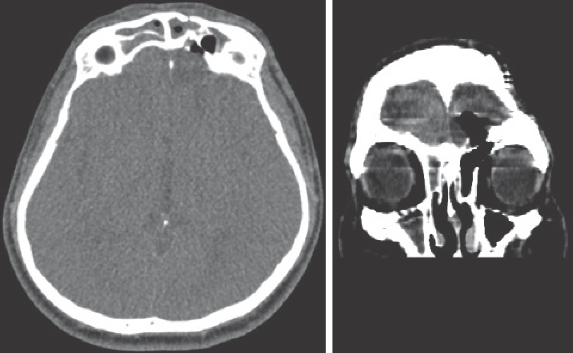 | FIGURE 1.Axial and coronal brain CT scan showing pneumocephalus with frontal sinus opening to the left frontal lobe. |
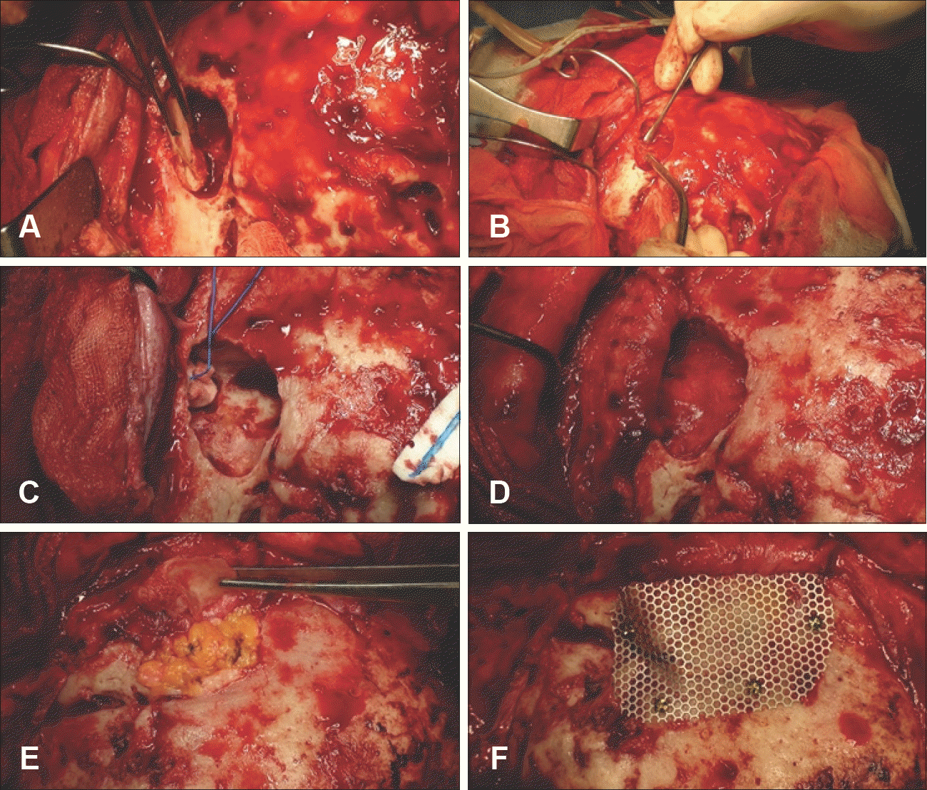 | FIGURE 2.Operative photographs demonstrating the cranialization procedure. A: Removal anterior wall of the frontal sinus. B: Removal of the granulated and inflamed wall of the frontal sinus. C: Meticulous extirpation of the frontal sinus mucosa. D: Flapping with the pericranial fascia to close the sinonasal tract. E: Packing the empty cavity with free fat tissue. F: Reconstruction of the anterior wall of the frontal sinus with a Mesh sheet. |
 XML Download