Abstract
Objective
Traumatic subdural hygroma (T-SDG) has been generally treated using conservative management rather than surgical methods. This study was performed to evaluate the clinical course of T-SDG with radiologic studies.
Methods
A retrospective study was conducted among patients diagnosed with T-SDG from January 2011 to December 2011. The patients were categorized into two groups. Group A has the widest width of T-SDG below 8 mm, Group B more than 8 mm. Computed tomography (CT) and magnetic resonance imaging (MRI) were carried out in both groups.
Results
Seventy-four patients were confirmed with T-SDG and were grouped as follows: 44 patients in Group A and 30 patients in Group B. There was no significant difference in age and sex ratio between group A and B. It took more time to resolve T-SDG in Group B (95.2±86.4 days) than Group A (14.4±6.7)(p<0.001). However, no significant difference was observed in the Glasgow Coma Scale (GCS) between the groups. In 10 patients of Group B, T-SDG developed into chronic subdural hematoma and one of these patients underwent surgery.
Go to : 
References
1. Borzone M, Capuzzo T, Perria C, Rivano C, Tercero E. Traumatic subdural hygromas: a report of 70 surgically treated cases. J Neurosurg Sci. 27:161–165. 1983.
2. French BN, Cobb CA 3rd, Corkill G, Youmans JR. Delayed evolution of posttraumatic subdural hygroma. Surg Neurol. 9:145–148. 1978.
3. Friede RL, Schachenmayr W. The origin ofsubdural neomembran-es. II. Fine structural of neomembranes. Am J Pathol. 92:69–84. 1978.
4. Hasegawa M, Yamashima T, Yamashita J, Suzuki M, Shimada S. Traumatic subdural hygroma: pathology and meningeal enhancement on magnetic resonance imaging. Neurosurgery. 31:580–585. 1992.
5. HillL NC, Goldstein NP, McKenzie BF, McGuckin WF, Svien HJ. Cerebrospinal-fluid proteins, glycoproteins, and lipoproteins in obstructive lesions of the central nervous system. Brain. 82:581–593. 1959.

6. Hoff J, Bates E, Barnes B, Glickman M, Margolis T. Traumatic subdural hygroma. J Trauma. 13:870–876. 1973.

7. Ju CI, Kim SW, Lee SM, Shin H. The surgical results of traumatic subdural hygroma treated with subduroperitoneal shunt. J Korean Neurosurg Soc. 37:436–442. 2005.
8. Koizumi H, Fukamachi A, Wakao T, Tasaki T, Nagaseki Y, Yanai Y. [Traumatic subdural hygromas in adults–on the possibility of development of chronic subdural hematoma (author's transl)]. Neurol Med Chir (Tokyo). 21:397–406. 1981.
9. Lee KS, Bae WK, Bae HG, Yun IG. The fate of traumatic subdural hygroma in serial computed tomographic scans. J Korean Med Sci. 15:560–568. 2000.

10. Lee KS, Bae WK, Park YT, Yun IG. The pathogenesis and fate of traumatic subdural hygroma. Br J Neurosurg. 8:551–558. 1994.

11. Lee KS, Doh JW, Bae HG, Yun IG. Relations among traumatic subdural lesions. J Korean Med Sci. 11:55–63. 1996.

12. Litofsky NS, Raffel C, McComb JG. Management of symptomat-ic chronic extra-axial fluid collections in pediatric patients. Neurosurgery. 31:445–450. 1992.

13. McCluney KW, Yeakley JW, Fenstermacher MJ, Baird SH, Bon-mati CM. Subdural hygroma versus atrophy on MR brain scans: “the cortical vein sign”. AJNR Am J Neuroradiol. 13:1335–1339. 1992.
Go to : 
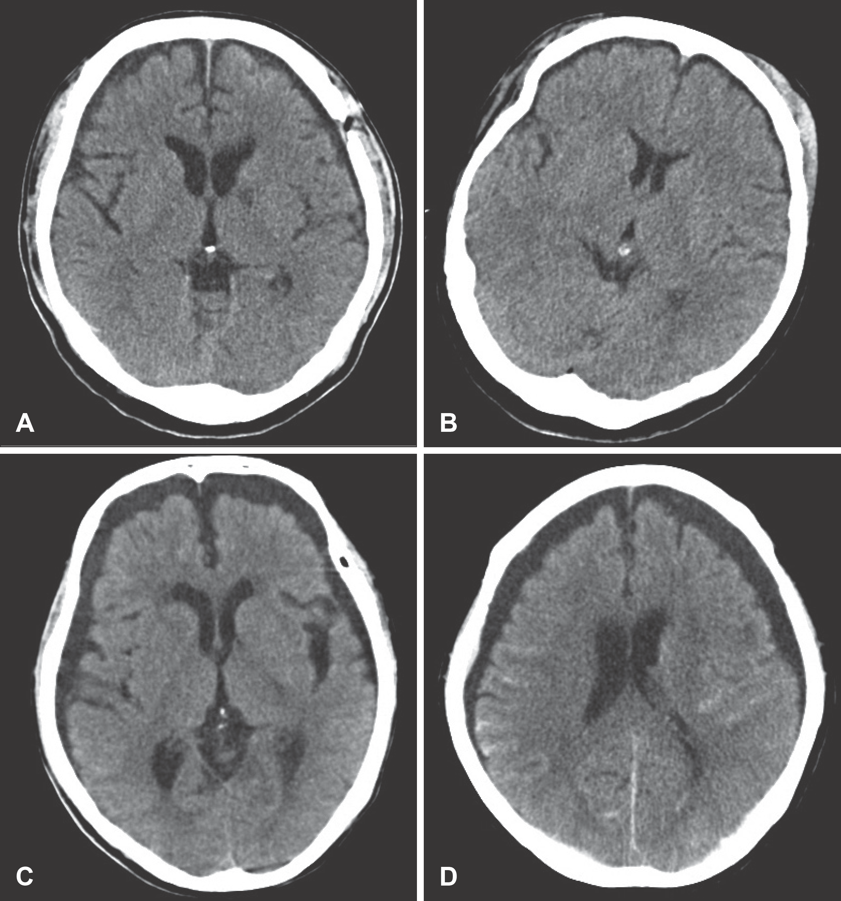
 | FIGURE 1.CT findings of representative cases. In a group A patient (A and B) the maximum width of T-SDG is below 8 mm whereas it is more than 8 mm in group B (C and D). T-SDG: traumatic subdural hematoma. |
 | FIGURE 2.The patient of T-SDG who changed to chronic subdural hematoma and was treated by surgical management. He had undergone surgical removal of epidural hematoma (EDH) in second hospital day. A: Post-operative day (POD) 1. B: POD 10. C: POD 27. D: POD 38. E: POD 47. F: POD 90. G: POD 118. |
TABLE 1.
Age distribution of patients with T-SDG
TABLE 2.
Main diagnosis of patients with T-SDG
TABLE 3.
Cause of trauma in patients with T-SDG
TABLE 4.
Sex, age, the time for the resolving T-SDG and GCS at admission between Group A and B


 PDF
PDF ePub
ePub Citation
 Print
Print


 XML Download