Abstract
Objective
The purpose of this study was to assess the diagnostic utility of the wrist ultrasonography (USG) in patients with and without carpal tunnel syndrome (CTS).
Methods
Individuals with electrodiagnostically proven CTS patients and healthy control subjects were enrolled prospectively. USG was done 60 wrists of 48 patients with CTS and 36 wrists of 18 controls. The USG analysis included median nerve cross sectional area (CSA) at the level of carpal tunnel inlet. We also evaluated the relationship between median nerve CSA at the level of carpal tunnel inlet and severity grade of nerve conduction test in CTS patients.
Results
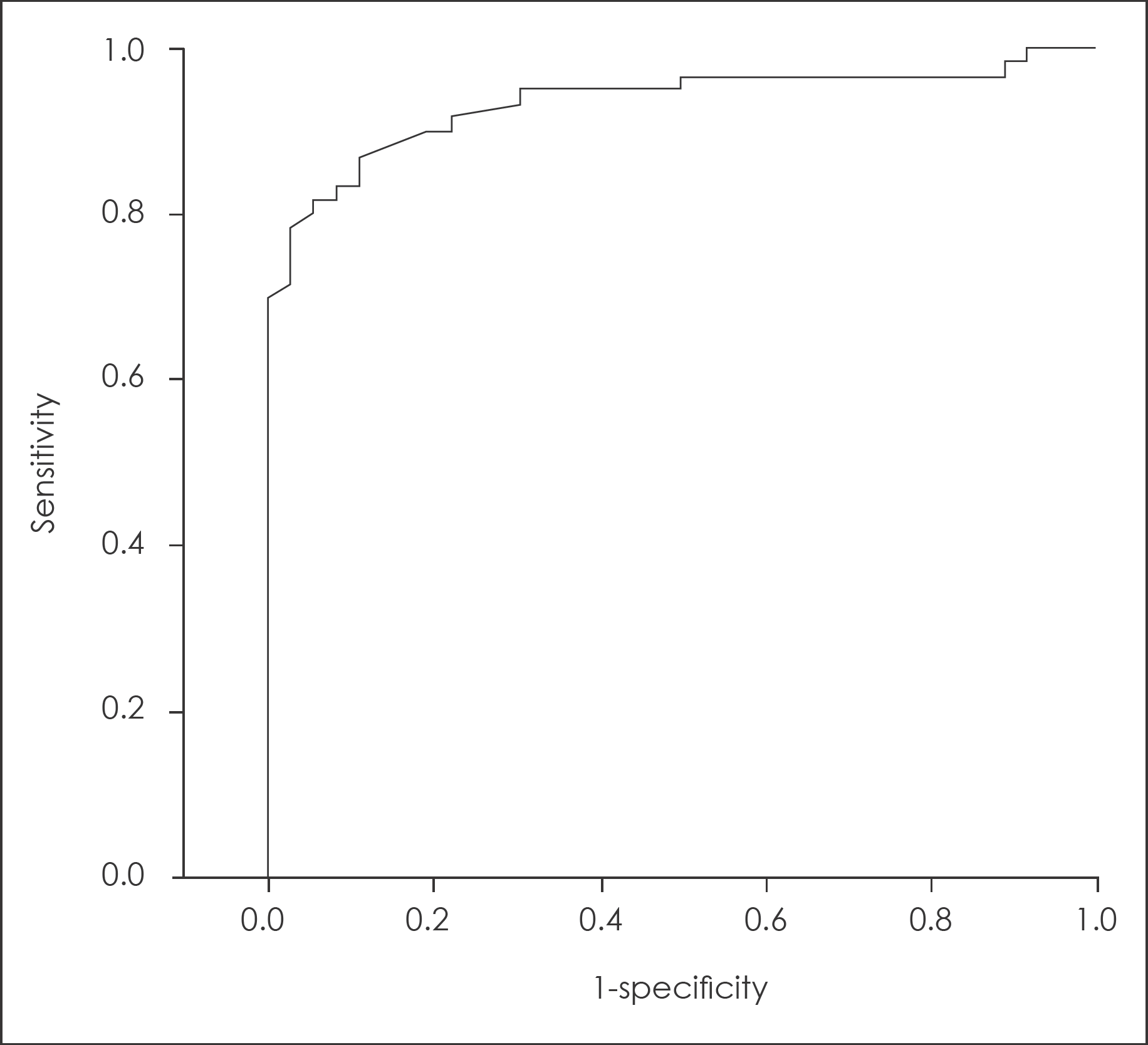
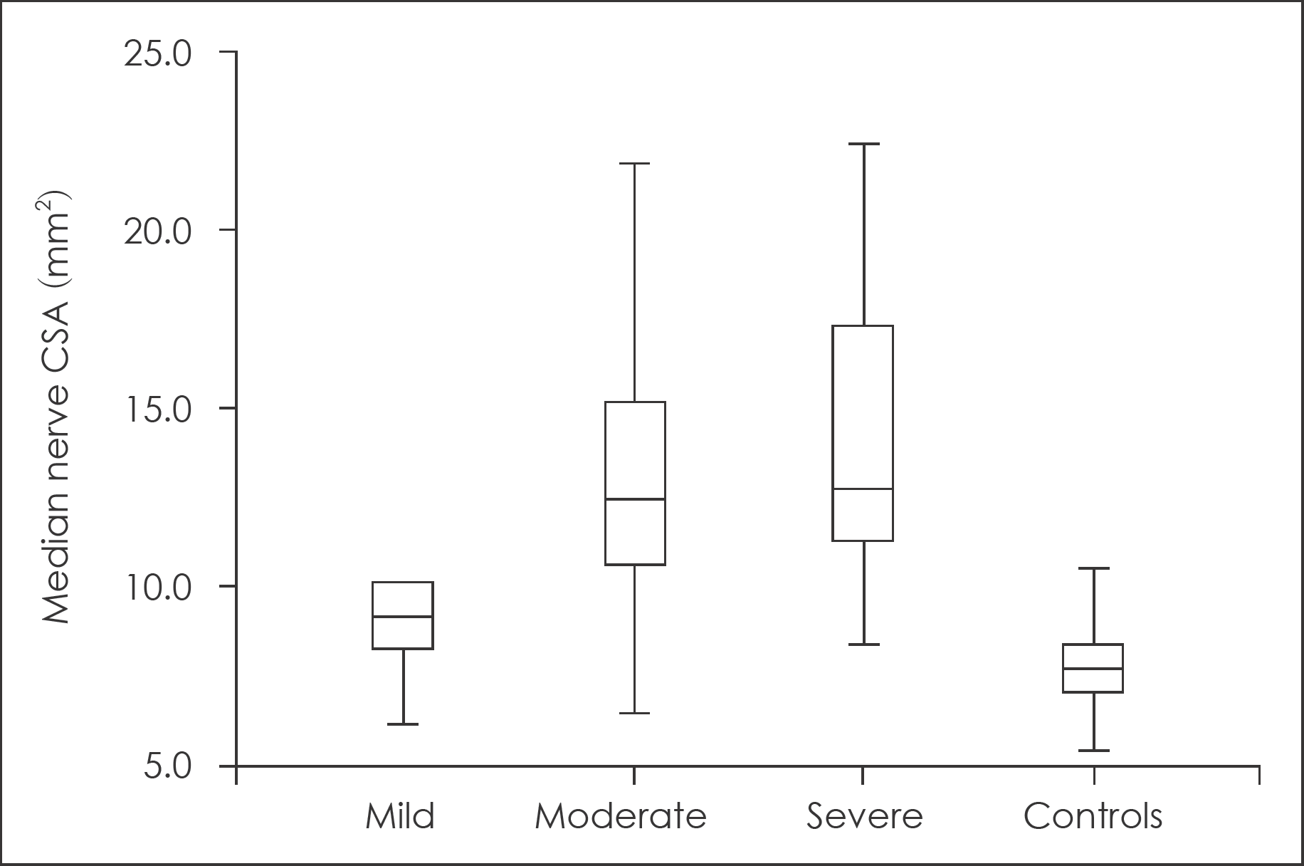
The median nerve CSA at the level of carpal tunnel inlet was significantly larger in CTS patients (13.6 mm2 versus 7.7 mm2, p<0.0001). And there was an association between median nerve CSA and severity grade of nerve conduction studies (p=0.036). Receiver operating characteristics (ROC) analysis yielded sensitivity of 86.7% and specificity of 88.9% using a cut-off value of 9 mm2. But the specificity was increased to 97.2%, although sensitivity was decreased to 78.3%, when using cut-off value at 10.1 mm2.
Go to : 
References
1. Bayrak IK, Bayrak AO, Kale M, Turker H, Diren B. Bifid median nerve in patients with carpal tunnel syndrome. J Ultrasound Med. 27:1129–1136. 2008.

2. Buchberger W, Judmaier W, Birbamer G, Lener M, Schmidauer C. Carpal tunnel syndrome: diagnosis with high-resolution sonography. AJR Am J Roentgenol. 159:793–798. 1992.

3. Cartwright MS, Shin HW, Passmore LV, Walker FO. Ultrasonographic reference values for assessing the normal median nerve in adults. J Neuroimaging. 19:47–51. 2009.

4. Chloros GD, Cartwright MS, Walker FO, Wiesler ER. Sonography and electrodiagnosis in carpal tunnel syndrome diagnosis, an analysis of the literature. Eur J Radiol. 71:141–143. 2009.

5. Duncan I, Sullivan P, Lomas F. Sonography in the diagnosis of carpal tunnel syndrome. AJR Am J Roentgenol. 173:681–684. 1999.

6. El Miedany YM, Aty SA, Ashour S. Ultrasonography versus nerve conduction study in patients with carpal tunnel syndrome: substantive or complementary tests? Rheumatology (Oxford). 43:887–895. 2004.

7. Grundberg AB. Carpal tunnel decompression in spite of normal electromyography. J Hand Surg Am. 8:348–349. 1983.
8. Hammer HB, Haavardsholm EA, Kvien TK. Ultrasonographic measurement of the median nerve in patients with rheumatoid arthritis without symptoms or signs of carpal tunnel syndrome. Ann Rheum Dis. 66:825–827. 2007.
9. Hunderfund AN, Boon AJ, Mandrekar JN, Sorenson EJ. Sonography in carpal tunnel syndrome. Muscle Nerve. 44:485–491. 2011.

10. Iyer VG. Understanding nerve conduction and electromyographic studies. Hand Clin. 9:273–287. 1993.

11. Kasius KM, Claes F, Verhagen WI, Meulstee J. Ultrasonography in severe carpal tunnel syndrome. Muscle Nerve. 45:334–337. 2012.

12. Klauser AS, Halpern EJ, De Zordo T, Feuchtner GM, Arora R, Gruber J, et al. Carpal tunnel syndrome assessment with US: value of additional cross-sectional area measurements of the median nerve in patients versus healthy volunteers. Radiology. 250:171–177. 2009.

13. Koyuncuoglu HR, Kutluhan S, Yesildag A, Oyar O, Guler K, Ozden A. The value of ultrasonographic measurement in carpal tunnel syndrome in patients with negative electrodiagnostic tests. Eur J Radiol. 56:365–369. 2005.

14. Nakamichi K, Tachibana S. Ultrasonographic measurement of median nerve cross-sectional area in idiopathic carpal tunnel syndrome: Diagnostic accuracy. Muscle Nerve. 26:798–803. 2002.

15. Nathan PA, Keniston RC, Meadows KD, Lockwood RS. Predictive value of nerve conduction measurements at the carpal tunnel. Muscle Nerve. 16:1377–1382. 1993.

16. Pastare D, Therimadasamy AK, Lee E, Wilder-Smith EP. Sonography versus nerve conduction studies in patients referred with a clinical diagnosis of carpal tunnel syndrome. J Clin Ultrasound. 37:389–393. 2009.

17. Phalen GS. The carpal-tunnel syndrome. Clinical evaluation of 598 hands. Clin Orthop Relat Res. 83:29–40. 1972.
18. Rempel D, Dahlin L, Lundborg G. Pathophysiology of nerve compression syndromes: response of peripheral nerves to loading. J Bone Joint Surg Am. 81:1600–1610. 1999.
19. Rempel D, Evanoff B, Amadio PC, de Krom M, Franklin G, Franzblau A, et al. Consensus criteria for the classification of carpal tunnel syndrome in epidemiologic studies. Am J Public Health. 88:1447–1451. 1998.

20. Rempel DM, Diao E. Entrapment neuropathies: pathophysiology and pathogenesis. J Electromyogr Kinesiol. 14:71–75. 2004.

21. Sarría L, Cabada T, Cozcolluela R, Martínez-Berganza T, García S. Carpal tunnel syndrome: usefulness of sonography. Eur Radiol. 10:1920–1925. 2000.

22. Seror P. Sonography and electrodiagnosis in carpal tunnel syndrome diagnosis, an analysis of the literature. Eur J Radiol. 67:146–152. 2008.

23. Stevens JC. AAEM minimonograph #26: the electrodiagnosis of carpal tunnel syndrome. American Association of Electrodiagnostic Medicine. Muscle Nerve. 20:1477–1486. 1997.
24. Swen WA, Jacobs JW, Bussemaker FE, de Waard JW, Bijlsma JW. Carpal tunnel sonography by the rheumatologist versus nerve conduction study by the neurologist. J Rheumatol. 28:62–69. 2001.
25. Werner RA, Andary M. Electrodiagnostic evaluation of carpal tunnel syndrome. Muscle Nerve. 44:597–607. 2011.

26. Wiesler ER, Chloros GD, Cartwright MS, Smith BP, Rushing J, Walker FO. The use of diagnostic ultrasound in carpal tunnel syndrome. J Hand Surg Am. 31:726–732. 2006.

27. Witt JC, Hentz JG, Stevens JC. Carpal tunnel syndrome with normal nerve conduction studies. Muscle Nerve. 29:515–522. 2004.

Go to : 
 | FIGURE 1.Receiver operating characteristics curve for ultrasonographic assessments of median nerve cross-sectional area at carpal tunnel inlet. The area under the curve is 0.935 (95% confidence interval, 0.86–0.98; p<0.0001). |
 | FIGURE 2.Box plot showing median nerve cross-sectional area (CSA) according to severity of nerve conduction study, and control subjects. |
TABLE 1.
Electrophysiological carpal tunnel syndrome grading scheme according to the classification reported by Stevens23)
TABLE 2.
Summary of carpal tunnel syndrome patients and healthy control subjects
| Patients (n=48) | Controls (n=18) | p | |
|---|---|---|---|
| Mean age±SD (years) | 58.5±12.1 | 54.3±12.9 | 0.114 |
| Female gender | 43 (89.5%)∗ | 10 (55.6%)∗ | 0.357 |
| Number of wrists (right/left) | 60 (29/31)∗ | 36 (18/18)∗ | |
| Median duration of symptoms (months) | 12.1 | N/A | |
| Clinically severe grade (wrists) | 18 (30.0%)∗ | N/A | |
| Atrophy of APBM | 31 (51.7%)∗ | N/A | |
| Electrodiagnostic grade | N/A | ||
| Mild | 06 (10.0%)∗ | ||
| Moderate | 28 (46.7%)∗ | ||
| Severe | 26 (43.3%)∗ |
TABLE 3.
Median nerve CSA according to NCS grade
| NCS grade | Median nerve CSA (mm2) | p |
|---|---|---|
| Mild | 09.4±2.4 | |
| Moderate | 13.3±4.2 | 0.036∗ |
| Severe | 14.9±5.4 |


 PDF
PDF ePub
ePub Citation
 Print
Print


 XML Download