

 PDF
PDF ePub
ePub Citation
 Print
Print


Abstract
Objective:
The purpose of this study is to identify risk factors related to the fusion failure after halo-vest immobilization of odontoid fracture type III.
Methods:
We retrospectively analyzed ten patients who underwent halo-vest immobilization for acute traumatic odontoid fracture between October 2002 and December 2011. All patients had type III odontoid fracture using the Anderson and D’Alonzo classification. We reviewed digital radiographs and analyzed the images during conservative treatment with halo-vest immobilization.
Results:
The patients consisted of nine men and one woman, with mean age of 40.2 years (range: 25-56), who had no history of medical comorbidity and significant neurologic deficit. The mean follow-up period was 6 months (range: 4-11). All patients were initially treated by halo-vest immobilization. Seven patients showed union of fractured site on radiologic findings after halo-vest immobilization only. However, other 3 patients underwent surgery for fixation due to fusion failure. Among the factors we analyzed such as, radiographic characteristics and clinical feature, presence of comminuted fracture, instability of fractured fragment and failed reduction of misalignment were the factors related to fusion failure.
Go to : 
REFERENCES
1). Althoff B. Fracture of the odontoid process. An experimental and clinical study. Acta Orthop Scand Suppl. 177:1–95. 1979.

2). Anderson LD., D'Alonzo RT. Fractures of the odontoid process of the axis. J Bone Joint Surg Am. 56:1663–1674. 1974.
3). Bucholz RD., Cheung KC. Halo vest versus spinal fusion for cervical injury: evidence from an outcome study. J Neurosurg. 70:884–892. 1989.
4). Clark CR., White AA 3rd. Fractures of the dens. A multicenter study. J Bone Joint Surg Am. 67:1340–1348. 1985.
5). Ekong CE., Schwartz ML., Tator CH., Rowed DW., Edmonds VE. Odontoid fracture: management with early mobilization using the halo device. Neurosurgery. 9:631–637. 1981.
6). Fujii E., Kobayashi K., Hirabayashi K. Treatment in fractures of the odontoid process. Spine (Phila Pa 1976). 13:604–609. 1988.
7). Glaser JA., Whitehill R., Stamp WG., Jane JA. Complications associated with the halo-vest. A review of 245 cases. J Neurosurg. 65:762–769. 1986.
8). Greene KA., Dickman CA., Marciano FF., Drabier JB., Hadley MN., Sonntag VK. Acute axis fractures. Analysis of management and outcome in 340 consecutive cases. Spine (Phila Pa 1976). 22:1843–1852. 1997.
9). Houtkin S., Levine DB. The halo yoke: a simplified device for attachment of the halo to a body cast. J Bone Joint Surg Am. 54:881–883. 1972.
10). Hsu WK., Anderson PA. Odontoid fractures: update on management. J Am Acad Orthop Surg. 18:383–394. 2010.
11). Jea A., Tatsui C., Farhat H., Vanni S., Levi AD. Vertically unstable type III odontoid fractures: case report. Neurosurgery 58: E797; discussion E797. 2006.
12). Kim DH., Vaccaro AR., Affonso J., Jenis L., Hilibrand AS., Albert TJ. Early predictive value of supine and upright X-ray films of odontoid fractures treated with halo-vest immobilization. Spine J. 8:612–618. 2008.
13). Koivikko MP., Kiuru MJ., Koskinen SK., Myllynen P., Santavirta S., Kivisaari L. Factors associated with nonunion in conservatively-treated type-II fractures of the odontoid process. J Bone Joint Surg Br. 86:1146–1151. 2004.
14). Lennarson PJ., Mostafavi H., Traynelis VC., Walters BC. Management of type II dens fractures: a case-control study. Spine (Phila Pa 1976). 25:1234–1237. 2000.
15). Lind B., Sihlbom H., Nordwall A. Halo-vest treatment of unstable traumatic cervical spine injuries. Spine (Phila Pa 1976). 13:425–432. 1988.
16). Maak TG., Grauer JN. The contemporary treatment of odontoid injuries. Spine (Phila Pa 1976) 31(11 Suppl): S53-S60; discussion S61. 2006.
17). Majercik S., Tashjian RZ., Biffl WL., Harrington DT., Cioffi WG. Halo vest immobilization in the elderly: a death sentence? J Trauma. 59:350–356. discussion 356-358. 2005.
18). Nickel VL., Perry J., Garrett A., Heppenstall M. The halo. A spinal skeletal traction fixation device. J Bone Joint Surg Am. 50:1400–1409. 1968.
19). Polin RS., Szabo T., Bogaev CA., Replogle RE., Jane JA. Nonoperative management of Types II and III odontoid fractures: the Philadelphia collar versus the halo vest. Neurosurgery. 38:450–456. discussion 456-457. 1996.
20). Przybylski GJ., Welch WC. Longitudinal atlantoaxial dislocation with type III odontoid fracture. Case report and review of the literature. J Neurosurg. 84:666–670. 1996.
21). Sasso RC. C2 dens fractures: treatment options. J Spinal Disord. 14:455–463. 2001.
22). Seybold EA., Bayley JC. Functional outcome of surgically and conservatively managed dens fractures. Spine (Phila Pa 1976). 23:1837–1845. discussion 1845-1846. 1998.
23). Shin JJ., Kim SJ., Kim TH., Shin HS., Hwang YS., Park SK. Optimal use of the halo-vest orthosis for upper cervical spine injuries. Yonsei Med J. 51:648–652. 2010.
24). Wang GJ., Mabie KN., Whitehill R., Stamp WG. The nonsurgical management of odontoid fractures in adults. Spine (Phila Pa 1976). 9:229–230. 1984.
Go to :
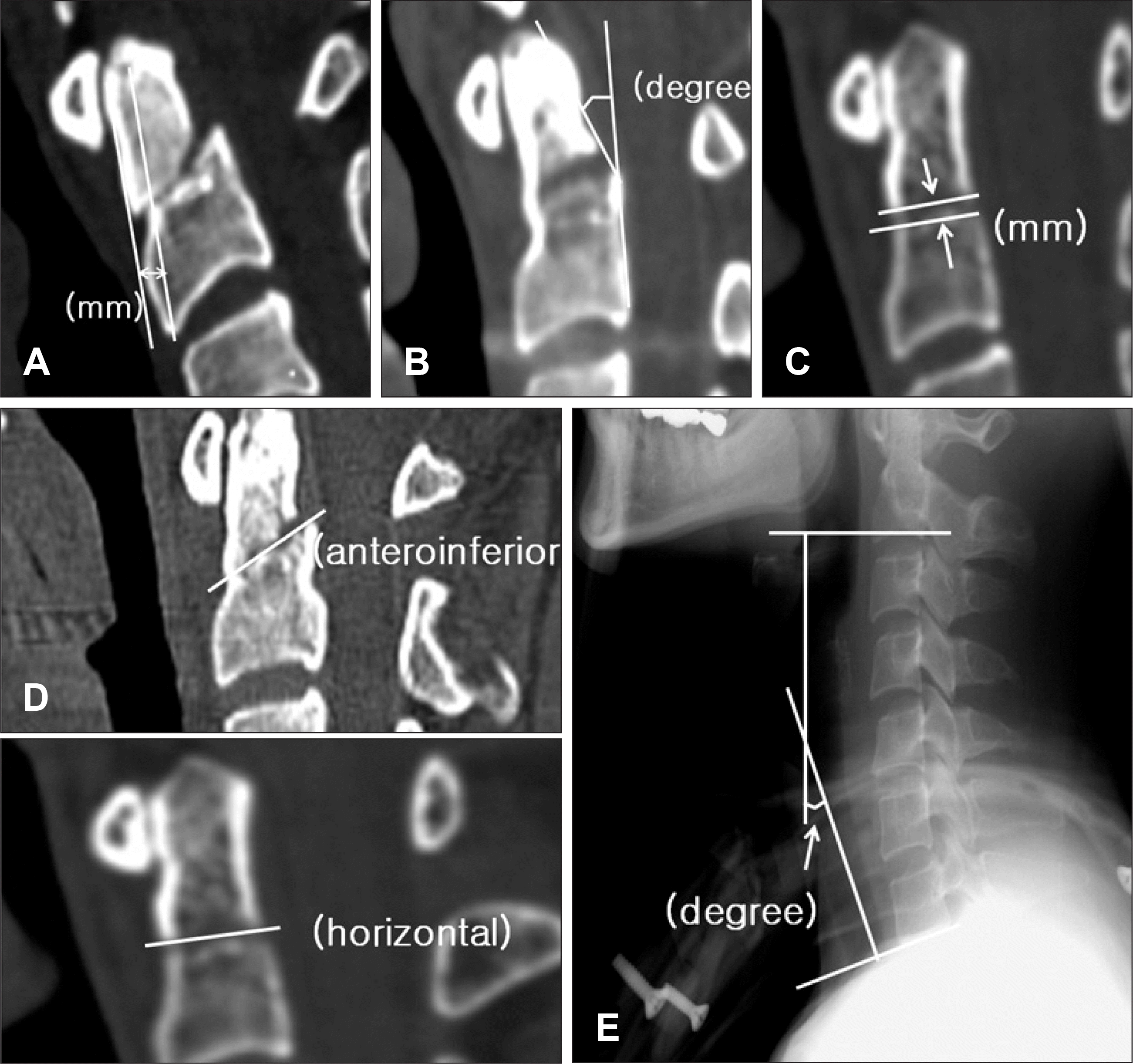 | FIGURE 1.Measurement of radiographic findings. A: A tangent line is drawn along the anterior aspect of the odontoid fragment and the anterior aspect of the C2 body. A transverse line is drawn connecting these two lines. This distance is measured as the displacement of fractured odontoid process. B: A tangent line is drawn along the posterior aspect of the odontoid fragment and the posterior aspect of the C2 body. The angle subtended by these lines is the degree of angulation of fractured odontoid process. C: The gap between fractured odontoid process and dens body was determined as measured distance between inferior border of fractured odontoid process and superior border of fractured C2 body. D: The type of fracture line of odontoid process was determined in the computed tomography lateral reconstruction image. E: Overall lordosis was determined as measured angle between inferior endplate of C2 and inferior endplate of C7 in the lateral view of cervical plain radiograph. |
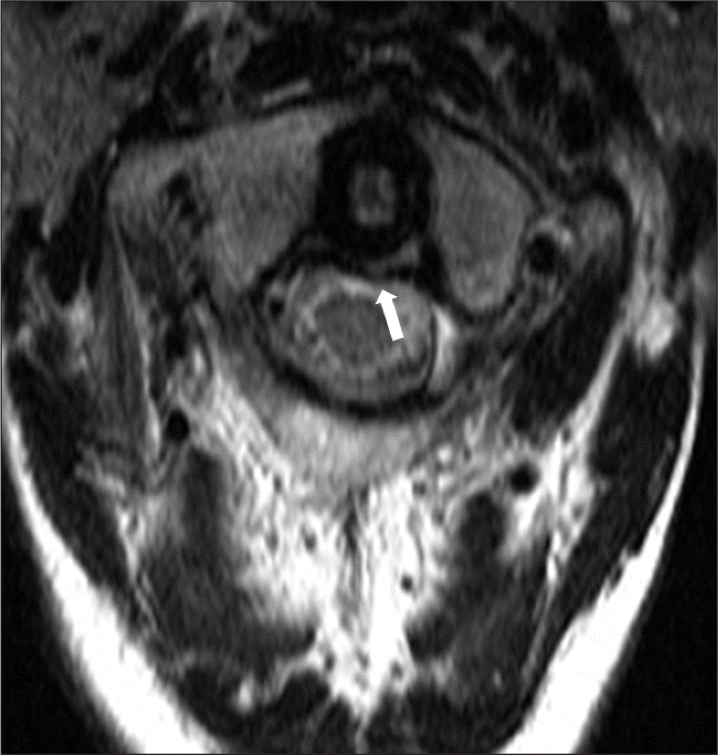 | FIGURE 2.Magnetic resonance imaging showed disruption of the transverse ligament of atlantoaxial joint (arrow). |
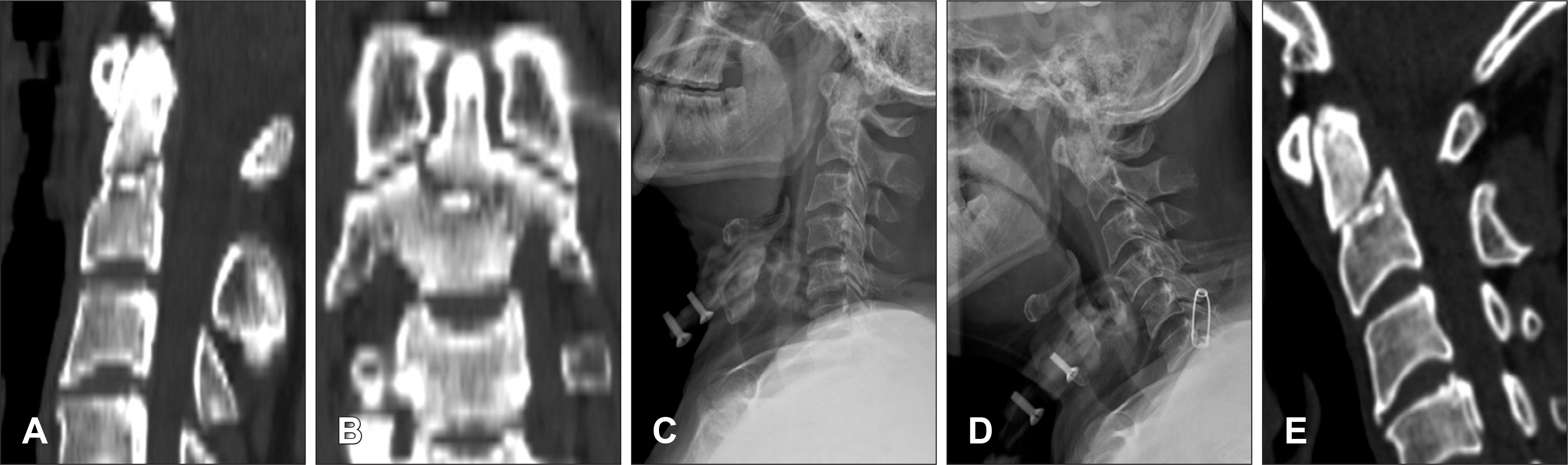 | FIGURE 3.Imaging studies from case 1. A: Computed tomography (CT) scans of the patient showed a type III odontoid fracture and there was ossification of posterior longitudinal ligament at the level of C3-C4. The fractured odontoid process was slightly displaced anteroinferiorly. B: A coronal reconstruction image revealed the comminuted fracture. C: The plain radiograph after 1 day of halo-vest immobilization. D: The plain radiograph on 3 weeks follow up showing the change of cervical lordotic curve. E: After 14 weeks, on the CT lateral reconstruction image, we found that the fracture site had not completely healed. |
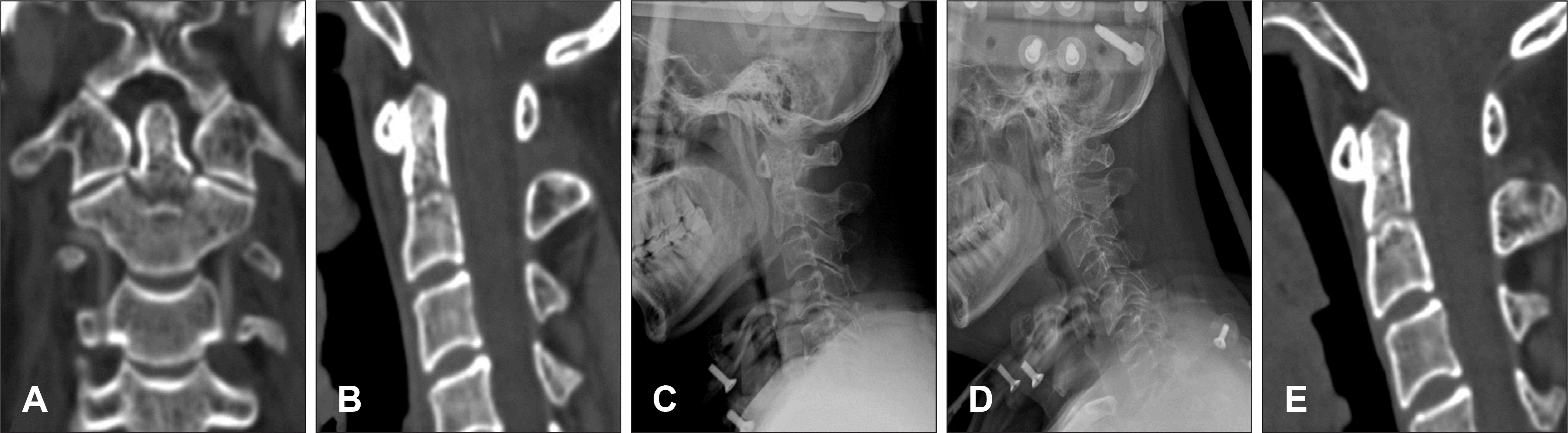 | FIGURE 4.Imaging studies from case 2. A, B: Computed tomography (CT) scans of the patient showed a type III odontoid fracture with horizontal line and the fractured odontoid process was not displaced. C: The plain radiograph on 1day after halo-vest immobilization. D: The plain radiograph on 5 weeks follow up showing the redisplacement of fractured odontoid process. E: The CT lateral reconstruction image on 10 weeks follow up showing the osteosclerotic margin of fractured site. |
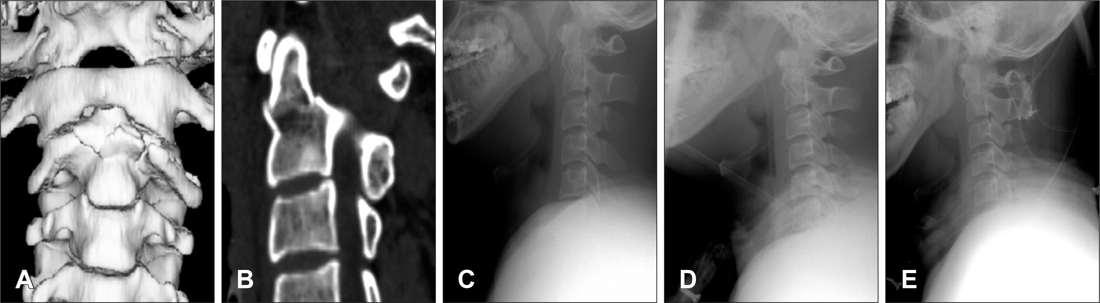 | FIGURE 5.Imaging studies from case 3. A, B: Computed tomography scans of the patient showed a type III odontoid fracture with irregular fracture line and the fractured odontoid process was displaced anteroinferiorly. C: The patient underwent the conservative treatment with halo-vest immobilization at his request, (D) but the displaced odontoid process was not corrected in alignment. E: Postoperative plain radiograph. Note that posterior interspinous fusion combined with sustained halo-vest immobilization. |
TABLE 1.
The clinical characteristics of 10 cases
| Case no. | Age (years)/ Sex | Trauma | Time from injury to HVI (days) | Periods of HVI (weeks) | Neurologic symptoms | Associated injury | F/U duration (months) | Result of HVI |
|---|---|---|---|---|---|---|---|---|
| 1 | 54/M | In-car TA | 1 | 14 | Both knee tingling sense | (-) | 4 | Failure |
| 2 | 49/M | In-car TA | 3 | 10 | (-) | (-) | 4 | Failure |
| 3 | 25/M | In-car TA | 2 | 3 day+postop 3 m† | (-) | (-) | 4 | Failure |
| 4 | 30/M | Fall down | 5 | 12 | (-) | (-) | 5 | Success |
| 5 | 49/M | In-car TA | 2 | 13 | Decreased sense on Rt. occiput | Compression fracture,T12& L1 | 5 | Success |
| 6 | 42/M | In-car TA | 5 | 12 | (-) | (-) | 10 | Success |
| 7 | 31/M | In-car TA | 7 | 25 | Delirium&transient memory impairment | Traumatic SAH | 7 | Success |
| 8 | 35/M | In-car TA | 4 | 14 | Paresthesia on Lt. arm | (-) | 4 | Success |
| 9 | 31/F | In-car TA | 9 | 8∗ | (-) | Fractures of Lt. femur & humerus | 7 | Success |
| 10 | 56/M | In-car TA | 6 | 14 | (-) | C5-6 ligamentous subluxation | 11 | Success |
TABLE 2.
The radiological characteristics of 10 cases
 XML Download