

 PDF
PDF ePub
ePub Citation
 Print
Print


Abstract
Currently, endovascular treatment of carotid cavernous fistula (CCF) is widely accepted and performed. However, a graft stent is rarely used for the treatment of high-flow CCF. Here we describe our experience using a graft stent to treat CCF and discuss the indications for its use. (Korean J Neurotrauma 2012;8:51-54)
REFERENCES
1). Ahn JY., Chung YS., Chung SS., Lee BH. Endovascular treatment of a traumatic carotid-jugular fistula by using stent-graft. J Korean Neurosurg Soc. 34:470–473. 2003.
2). Ahn JY., Lee BH., Joo JY. Stent-assisted Guglielmi detachable coils embolisation for the treatment of a traumatic carotid cavernous fistula. J Clin Neurosci. 10:96–98. 2003.

3). Archondakis E., Pero G., Valvassori L., Boccardi E., Scialfa G. Angiographic follow-up of traumatic carotid cavernous fistulas treated with endovascular stent graft placement. AJNR Am J Neuroradiol. 28:342–347. 2007.
4). Barrow DL., Spector RH., Braun IF., Landman JA., Tindall SC., Tindall GT. Classification and treatment of spontaneous carotid-cavernous sinus fistulas. J Neurosurg. 62:248–256. 1985.
5). Bartorelli AL., Trabattoni D., Reali M. Percutaneous obliteration of an iatrogenic pseudoaneurysm of the right subclavian artery with a PTFE-covered stent-graft. Int J Cardiovasc Intervent. 4:195–196. 2001.
6). Briganti F., Tortora F., Marseglia M., Napoli M., Cirillo L. Covered Stent Implantation for the Treatment of Direct Carotid-Cavernous Fistula and Its Mid-Term Follow-up. Interv Neuroradiol. 15:185–190. 2009.
7). Choi BJ., Lee TH., Kim CW., Choi CH. Endovascular graft-stent placement for treatment of traumatic carotid cavernous fistulas. J Korean Neurosurg Soc. 46:572–576. 2009.
8). Eddleman CS., Surdell D., Miller J., Shaibani A., Bendok BR. Endovascular management of a ruptured cavernous carotid artery aneurysm associated with a carotid cavernous fistula with an intracranial self-expanding microstent and hydrogel-coated coil embolization: case report and review of the literature. Surg Neurol. 68:562–567. discussion 567. 2007.
9). Elsner M., Auch-Schwelk W., Britten M., Walter DH., Schächinger V., Zeiher AM. Coronary stent grafts covered by a polytetrafluoroethylene membrane. Am J Cardiol. 84:335–338. A8. 1999.
10). Gomez F., Escobar W., Gomez AM., Gomez JF., Anaya CA. Treatment of carotid cavernous fistulas using covered stents: midterm results in seven patients. AJNR Am J Neuroradiol. 28:1762–1768. 2007.
11). Hoit DA., Schirmer CM., Malek AM. Stent graft treatment of cerebrovascular wall defects: intermediate-term clinical and angiographic results. Neurosurgery 62: ONS380-ONS388; discussion ONS388-ONS389. 2008.
12). La Tessa G., Pasqualetto L., Catalano G., Marino M., Gargano C., Cirillo L, et al. Traumatic carotid cavernous fistula: failure of endovascular treatment with two stent-grafts. Interv Neuroradiol. 11:369–375. 2005.
13). Meijer FJ., Van Der Vliet AM. Treatment of a traumatic carotid-cavernous fistula with a covered stent: some considerations. Case report. Interv Neuroradiol. 15:191–196. 2009.
14). Men S., Oztürk H., Hekimoğlu B., Sekerci Z. Traumatic carotid-cavernous fistula treated by combined transarterial and transvenous coil embolization and associated cavernous internal carotid artery dissection treated with stent placement. Case report. J Neurosurg. 99:584–586. 2003.
15). Morón FE., Klucznik RP., Mawad ME., Strother CM. Endovascular treatment of high-flow carotid cavernous fistulas by stent-assisted coil placement. AJNR Am J Neuroradiol. 26:1399–1404. 2005.
16). Serbinenko FA. Balloon catheterization and occlusion of major cerebral vessels. J Neurosurg. 41:125–145. 1974.
17). Tiewei Q., Ali A., Shaolei G., Feng L., Zhongsong S., Xuesong L, et al. Carotid cavernous fistulas treated by endovascular covered stent grafts with follow-up results. Br J Neurosurg. 24:435–440. 2010.
FIGURE 2.
Lateral angiogram of the left internal carotid artery showing total steal of flow toward the superior ophthalmic vein, pterygoid plexus, contralateral cavernous sinus, and petrosal sinus.
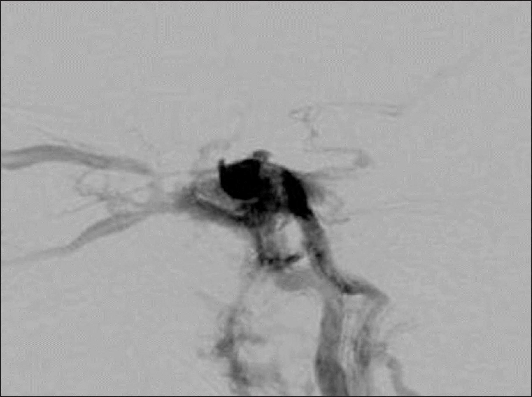
FIGURE 3.
Immediate post-procedural left internal carotid angiogram, left oblique (A) and lateral views (B), demonstrating complete occlusion of carotid cavernous fistula and normal patency of internal carotid artery.
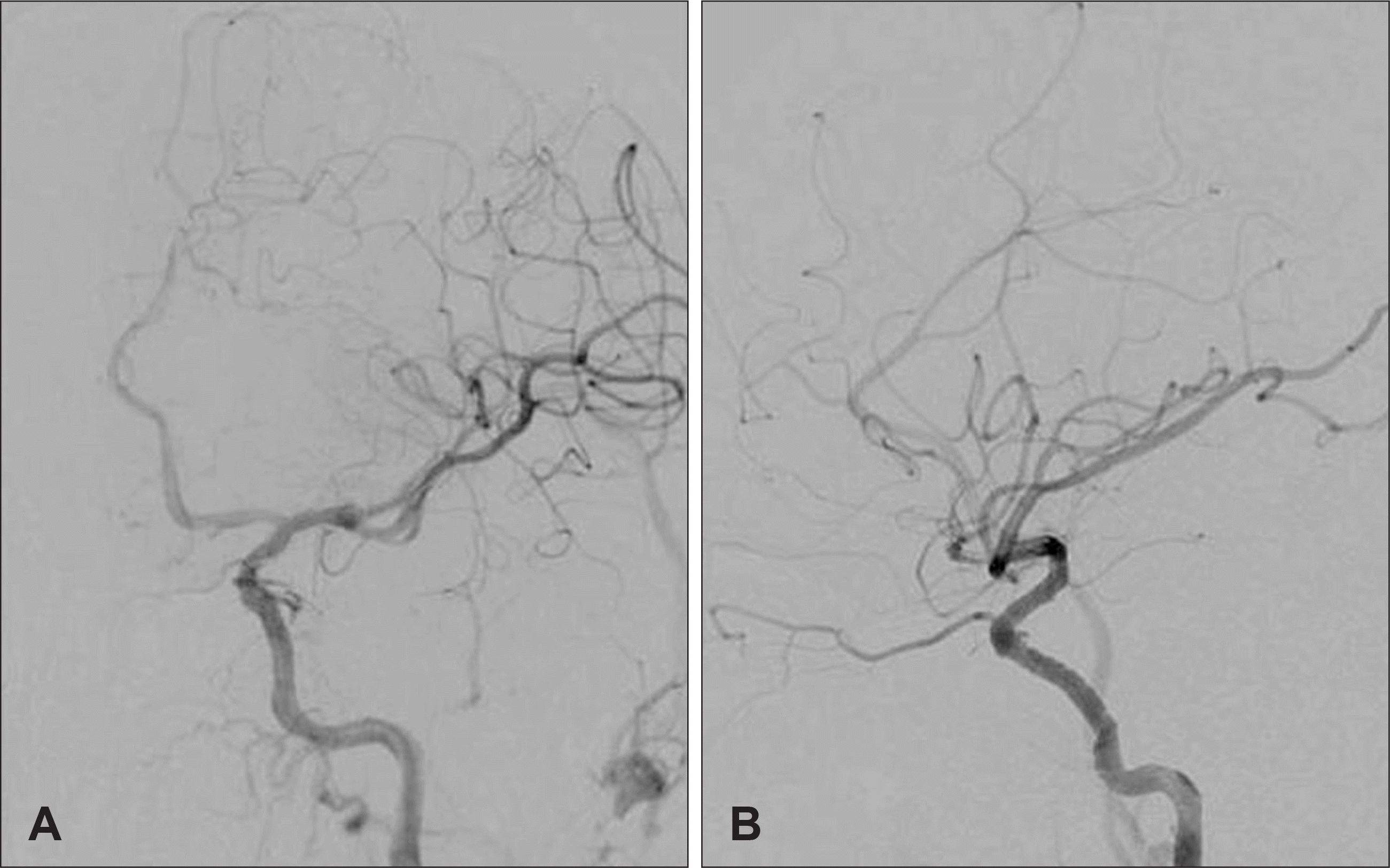
FIGURE 4.
Left lateral internal carotid angiogram on day 14 after treatment demonstrating the partial reopening of the fistula (A) and endoleak (arrow) after angioplasty with a coronary balloon (B).
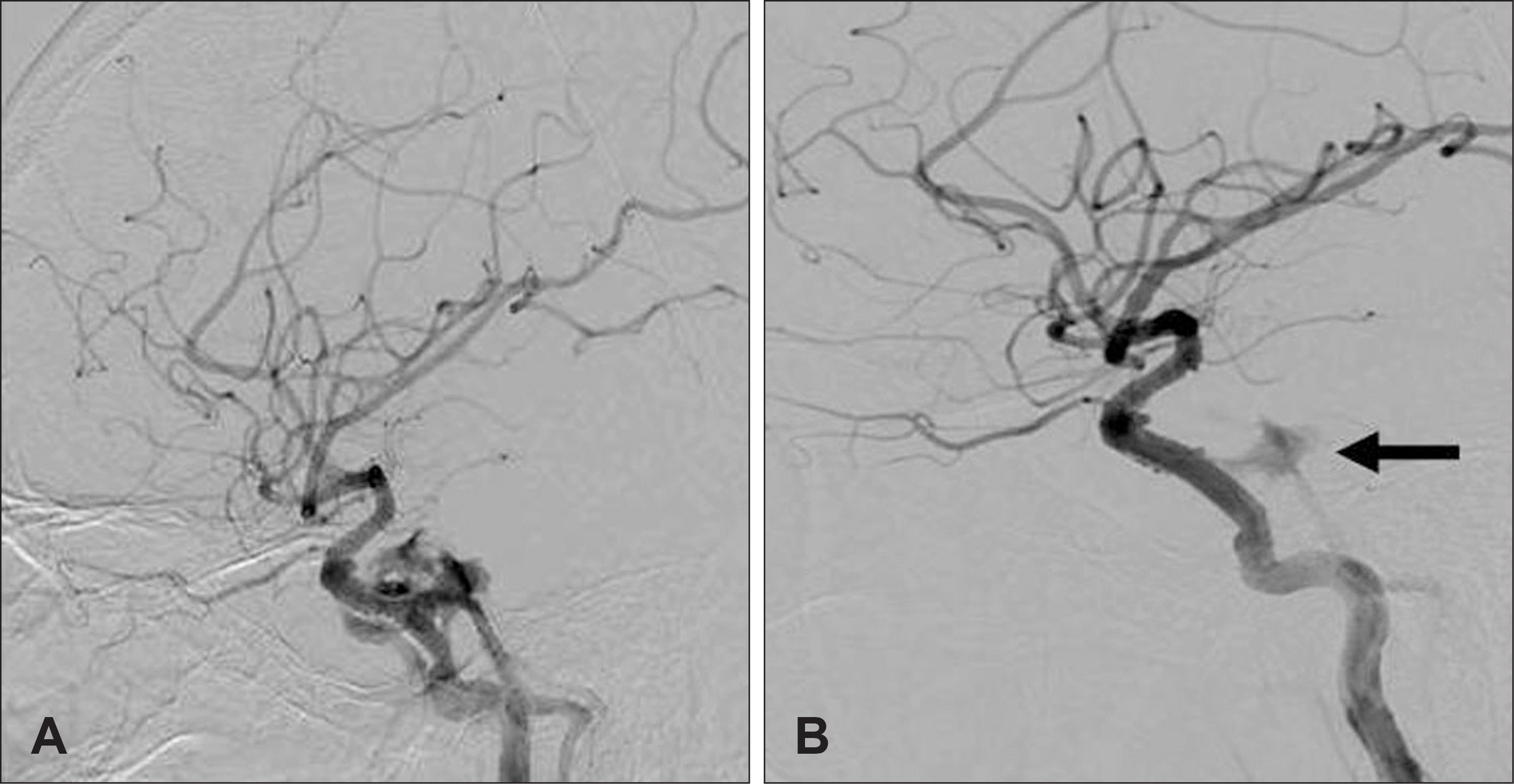
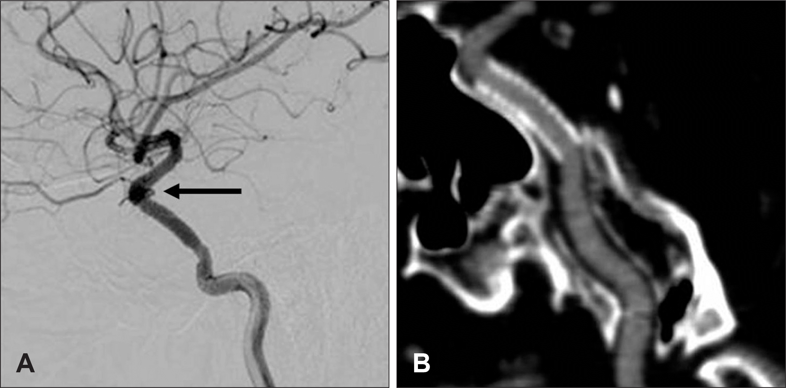
FIGURE 5.
Lateral angiogram of left internal carotid artery after 8 months showing a complete obliteration of carotid cavernous fistula and pseudoaneurysm of the distal end of the stent (arrow) (A). Computed tomographic angiography 8 months later reveals normal patency of the internal carotid artery (B).
 XML Download