

 PDF
PDF ePub
ePub Citation
 Print
Print


Abstract
Objective:
There is a broad spectrum of compensated hydrocephalus. Various terms such as long-standing overt ventriculomegaly in adult (LOVA) has been coined, however, even such terms leave diverse aspect of this condition out of account. We have experienced compensated hydrocephalus cases which were considered to be activated after a long time period of quiescent state, and tried to compare their clinical characteristics with the relatively well described entity of LOVA.
Methods:
We conducted a retrospective review of 206 patients who underwent ventriculoperitoneal shunt (VPS) between February 2001 and May 2012. Of these, 6 patients had chronic compensated hydrocephalus. The clinical and radiological characteristics are evaluated.
Results:
Definite triventriculomegaly was observed in two patients. Macrocephaly was observed in two cases, one with aqueductal stenosis (AS), the other with unknown status of aqueduct. All of the cases with triventriculomegaly were normoce-phalic. Spinal causes were thought as aggravating factor in two. Two endoscopic third ventriculostomy and eight VPS were performed in five patients. Four patients responded well but one took a very complicated course.
Conclusion:
The relationships between macrocephaly, triventriculomegaly, and AS suggested in other studies were inconsistent. Blockage or narrowing of cerebrospinal fluid pathways were observed at various sites. Disturbances of spinal arachnoid pathways were related to the activation in some cases. Treatment is to be tailored individually considering various reigniting event. It is suggested that this entity is to be evaluated for better nomenclature reflecting diverse aspects of this condition. Further study is needed to elucidate underlying pathophysiology and effective management.
REFERENCES
1). Bradley WG Jr., Bahl G., Alksne JF. Idiopathic normal pressure hydrocephalus may be a “two hit” disease: benign external hydrocephalus in infancy followed by deep white matter ischemia in late adulthood. J Magn Reson Imaging. 24:747–755. 2006.

2). Canu ED., Magnano I., Paulus KS., Piras MR., Conti M., Costantino S, et al. Neuropsychophysiological findings in a case of long-standing overt ventriculomegaly (LOVA). Neurosci Lett. 385:24–29. 2005.
3). Cowan JA., McGirt MJ., Woodworth G., Rigamonti D., Williams MA. The syndrome of hydrocephalus in young and middle-aged adults (SHYMA). Neurol Res. 27:540–547. 2005.
4). Edwards RJ., Britz GW., Marsh H. Chronic headaches due to occult hydrocephalus. J R Soc Med. 96:77–78. 2003.
5). Fukuhara T., Luciano MG. Clinical features of late-onset idiopathic aqueductal stenosis. Surg Neurol. 55:132–136. discussion 136-137. 2001.
6). Hamada H., Hayashi N., Kurimoto M., Takaiwa A., Kurosaki K., Endo S. Neuropsychological changes after endoscopic third ventriculostomy for long-standing overt ventriculomegaly in adults. Case report. Neurol Med Chir (Tokyo). 49:362–364. 2009.
7). Kaestner S., Kruschat T., Nitzsche N., Deinsberger W. Gravitational shunt units may cause under-drainage in bedridden patients. Acta Neurochir (Wien). 151:217–221. discussion 221. 2009.
8). Kiefer M., Eymann R., Steudel WI., Strowitzki M. Gravitational shunt management of long-standing overt ventriculomegaly in adult (LOVA) hydrocephalus. J Clin Neurosci. 12:21–26. 2005.
9). Knol DS., van Gijn J., Kruitwagen CL., Rinkel GJ. Size of third and fourth ventricle in obstructive and communicating acute hydrocephalus after aneurysmal subarachnoid hemorrhage. J Neurol. 258:44–49. 2011.
10). Larsson A., Stephensen H., Wikkelsø C. Adult patients with “asymptomatic” and “compensated” hydrocephalus benefit from surgery. Acta Neurol Scand. 99:81–90. 1999.
11). Lee WC., Seo DH., Choe IS., Park SC., Ha YS., Lee KC. A comparative result of ventriculoperitoneal shunt, focusing mainly on gravity-assisted valve and programmable valve. J Korean Neurosurg Soc. 48:251–258. 2010.
12). Nugent GR., Al-Mefty O., Chou S. Communicating hydrocephalus as a cause of aqueductal stenosis. J Neurosurg. 51:812–818. 1979.
13). Oi S., Shimoda M., Shibata M., Honda Y., Togo K., Shinoda M, et al. Pathophysiology of long-standing overt ventriculomegaly in adults. J Neurosurg. 92:933–940. 2000.
14). Ono K., Hatada J., Yamada M. [Long-standing overt ventriculomegaly in adults (LOVA) needing ventriculo-peritoneal shunt with double programmable pressure valves]. No Shinkei Geka. 40:37–42. 2012.
15). Rekate HL. Longstanding overt ventriculomegaly in adults: pitfalls in treatment with endoscopic third ventriculostomy. Neurosurg Focus 22: E6. 2007.
16). Rekate HL. Selecting patients for endoscopic third ventriculostomy. Neurosurg Clin N Am. 15:39–49. 2004.
17). Wilson RK., Williams MA. Evidence that congenital hydrocephalus is a precursor to idiopathic normal pressure hydrocephalus in only a subset of patients. J Neurol Neurosurg Psychiatry. 78:508–511. 2007.
FIGURE 1.
CT/MRI axial scans demonstrating ventriculomegaly with relatively well-identified sulci. Periventricular edema is observed in case 4 which suggest acute aggravation of quiescent long-standing hydrocephlalic process. CT: computed tomography, MRI: magnetic resonance imaging
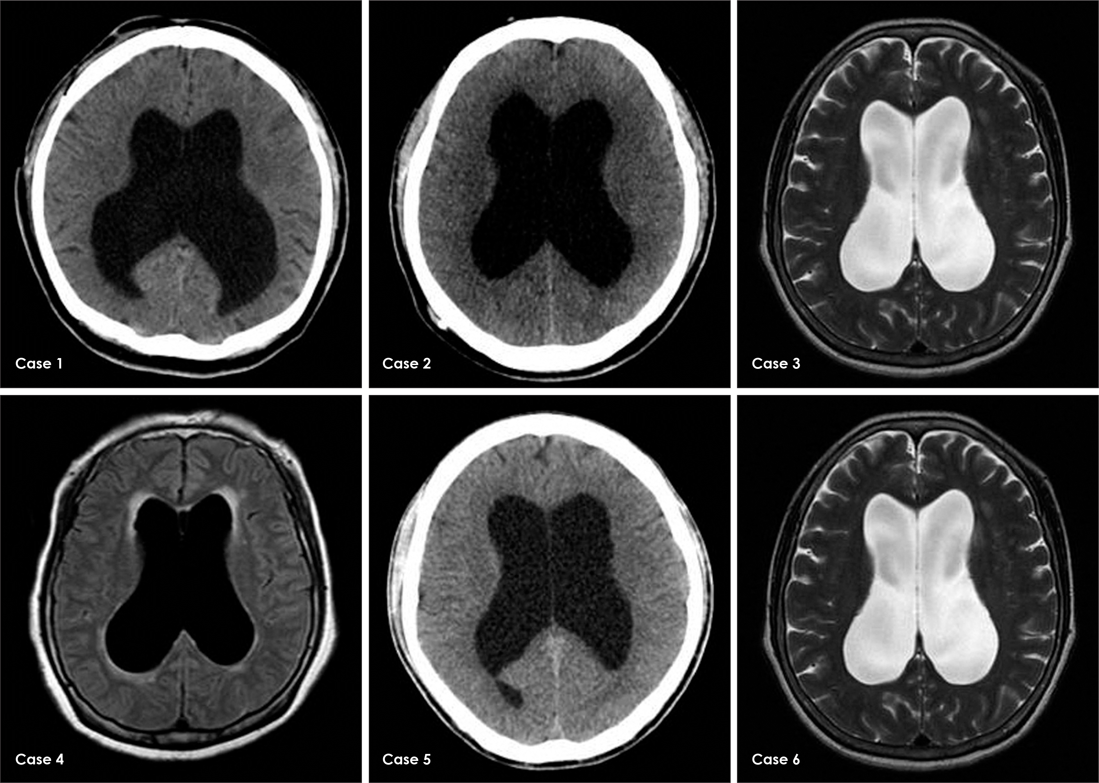
FIGURE 2.
T2-weighted sagittal MRI showing persistent T10-L2 segmental arachnoid cyst (A), MRI taken before ETV show AS, sellar enlargement, upwardly stretched corpus callosum and periventricular edema (B), ETV was performed and the fenestration was enlarged using a 3-French Fogarty balloon catheter. Unexpectedly, third ventricular floor was very narrow and the membrane was thick (C, arrow). AS: aqueductal stenosis, ETV: endoscopic third ventriculostomy, MRI: magnetic resonance imaging.
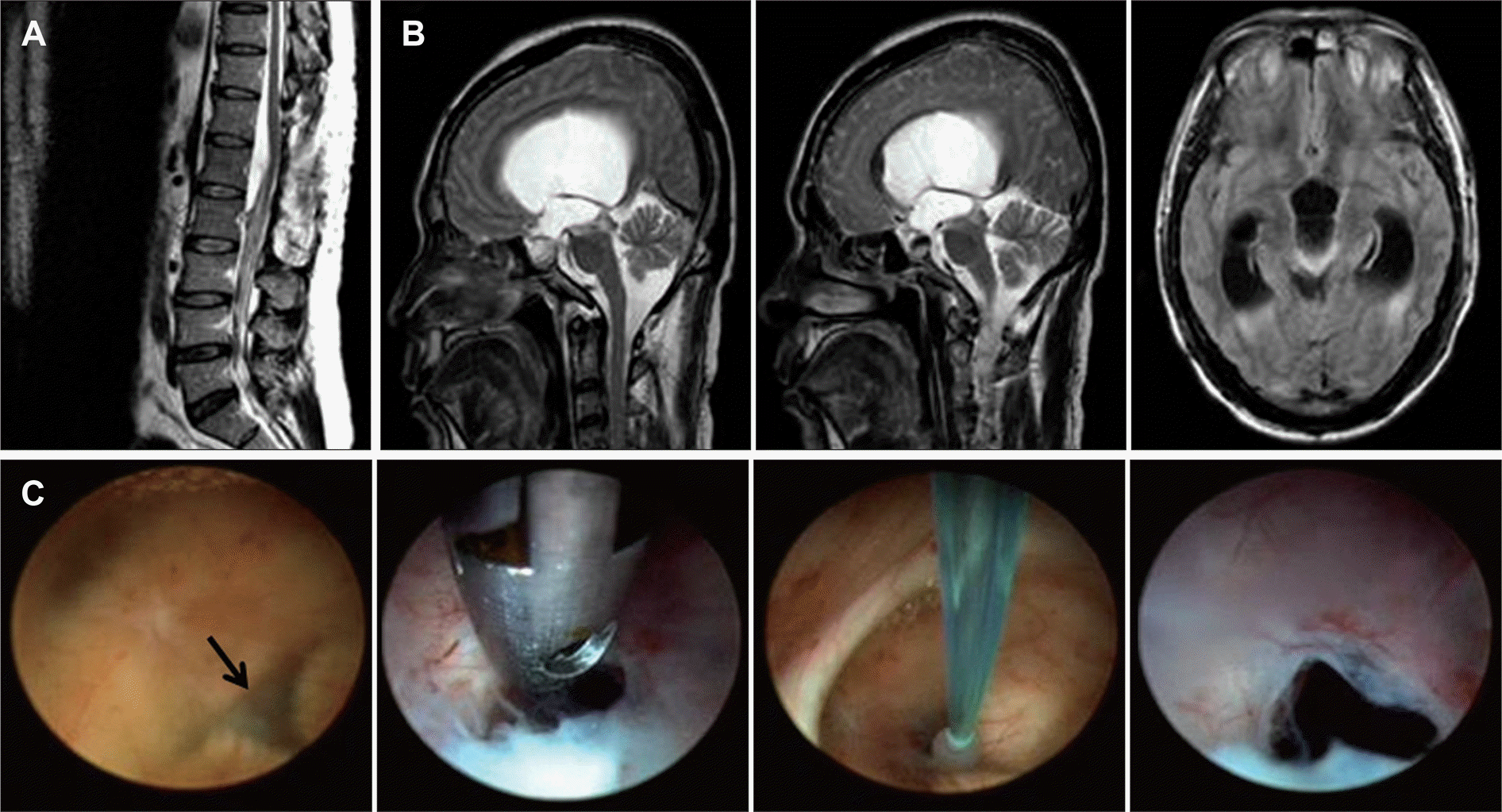
FIGURE 3.
Preoperative MRI demonstrating triventriculomegaly, empty sella and abrupt narrowing distal to the aqueduct (A, arrow). Thoracic spine MRI revealed about 18 cm length epidural lesion (B). MRI: magnetic resonance imaging.
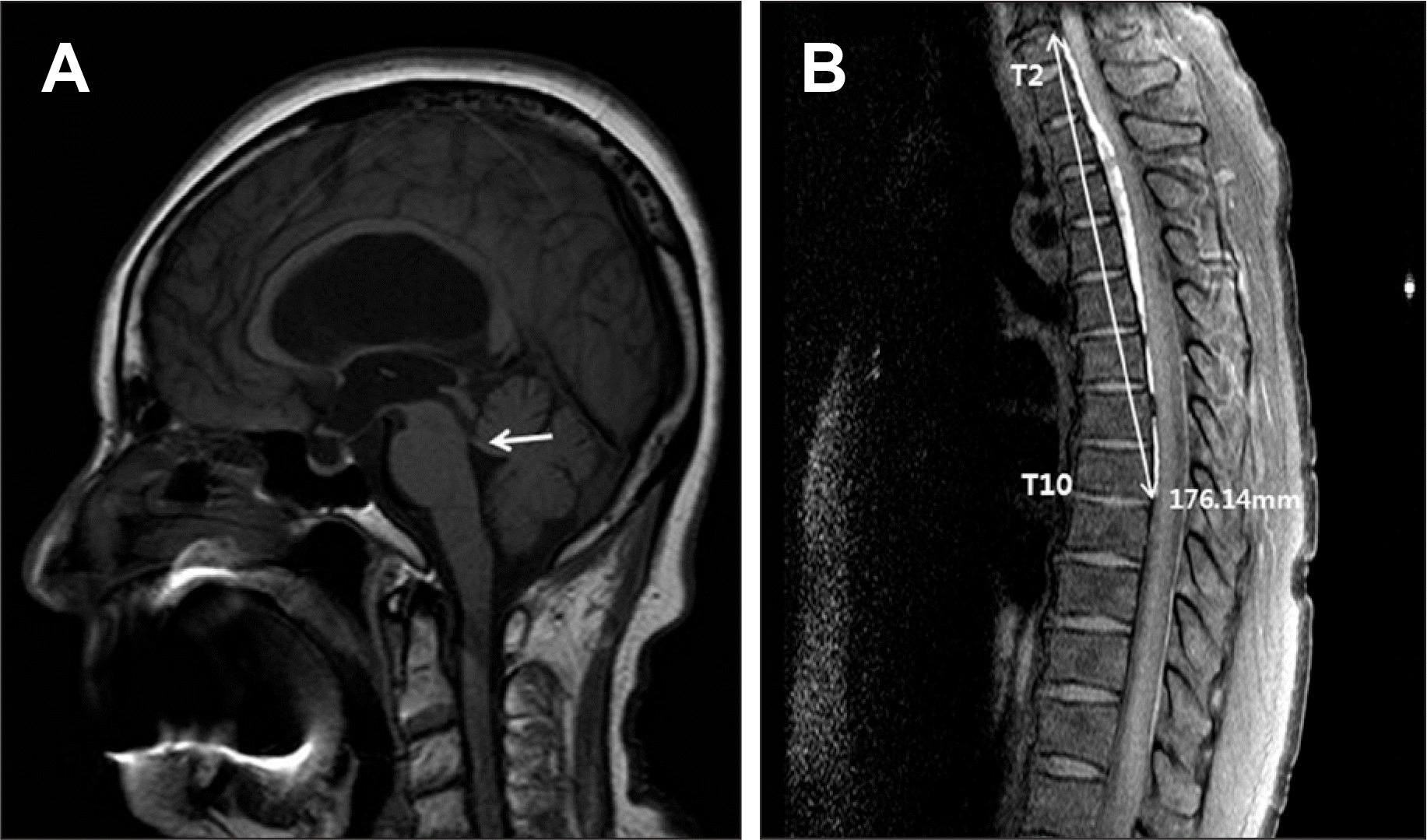
FIGURE 4.
MRI showing ventriculomegaly and abrupt narrowing at the foramen of Magendie level (arrow) suggesting obstructive hydrocephalus (A). Radioisotope cisternography shows communicating type hydrocephalus (B).
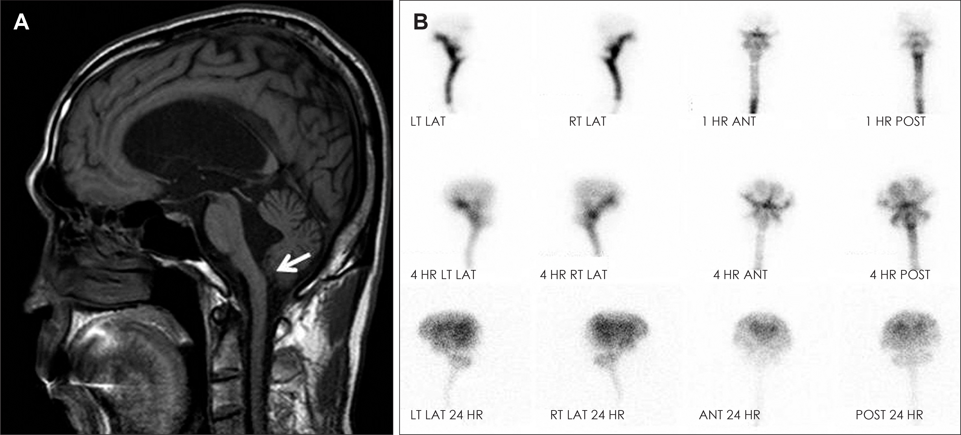
TABLE 1.
Summary of cases
AS: aqueductal stenosis, EDH: epidural hematoma, ETV: endoscopic third ventriculostomy, F: female, GAV: gravity assisted valve, ICP: intracranial pressure, IQ: intelligence quotient, M: male, mRS: modified Rankin score, N: no, NA: not available, No: number, op: operation, PHx: past history, pr-valve: pressure regulated valve, ProGAV: programmable GAV, Sx: symptom, Y: yes, YO: years old, ±: indefinite, SAH : subarachnoid hemorrhage
 XML Download